Autism spectrum disorder (ASD) is a phenotypically heterogeneous neurodevelopmental disorder that affects an estimated 1 in 44 children in the United States (
1). ASD prevalence rates show a marked sex bias, with males diagnosed 3 to 4 times more frequently than females (
1,
2). Although sex differences in ASD prevalence rates may be influenced by underrecognition of ASD symptoms in females, studies using a range of case ascertainment methods have consistently reported a higher male-to-female ratio for the disorder (
2,
3), suggesting a potential biological basis for sex differences in ASD. Indeed, biological factors affecting sexual differentiation (i.e., sexually dimorphic patterns of gene expression, sex hormones) are known to influence brain development and may be associated with brain-based sex differences in autism. However, there have been few well-powered studies investigating sex-based differences in brain structure in ASD, and the neurobiological mechanisms underlying the male bias in ASD remain unclear. A better understanding of the relationship between neurotypical sex differences and sex-differential neuroendophenotypes in ASD is crucial for informing our understanding of sex-related susceptibility to the disorder.
In this issue, Floris et al. (
4) examine sex-differential neuroanatomy in males and females with and without ASD to determine whether ASD is characterized by a shift toward male-typical brain structure. To do so, the authors used a machine learning approach, leveraging convolutional neural networks to predict male sex based on structural brain images from males and females in the UK Biobank, Autism Brain Imaging Data Exchange (ABIDE), and EU-AIMS/AIMS-2-TRIALS Longitudinal European Autism Project (LEAP) cohorts. In the validation and testing data sets, the authors found that sex prediction accuracy was higher for autistic males relative to both neurotypical males and autistic females. Importantly, these findings were specific to ASD; testing the sex prediction classifier in a large sample of individuals with and without attention deficit hyperactivity disorder failed to show any differences in sex prediction accuracies. Furthermore, sex prediction accuracy differed as a function of age, such that the classifier showed lower accuracy in children relative to adults. The authors conclude that features associated with male-characteristic brain structure are more predictive of autistic males, while features associated with female-characteristic brain structure are less predictive of autistic females.
Sex differences in ASD have previously been explained in terms of a female protective effect, such that a greater genetic and environmental etiological load may be required for females to develop ASD relative to males (
5,
6). Under this model, liability for ASD follows a normal distribution at the population level, with males requiring a relatively lower cumulative liability to develop ASD compared with females, who, because of sex-related protective factors, require higher liability to develop the ASD phenotype. This theory is supported by genetics research, which indicates that autistic females carry a higher burden of rare and de novo copy number variants compared with autistic males (
7,
8), and that genes more highly expressed in the male brain are upregulated in ASD postmortem brain samples (
9). Here, Floris et al. examine how patterns of sex-differential neuroanatomy relate to brain gene expression by comparing patterns of male-shifted neuroanatomy in structural brain images to spatial patterns of gene expression from the Allen Human Brain Atlas (
10). Most notably, brain regions that differentiated ASD from neurotypical individuals were enriched for genes expressed prenatally in glia and excitatory neurons, as well as genes upregulated in ASD postmortem cortex. These findings are in agreement with transcriptomic studies of ASD pathophysiology, which have reported upregulation of glial gene expression and downregulation of neuronal and synaptic genes in ASD (
9,
11).
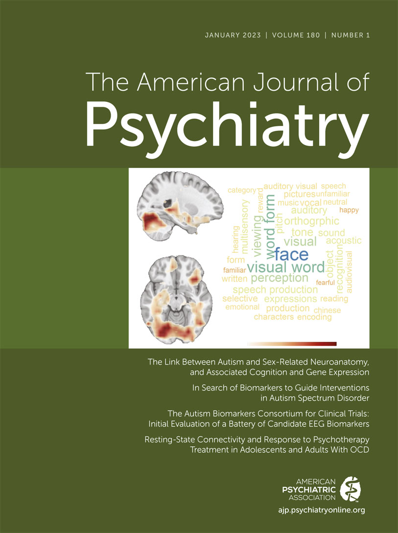
A unique aspect of the Floris et al. investigation is the use of region-aligned prediction (RAP) (
12) to extract a feature matrix for each spatial location for each individual, which allows for the prediction of sex at the brain region level in each participant. Comparing group-level RAP maps, the authors found that auditory and visual processing regions associated with speech and face perception showed a shift toward male-typical brain structure, as these regions robustly differentiated neurotypical males from females. These findings were mirrored when comparing maps between autistic and neurotypical females, suggesting that autistic females show a spatial pattern of male differentiation in sensory pathways similar to that of neurotypical males. In contrast, comparing autistic and neurotypical males revealed higher male prediction probabilities in brain regions involved in motor and reward-related behavior. These findings comport with two major theories on the neurobiological etiology of ASD: 1) the social motivation theory, which suggests that reduced reward representation of social stimuli reduces the likelihood of allocating attention to them across development (
13), and 2) sensory-based theories suggesting that the core social symptoms observed in ASD are a downstream consequence of very early disruptions to sensory and motor processing (
14). The findings of the Floris et al. study raise the question of whether region-specific sexual differentiation may affect expression of the ASD phenotype at the individual level. For example, might the extent to which individuals show a shift toward maleness in reward-related brain areas be associated with difficulties with reward processing? And similarly, does predicted maleness in sensory brain regions predict the presence of sensory symptoms? In line with this hypothesis, Floris et al. found that in autistic females, higher predicted maleness in brain regions associated with face processing and communication was related to lower accuracy in an emotional faces task. While current and previous work has attempted to parse neurobiological and phenotypic heterogeneity in ASD on the basis of, for example, age, sex, and genetic liability, the extent to which region-specific sex-related neuroanatomy may impact the development of specific ASD symptoms and co-occurring conditions remains unresolved.
By comparing RAP maps to spatial patterns of gene expression, Floris et al. produced a list of genes whose expression patterns in the adult brain mirror brain regions predictive of male sex. To better understand the functional significance of the resulting gene lists, the authors also tested enrichment against curated lists of autism-associated genes, genes expressed during prenatal neocortical development, and genes that show sex-differential expression patterns. Here, brain regions that differentiated autistic from neurotypical males were enriched for prenatally expressed glial genes, in agreement with postmortem studies that show higher glial gene expression in males and in ASD (
9). In support of the hypothesized female protective effect, brain regions that differentiated neurotypical males and females were enriched for genes that are downregulated in autism. Remarkably, the authors also found that male-shifted brain regions in autistic females (relative to neurotypical females) showed a transcriptional pattern similar to that of brain regions that showed sex differences in neurotypical males versus females, including enrichments for genes expressed in prenatal excitatory neurons and sex-differentially expressed genes acting prenatally. Importantly, the brain regions that differentiated autistic and neurotypical females also showed enrichment for genes that are upregulated in ASD, as well as for genes that are expressed in prenatal excitatory neurons. These findings of an association with genes expressed in glia and excitatory neurons in male-shifted brain regions are intriguing given that imbalances in excitation and inhibition (E:I) of neuronal circuits have been implicated in the pathophysiology of ASD (
15). Furthermore, there is evidence that cortical hyperactivity may be related at the phenotypic level to sensory-related atypicalities in ASD (
16). How might sexual differentiation of the human brain be related to E:I balance in a brain-region-specific manner? Previous work suggests that biological factors affecting sexual differentiation, including autism-associated X-linked genes, may affect E:I balance in the cortex (
15), potentially producing sex differences in the biological pathways underlying ASD in males and females. It will be important for future studies to examine the relationship between autism-associated sex differences in neuroanatomy and sensory phenotypes in males and females, as well as in vivo neurotransmitter concentrations relevant to E:I in individuals with and without ASD.
The findings by Floris et al. suggest some possible avenues for future research. First, sex prediction accuracy showed marked differences by age, with the largest differences in sex classification accuracy observed between younger males and females. These findings suggest that the neurobiological mechanisms underlying sex-differential neuroanatomy may change over the course of development. Integrative analyses examining the relationship between region-specific gene expression profiles in the developing brain and sex-related neuroanatomy may help to elucidate age-related effects of genes important for autism-associated sexual differentiation. As stated by Floris et al., such analyses will be dependent on the development of high-resolution gene expression maps in developmental populations, analogous to the gene expression data provided by the Allen Human Brain Atlas. Second, the age and sex differences in classifier accuracy reported by Floris et al. raise the intriguing question of what effect puberty may have on sex-differential neuroanatomy. There has been little work examining the impact of sex hormones on adolescent ASD symptoms or brain structure in males and females with ASD. Similarly, little is known about the potential impact of adult hormonal transitions (e.g., menopause) on sex-differential neuroanatomy (
17). Third, the regional gene expression profiles provided by the Allen Human Brain Atlas may not accurately reflect gene expression gradients in the ASD brain. Indeed, recent work indicates that there are widespread transcriptomic changes across the cortex in ASD, with the most robust dysregulation observed in sensory cortices (
18). It will therefore be crucial for future work to link sex-differential neuroimaging data in autistic and neurotypical samples to gene expression patterns observed in ASD postmortem cortex. Finally, as with the vast majority of MRI studies, it remains to be seen whether these findings in individuals with IQ in the normal range replicate in samples of individuals with more severe ASD symptoms.
In summary, the study by Floris et al. presents evidence that autism is characterized by a shift toward male-characteristic patterns of brain structure. As ASD prevalence rates show a striking sex bias, this work sheds light on potential neuroanatomical correlates of autism in males and females and informs our understanding of the biological mechanisms underlying sex differences in ASD.
Acknowledgments
The author thanks Dr. Daniel Geschwind and Dr. Michael Gandal for their helpful suggestions on this editorial.

