Full access
Regular Articles
Published Online: 1 October 2012

What Are the Opportunities for EEG-Based Monitoring of Delirium in the ICU?

Publication: The Journal of Neuropsychiatry and Clinical Neurosciences

Abstract

Recognition of delirium in intensive care unit (ICU) patients is poor, despite the use of screening tools. Electroencephalography (EEG) with a limited number of electrodes and automatic processing may be a more sensitive approach for delirium monitoring. The authors conducted a systematic literature search on EEG characteristics that define delirium, finding 14 studies, which were predominantly conducted in elderly patients. The relative power of the theta and alpha frequency band most often (7/14 studies) distinguished delirium from non-delirium subjects. Given the feasibility for continuous EEG monitoring in ICU, EEG delirium monitoring in ICU patients is promising.
Delirium is an acute or subacute disturbance of consciousness, cognition, and attention that usually fluctuates over time.1 It is a common disorder in the intensive care unit (ICU), with a reported incidence up to 80% during the ICU stay.2,3 Delirium is associated with higher mortality, longer hospital stay, more long-term cognitive impairment, and increased costs.48 Despite its frequency and impact, recognition of delirium by ICU physicians is poor (sensitivity: 29%).9 Therefore, the Society of Critical Care Medicine (SCCM) and the American Psychiatric Association (APA) recommend daily monitoring of delirium in ICU patients to improve early diagnosis and treatment.10,11 Various delirium assessment tools have been developed for use by nonpsychiatric personnel. Of these tools, the Confusion Assessment Method for the ICU (CAM-ICU) showed highest sensitivity in a research setting, ranging from 64% to 97%.9,12 However, in routine, daily practice the sensitivity of the CAM-ICU appeared to be much lower (47%).13 Sensitivity for hypoactive delirium was particularly poor (31%), which is the subtype most difficult to recognize by ICU physicians.9,13 In this multicenter study, 282 patients were assessed by teams of three delirium experts, including psychiatrists, geriatricians, and neurologists, who based their assessment on cognitive examination, inspection of medical files, and established criteria1 for delirium.13 The low sensitivity of the CAM-ICU in daily practice hampers early detection of delirium. Other drawbacks of the CAM-ICU are that it cannot quantify delirium severity and that it assesses delirium at a certain moment in time,12 whereas delirium may fluctuate considerably over the day.1 These factors impede recognition and thereby delay treatment. Delayed treatment of delirium was found to be related to mortality.14 Therefore, an objective detection tool for continuous monitoring of delirium is needed.
Another approach to detecting delirium is by monitoring physiological alterations. Delirium is a manifestation of encephalopathy; however, sometimes these terms are used interchangably. For decades, it has been known that during delirium, electroencephalography (EEG) shows generalized slowing of background activity.15 Recently, continuous EEG monitoring with automatic processing has become technically feasible.16 An example is the assessment of depth of anesthesia, which can be monitored with a limited number of EEG electrodes.17 In ICU patients, continuous EEG monitoring is increasingly used to detect nonconvulsive epileptic seizures.18 EEG-based encephalopathy monitoring with a limited number of electrodes and automatic processing, is therefore feasible. It is, however, currently unclear which EEG characteristic is particularly altered in delirium, and which EEG lead is most informative. In this systematic review, we aimed to explore which parameter and which EEG lead show the largest difference between patients with and without delirium.

Methods

Search Strategy

We systematically searched literature on delirium and EEG on November 25th, 2011, using the databases of EMBASE (January 1980 to November 2011) and PubMed (January 1966 to November 2011). Synonyms for delirium and corresponding Mesh terms were combined with search terms for EEG synonyms, and Mesh terms as can be seen in the online data supplement. Resulting English, French, German, and Dutch scientific articles on humans were imported into Reference Manager (Reference Manager Professional Edition, Version 11; Thomson ISI ResearchSoft). The reference lists of all full-text articles were also screened, and relevant publications were imported into the database.

Selection of Studies

The results of the literature search were evaluated independently by two authors (AWvdK, RJvdW). Based on title and abstract, we excluded case reports, animal studies, reviews, letters, and studies in children. Furthermore, articles that described the EEG of delirium subjects only in a descriptive way were excluded. Moreover, when the statistical significance of the difference in quantitative EEG characteristics between delirium and non-delirium subjects was not tested; articles were also excluded unless the statistical significance could be determined on the basis of the published data. All eligible studies were retrieved in full text. The reference lists of all full-text articles were also screened, following the same criteria used for screening titles and abstracts. In case of disagreement between the two authors, references were evaluated by three authors (AWvdK, RJvdW, AJCS) to reach consensus on inclusion for this systematic review.

Data Extraction

The selected full-text articles were closely reviewed, and the first author, title, and year of publication were noted. Furthermore, the following features were tabulated: size and characteristics of the study population, methods of diagnosing delirium, time between delirium diagnosis and EEG recording, EEG derivations for quantitative EEG analysis, use of artifact-free or raw data for quantitative EEG analysis, length of EEG period used for quantitative analysis, studied EEG parameters, and differences between patients with and without delirium with regard to these EEG parameters that reached statistical significance (p <0.05). Background information concerning quantitative EEG parameters can be found in the online data supplement.
For those articles in which the statistical significance had to be determined on the basis of the published data, a Student’s t-test was used to compare delirium subjects to non-delirium subjects. When data were not normally distributed, a Mann-Whitney U test was used.

Results

Literature Search

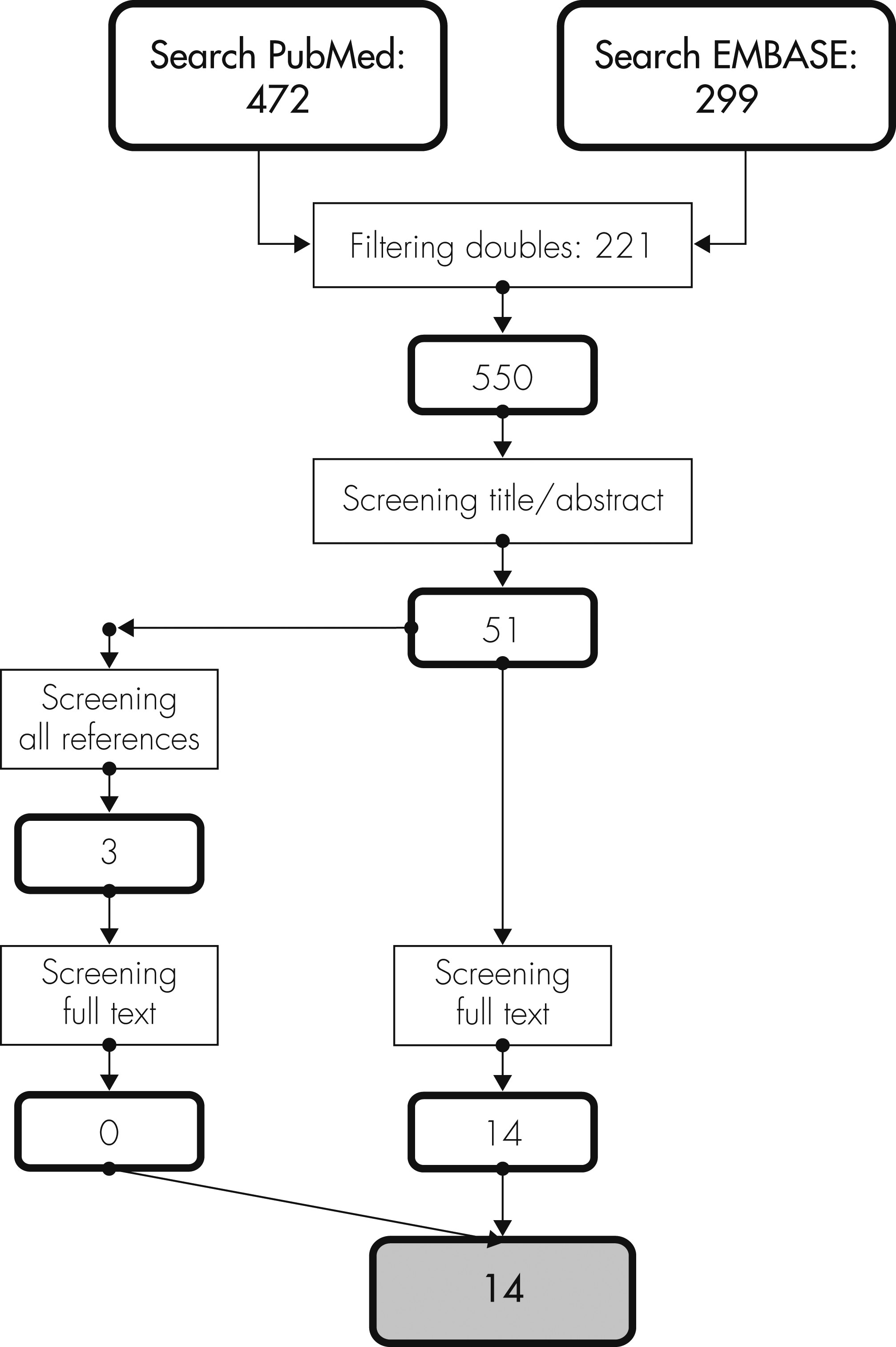
The literature search resulted in 550 eligible publications (Figure 1). The search term “encephalopathy” combined with “EEG” was also considered, and resulted in a large number of articles (2,420 in PubMed and 1,627 in EMBASE). When we screened in PubMed as in EMBASE 100 articles on EEG and encephalopathy, we did not find any additional publications. Therefore, encephalopathy was not further used as a search term. After excluding 499 articles based on title and abstract, 51 papers were selected and retrieved in full text. By screening the references of these articles, 3 extra full-text articles were selected and retrieved. As we excluded another 40 publications, 14 articles fulfilled inclusion—but not exclusion—criteria. Two of these did not report the statistical significance of the differences between delirium and non-delirium subjects. This was computed using the published data.15,19
FIGURE 1. Summary of Literature Search on Delirium and Quantitative EEG

Study Characteristics

The study populations of the included publications are described in the online data supplement. The number of included delirium patients per publication varied between 10 and 51 subjects, whereas one article did not mention which fraction of the population had delirium.20 Six of the 14 studies were conducted in elderly patients (mean age over 75 years) and 3 in ICU patients. Four studies compared dementia patients with delirium and dementia patients without delirium to patients without dementia and delirium.15,2123 Delirium was diagnosed in the majority of studies (9 of 14) by a psychiatrist, using criteria of various editions of the Diagnostic and Statistical Manual of Mental Disorders.2129 In three publications, the CAM-ICU was used to diagnose delirium.20,30,31 One study used patient records to diagnose delirium,15 and another one used the diagnosis of the ward physician.19
The timeframe between the delirium diagnosis and EEG recording varied considerably among the different investigations. In three of these, the delirium diagnosis and EEG recording were performed at almost the same time.20,27,31 Three other investigations mentioned a timeframe of 4–5 hours,22,23,30 and another three, of maximal 24 hours.21,26,29 In the other five investigations, the timeframe was not reported.15,19,24,25,28 The duration of EEG recording for quantitative analyses varied between 30 seconds and 600 seconds (not specified in one publication).28 The EEG derivations used for recording were, in two publications, not specified.28,30 In five studies, only posterior (parietal–occipital) derivations were used.2125

EEG Characteristics

As shown in the online data supplement, the most frequently studied variable was the relative power of the alpha frequency band (8 of 14 studies); next, the relative power of the theta frequency band (7 of 14), the relative power of the delta frequency band (5 of 14), and the peak frequency (5 of 14).
The relative power of the theta frequency band was significantly different between delirium and non-delirium patients in all seven studies that investigated this variable. However, in two of these seven publications, only the lower part of the theta frequency band, and not the higher part of the frequency band, was significantly increased in delirium.22,23 The relative power of the alpha frequency band was related to delirium in 7 out of 8 studies. One of these studies showed an increase in the relative power of the theta and a decrease in the relative power of the alpha frequency band in several groups of delirium patients, but not in delirium/Parkinson’s disease patients.21 Two studies showed that the relative power of the theta, the alpha, and also the delta frequency band, were not only different between delirium and non-delirium patients, but also within patients before and during an episode of delirium.24,25
Other variables that often differed between delirium and non-delirium patients included the relative power of the delta frequency band (increased in delirium in 5 of 5 investigations that studied this) and the peak frequency (decreased in delirium in 4 of 5 investigations that studied this; online supplement). Two studies focused on bispectral index (BIS) EEG monitoring during delirium. In both studies, the BIS was significantly decreased in delirium patients as compared with non-delirium controls.20,31 Unfortunately, one study did not specify the number of delirium patients included.20

Discussion

To develop EEG-based delirium monitoring with a limited number of electrodes and automatic processing, it is essential to know which parameter and which EEG lead show the largest difference between delirium and non-delirium patients. We evaluated various EEG parameters. Of these, the relative power of the theta frequency band was most frequently studied, and, without exception, different between delirium and non-delirium subjects. Investigations on quantitative EEG and delirium were mostly conducted in elderly patients and often used posterior EEG derivations only.
In order to perform continuous monitoring of delirium, a minimal number of electrodes are needed to increase feasibility and minimize the burden for the patient. Several publications mention that EEG changes in delirium are most prominent in the posterior regions, although this was not supported by original studies22,23,25 Furthermore, none of the 14 included studies determined at which EEG derivation the EEG changes in delirium were strongest.
All 14 included studies used rather short periods of artifact-free EEG data to determine EEG characteristics. Fluctuation of EEG characteristics during these periods was not considered, although fluctuation of EEG characteristics over several days was studied by a number of authors.19,2427 Therefore, the exact relationship between the clinical fluctuation of delirium and the EEG characteristics of delirium is still uncertain.
Because of these fluctuations,1 it is important to diagnose delirium soon before or after the EEG recording. Therefore, a long timeframe between delirium diagnosis and recording of EEG is a serious shortcoming. In only three studies were the delirium diagnosis and EEG recording performed at almost the same time.20,27,31
To allow continuous EEG monitoring of delirium, it is necessary to distinguish delirium from other conditions. Slowing of background activity occurs not only in delirium, but also in dementia and sleep.22,32 One article on dementia in intensive care unit (ICU) patients reported the prevalence of dementia to be 17% in patients older than 65 years.33 However, studies on the prevalence of dementia in the ICU are limited. In a recent multicenter study including 2,116 ICU patients, not more than 1.7% of the patients were reported to suffer from dementia.34 Patients with dementia are usually treated substantially less aggressively than patients without dementia and are therefore less likely to be admitted to the ICU.35 Therefore, we suspect that, in a general ICU population, the prevalence is much lower than 17%. However, even if a substantial proportion of ICU patients were suffering from dementia, delirium can be distinguished from dementia by recording of an active (eyes constantly open) EEG. The relative power in the delta and upper half of the alpha frequency band in an active EEG differ significantly between delirium and dementia patients.22
Nevertheless, deep sleep is also characterized by slow-wave activity.32 The 14 studies included in this review all measured EEG during waking or light sedation and did not compare EEG characteristics of delirium with EEG characteristics of physiological sleep. EEG in sleep, however, has certain characteristics such as k-complexes and sleep-spindles,32 which could be used to distinguish sleep from delirium, but this has not been investigated yet.
Despite an extensive literature search, only 14 articles could be found on quantitative EEG in delirium patients. Delirium is a frequent and serious problem in the ICU, but almost half of the quantitative EEG studies in delirium focus on elderly non-ICU subjects. Furthermore, we could not consider medication use in the different studies, although medication as haloperidol, which is often used during delirium,10 may influence the EEG. Several studies reported EEG changes in healthy volunteers during haloperidol use when compared with placebo. These changes were, however, not uniform.3638 Therefore, the exact effect of haloperidol at a given dose on the EEG is unknown.
A future study should determine whether the relative power of the theta frequency band is indeed the most affected EEG characteristic. Furthermore, it is important to determine which EEG deviation is most sensitive for EEG changes related to delirium. If the most informative electrode deviations are known, one can limit the number of electrodes needed for continuous monitoring. Next, we should focus on continuous monitoring, with a limited number of electrodes, and determine the sensitivity of EEG-based delirium monitoring in a random sample of ICU patients over time. Given the feasibility of continuous EEG monitoring for epilepsy in ICU patients, this seems to be a promising approach to overcoming the underdiagnosis of current delirium screening tools.
Although some aspects of EEG in relation to delirium still have to be addressed, continuous EEG monitoring offers opportunities. Delirium can be adequately diagnosed in various populations by use of EEG. The relative power of the theta frequency band is the most promising parameter to study in a future prospective investigation in ICU patients.
Supplementary Material

Supplementary Material (472_ds001.pdf)

References

1.
American Psychiatric Association: Diagnostic and Statistical Manual of Mental Disorders, 4th Edition, Text Revision. Washington, DC, American Psychiatric Association, 2000
2.
Ely EW, Shintani AK, Truman B, et al.: Delirium as a predictor of mortality in mechanically ventilated patients in the intensive care unit. JAMA 2004; 291:1753–1762
3.
Girard TD, Kress JP, Fuchs BD, et al.: Efficacy and safety of a paired sedation and ventilator weaning protocol for mechanically ventilated patients in intensive care (Awakening and Breathing Controlled Trial): a randomised, controlled trial. Lancet 2008; 371:126–134
4.
Jackson JC, Hart RP, Gordon SM, et al.: Six-month neuropsychological outcome of medical intensive care unit patients. Crit Care Med 2003; 31:1226–1234
5.
Pisani MA, Kong SYJ, Kasl SV, et al.: Days of delirium are associated with 1-year mortality in an older intensive care unit population. Am J Respir Crit Care Med 2009; 180:1092–1097
6.
Thomason JW, Shintani A, Peterson JF, et al.: Intensive care unit delirium is an independent predictor of longer hospital stay: a prospective analysis of 261 non-ventilated patients. Crit Care 2005; 9:R375–R381
7.
Milbrandt EB, Deppen S, Harrison PL, et al.: Costs associated with delirium in mechanically ventilated patients. Crit Care Med 2004; 32:955–962
8.
van den Boogaard M, Schoonhoven L, Evers AW, et al.: Delirium in critically ill patients: impact on long-term health-related quality of life and cognitive functioning. Crit Care Med 2012; 40:112–118
9.
van Eijk MMJ, van Marum RJ, Klijn IAM, et al.: Comparison of delirium assessment tools in a mixed intensive care unit. Crit Care Med 2009; 37:1881–1885
10.
American Psychiatric Association: Practice guideline for the treatment of patients with delirium. Am J Psychiatry 1999; 156(Suppl):1–20
11.
Jacobi J, Fraser GL, Coursin DB, et al.Task Force of the American College of Critical Care Medicine (ACCM) of the Society of Critical Care Medicine (SCCM), American Society of Health-System Pharmacists (ASHP), American College of Chest Physicians: Clinical practice guidelines for the sustained use of sedatives and analgesics in the critically ill adult. Crit Care Med 2002; 30:119–141
12.
Ely EW, Margolin R, Francis J, et al.: Evaluation of delirium in critically ill patients: validation of the Confusion Assessment Method for the Intensive Care Unit (CAM-ICU). Crit Care Med 2001; 29:1370–1379
13.
van Eijk MM, van den Boogaard M, van Marum RJ, et al.: Routine use of the Confusion Assessment Method for the intensive care unit: a multicenter study. Am J Respir Crit Care Med 2011; 184:340–344
14.
Heymann A, Radtke F, Schiemann A, et al.: Delayed treatment of delirium increases mortality rate in intensive care unit patients. J Int Med Res 2010; 38:1584–1595
15.
Jacobson SA, Leuchter AF, Walter DO: Conventional and quantitative EEG in the diagnosis of delirium among the elderly. J Neurol Neurosurg Psychiatry 1993; 56:153–158
16.
Hirsch LJ: Continuous EEG monitoring in the intensive care unit: an overview. J Clin Neurophysiol 2004; 21:332–340
17.
Palanca BJ, Mashour GA, Avidan MS: Processed electroencephalogram in depth of anesthesia monitoring. Curr Opin Anaesthesiol 2009; 22:553–559
18.
Friedman D, Hirsch LJ: Diagnosing and monitoring seizures in the ICU: the role of continuous EEG for detection and management of seizures in critically ill patients, in Seizures in Critical Care. Edited by, Varelas P. New York, Humana Press, 2010, pp 21–47
19.
Jacobson SA, Leuchter AF, Walter DO, et al.: Serial quantitative EEG among elderly subjects with delirium. Biol Psychiatry 1993; 34:135–140
20.
Ely EW, Truman B, Manzi DJ, et al.: Consciousness monitoring in ventilated patients: bispectral EEG monitors arousal not delirium. Intensive Care Med 2004; 30:1537–1543
21.
Koponen H, Partanen J, Pääkkönen A, et al.: EEG spectral analysis in delirium. J Neurol Neurosurg Psychiatry 1989; 52:980–985
22.
Thomas C, Hestermann U, Walther S, et al.: Prolonged activation EEG differentiates dementia with and without delirium in frail elderly patients. J Neurol Neurosurg Psychiatry 2008; 79:119–125
23.
Thomas C, Hestermann U, Kopitz J, et al.: Serum anticholinergic activity and cerebral cholinergic dysfunction: an EEG study in frail elderly with and without delirium. BMC Neurosci 2008; 9:86–95
24.
Katz IR, Mossey J, Sussman N, et al.: Bedside clinical and electrophysiological assessment: assessment of change in vulnerable patients. Int Psychogeriatr 1991; 3:289–300
25.
Katz IR, Curyto KJ, TenHave T, et al.: Validating the diagnosis of delirium and evaluating its association with deterioration over a one-year period. Am J Geriatr Psychiatry 2001; 9:148–159
26.
Matsushima E, Nakajima K, Moriya H, et al.: A psychophysiological study of the development of delirium in coronary care units. Biol Psychiatry 1997; 41:1211–1217
27.
Reischies FM, Neuhaus AH, Hansen ML, et al.: Electrophysiological and neuropsychological analysis of a delirious state: the role of the anterior cingulate gyrus. Psychiatry Res 2005; 138:171–181
28.
Trzepacz PT, Brenner RP, Coffman G, et al.: Delirium in liver transplantation candidates: discriminant analysis of multiple test variables. Biol Psychiatry 1988; 24:3–14
29.
Trzepacz PT, Sclabassi RJ, Van Thiel DH: Delirium: a subcortical phenomenon? J Neuropsychiatry Clin Neurosci 1989; 1:283–290
30.
Plaschke K, Hill H, Engelhardt R, et al.: EEG changes and serum anticholinergic activity measured in patients with delirium in the intensive care unit. Anaesthesia 2007; 62:1217–1223
31.
Plaschke K, Fichtenkamm P, Schramm C, et al.: Early postoperative delirium after open-heart cardiac surgery is associated with decreased bispectral EEG and increased cortisol and interleukin-6. Intensive Care Med 2010; 36:2081–2089
32.
Rechtschaffen A, Kales A: A Manual of Standardized Terminology, Techniques, and Scoring System for Sleep Stages of Human Subjects. Washington, DC, U.S. Government Printing Office, 1968
33.
Pisani MA, Redlich CA, McNicoll L, et al.: Short-term outcomes in older intensive care unit patients with dementia. Crit Care Med 2005; 33:1371–1376
34.
van den Boogaard M, Pickkers P, Slooter AJC, et al.: Development and validation of PRE-DELIRIC (PREdiction of DELIRium in ICu patients) delirium prediction model for intensive care patients: observational multicentre study. BMJ 2012; 344:e420
35.
Richardson SS, Sullivan G, Hill A, et al.: Use of aggressive medical treatments near the end of life: differences between patients with and without dementia. Health Serv Res 2007; 42:183–200
36.
Liem-Moolenaar M, Gray FA, de Visser SJ, et al.: Psychomotor and cognitive effects of a single oral dose of talnetant (SB223412) in healthy volunteers compared with placebo or haloperidol. J Psychopharmacol 2010; 24:73–82
37.
Yoshimura M, Koenig T, Irisawa S, et al.: A pharmaco-EEG study on antipsychotic drugs in healthy volunteers. Psychopharmacology (Berl) 2007; 191:995–1004
38.
McClelland GR, Cooper SM, Pilgrim AJ: A comparison of the central nervous system effects of haloperidol, chlorpromazine, and sulpiride in normal volunteers. Br J Clin Pharmacol 1990; 30:795–803

Information & Authors

Information

Published In

Go to The Journal of Neuropsychiatry and Clinical Neurosciences
The Journal of Neuropsychiatry and Clinical Neurosciences
Pages: 472 - 477
PubMed: 23224454

History

Received: 25 November 2011
Revision received: 8 March 2012
Accepted: 19 March 2012
Published online: 1 October 2012
Published in print: Fall 2012

Authors

Details

Arendina W. van der Kooi, M.Sc.
From the Department of Intensive Care Medicine, University Medical Centre Utrecht, Utrecht, The Netherlands (AWvdK, RJvdW, AJCS) and the Department of Clinical Neurophysiology, University Medical Centre Utrecht, Utrecht, The Netherlands (FSSL).
Frans S.S. Leijten, M.D., Ph.D.
From the Department of Intensive Care Medicine, University Medical Centre Utrecht, Utrecht, The Netherlands (AWvdK, RJvdW, AJCS) and the Department of Clinical Neurophysiology, University Medical Centre Utrecht, Utrecht, The Netherlands (FSSL).
Ruben J. van der Wekken, M.D.
From the Department of Intensive Care Medicine, University Medical Centre Utrecht, Utrecht, The Netherlands (AWvdK, RJvdW, AJCS) and the Department of Clinical Neurophysiology, University Medical Centre Utrecht, Utrecht, The Netherlands (FSSL).
Arjen J.C. Slooter, M.D., Ph.D.
From the Department of Intensive Care Medicine, University Medical Centre Utrecht, Utrecht, The Netherlands (AWvdK, RJvdW, AJCS) and the Department of Clinical Neurophysiology, University Medical Centre Utrecht, Utrecht, The Netherlands (FSSL).

Notes

Correspondence: A.J.C. Slooter, M.D., Ph.D., Dept. of Intensive Care Medicine, University Medical Centre Utrecht, the Netherlands; e-mail: [email protected]

Metrics & Citations

Metrics

Citations

Export Citations

If you have the appropriate software installed, you can download article citation data to the citation manager of your choice. Simply select your manager software from the list below and click Download.

For more information or tips please see 'Downloading to a citation manager' in the Help menu.

Format
Citation style
Style
Copy to clipboard

View Options

View options

PDF/EPUB

View PDF/EPUB

Get Access

Login options

Already a subscriber? Access your subscription through your login credentials or your institution for full access to this article.

Personal login Institutional Login Open Athens login
Purchase Options

Purchase this article to access the full text.

PPV Articles - Journal of Neuropsychiatry and Clinical Neurosciences

PPV Articles - Journal of Neuropsychiatry and Clinical Neurosciences

Not a subscriber?

Subscribe Now / Learn More

PsychiatryOnline subscription options offer access to the DSM-5-TR® library, books, journals, CME, and patient resources. This all-in-one virtual library provides psychiatrists and mental health professionals with key resources for diagnosis, treatment, research, and professional development.

Need more help? PsychiatryOnline Customer Service may be reached by emailing [email protected] or by calling 800-368-5777 (in the U.S.) or 703-907-7322 (outside the U.S.).

Media

Figures

Other

Tables

Share

Share

Share article link

Share