Full access
Departments
Published Online: 29 April 2015

Neurological and Psychiatric Findings of Marchiafava-Bignami Disease in a Nonalcoholic Diabetic Patient With High Blood Glucose Levels

Publication: The Journal of Neuropsychiatry and Clinical Neurosciences
To the Editor: Marchiafava-Bignami disease (MBD) is a rare disorder of unknown etiology. MBD is often seen in association with chronic alcoholism, but can also occur in patients with metabolic disorders and malnutrition.1 The disease is characterized by demyelination and necrosis of corpus callosum. MBD may present with various clinical manifestations, including confusion, psychotic and emotional symptoms, depression, apathy, interhemispheric disconnection syndrome, seizures, and dementia.13 Here the authors report the clinical and radiological findings of MBD in a 63-year-old diabetic woman without a history of alcohol consumption and emphasize the key imaging characteristics and neurological and psychiatric symptoms of MBD.

Case Report

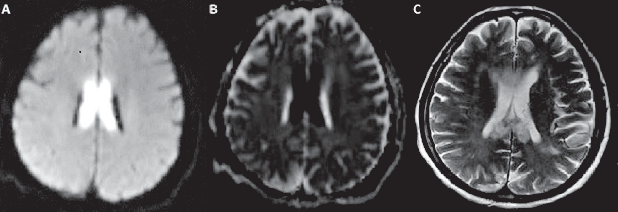
A 63-year-old woman was being followed in the internal medicine clinic for unstable high blood glucose levels and the neurology department received a consultation due to the fluctuation in the level of consciousness and agitation. Her medical history included diabetes mellitus, chronic renal failure, and hypertension, with no history of alcohol consumption or smoking. The initial symptoms began 6 days earlier, with severe nausea and vomiting only a few times. Her neurological examination revealed reduction of word expression, impaired memory, and attention with agitation. Brain MRI scans revealed symmetrical and bilateral hyperintense lesions on diffusion-weighted images and T2-weighted images throughout the entire corpus callosum (Figure 1 [A and C]). These areas were hypointense on apparent diffusion coefficient sequences (Figure 1 [B]). No pathological contrast enhancement was detected. Electroencephalography demonstrated active epileptiform discharges in the right frontocentral region extending to the homologous areas of the left hemisphere. Laboratory findings were as follows: blood glucose, 557.49 mg/dL; sodium, 124.39 mmol/L; potassium, 6.11 mmol/L; BUN, 70.5 mg/dL; and creatinine, 3.72 mg/dL (chronic renal failure: mean creatinine 3.2 mg/dL). Plasma osmolarity was 304.9 mOsm/L. Cerebrospinal fluid findings were normal.
FIGURE 1. Widespread, Bilateral Symmetrical Hyperintense Lesions Were Detected in Diffusion-Weighted and T2-Weighted Images [A and C] and the Lesions Appear Hypointense in Apparent Diffusion Coefficient Sequences [B]
The appearance of the observed lesions was compatible with MBD. There was no pathology in the paraneoplastic panel. On the 10th day of follow-up with blood glucose stabilization, correction of metabolic disturbances, and intravenous nutritional support, the patient’s neurological examination revealed marked improvement of memory and attention, with a significant increase in perception. Cranial MRI scans also showed partial normalization. On the 15th day, the patient had an intact neurological examination and was discharged.

Discussion

MBD is a rare but frequently fatal disease, and is often characterized by corpus callosum demyelination and necrosis.1 The disease frequently develops after chronic alcohol consumption, but nonalcoholic MBD may also occur, even in patients with metabolic disorders with or without malnutrition.1,3 The acute form of MBD is characterized by impairment of consciousness, epileptic seizures, and coma.1 The subacute form may cause mental confusion, psychosis, emotional distress, depression, apathy, and interhemispheric disconnection.1,4 Progressive dementia dominates the clinical course of the chronic form of the disease.1 Death often occurs in association with sudden cardiorespiratory failure after necrosis of some parts of the limbic regions.5
Yadala and Luo6 recently reported a nonalcoholic diabetic case of MBD in a 38-year-old man with a wide range of unstable and fluctuant blood glucose levels. The authors suggested the possibility of energy production disarrangement occurring in the corpus callosum. In our presented case, a history of vomiting a few times caused only mild malnutrition. Therefore, this suggests that long-standing untreated diabetes mellitus and high blood glucose levels as well as related hyperosmolarity may have triggered MBD.
Early recognition and treatment of the disease may provide favorable clinical outcomes.1,5 However, until now, there have been no reports of a specific proven treatment for MBD. Nutritional support and cessation of alcohol intake are important during treatment.1
In conclusion, acute onset MBD is not always fatal, and complete recovery is possible after timely diagnosis and appropriate treatment.1,3,5 In addition, it should be noted that nonalcoholic MBD may occur even in metabolic disorders with or without malnutrition, and advanced neuroimaging techniques such as MRI facilitate early diagnosis of MBD.

References

1.
Celik Y: Marchiafava-Bignami disease in alcoholism, in Comprehensive Handbook of Alcohol Related Pathology. Edited by Preedy VR, Watson RR. San Diego, Elsevier Academic Press, 2004, pp 713–718
2.
Berek K, Wagner M, Chemelli AP, et al: Hemispheric disconnection in Marchiafava-Bignami disease: clinical, neuropsychological and MRI findings. J Neurol Sci 1994; 123:2–5
3.
Suzuki Y, Oishi M, Ogawa K, et al: A patient with Marchiafava-Bignami disease as a complication of diabetes mellitus treated effectively with corticosteroid. J Clin Neurosci 2012; 19:761–762
4.
Lechevalier B, Andersson JC, Morin P: Hemispheric disconnection syndrome with a “crossed avoiding” reaction in a case of Marchiafava-Bignami disease. J Neurol Neurosurg Psychiatry 1977; 40:483–497
5.
Wenz H, Eisele P, Artemis D, et al: Acute Marchiafava-Bignami disease with extensive diffusion restriction and early recovery: case report and review of the literature. J Neuroimaging 2014; 24:421–424.
6.
Yadala S, Luo JJ: Marchiafava-Bignami disease in a nonalcoholic diabetic patient. Case Rep Neurol Med 2013; 2013:979383

Information & Authors

Information

Published In

Go to The Journal of Neuropsychiatry and Clinical Neurosciences
The Journal of Neuropsychiatry and Clinical Neurosciences
Pages: e149 - e150
PubMed: 25923862

History

Published in print: Spring 2015
Published online: 29 April 2015

Authors

Details

Ozden Kilinc, M.D.
Dept. of Neurology, Marmara University Hospital, Istanbul, Turkey
Damla Ozbek, M.D.
Dept. of Neurology, Marmara University Hospital, Istanbul, Turkey
Esra Ozkan, M.D.
Dept. of Neurology, Marmara University Hospital, Istanbul, Turkey
Ipek Midi, M.D.
Dept. of Neurology, Marmara University Hospital, Istanbul, Turkey

Notes

Correspondence: Ozden Kilinc, M.D.; e-mail: [email protected]

Metrics & Citations

Metrics

Citations

Export Citations

If you have the appropriate software installed, you can download article citation data to the citation manager of your choice. Simply select your manager software from the list below and click Download.

For more information or tips please see 'Downloading to a citation manager' in the Help menu.

Format
Citation style
Style
Copy to clipboard

View Options

View options

PDF/EPUB

View PDF/EPUB

Login options

Already a subscriber? Access your subscription through your login credentials or your institution for full access to this article.

Personal login Institutional Login Open Athens login
Purchase Options

Purchase this article to access the full text.

PPV Articles - Journal of Neuropsychiatry and Clinical Neurosciences

PPV Articles - Journal of Neuropsychiatry and Clinical Neurosciences

Not a subscriber?

Subscribe Now / Learn More

PsychiatryOnline subscription options offer access to the DSM-5-TR® library, books, journals, CME, and patient resources. This all-in-one virtual library provides psychiatrists and mental health professionals with key resources for diagnosis, treatment, research, and professional development.

Need more help? PsychiatryOnline Customer Service may be reached by emailing [email protected] or by calling 800-368-5777 (in the U.S.) or 703-907-7322 (outside the U.S.).

Media

Figures

Other

Tables

Share

Share

Share article link

Share