Full access
Windows to the Brain
Published Online: 23 October 2018

Poststroke Depression: Contributions From Network Science

Publication: The Journal of Neuropsychiatry and Clinical Neurosciences
Worldwide, stroke is the third most common cause of disability.10 Although the mortality rate and incidence of stroke has decreased in much of the world, the overall stroke burden has increased.11,12 Most worrisome, the rates of stroke in younger adults (20–64 years of age) may actually be increasing.10 Stroke-related disability burden is considerable due to long-term physical, cognitive, and psychological sequelae. Development of depression is one of the more common sequelae and is associated with multiple detrimental outcomes, making both prevention and prompt identification and treatment high priorities.
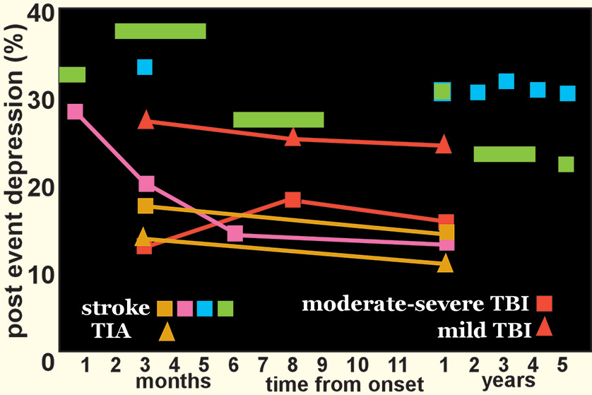
13,14
A recent meta-analysis found that within the initial 5 years after a stroke, 31% of patients developed poststroke depression (PSD) (Figure 1).2 A similar prevalence (34%) of PSD was reported in a recent study at 2–5 years poststroke.15 A longitudinal study found that one-third of cases of PSD developed within the first 3 months after stroke, and half of these remitted by 1 year.1 A similar pattern was reported in a study of first-ever ischemic strokes (mostly mild) (Figure 1).3 It is noteworthy that two studies using data from stroke registries that included patients with transient ischemic attacks (TIAs) reported similar rates of depression following stroke and TIA (Figure 1).4,16 Relatively similar rates were also reported in a study limited to patients that were younger (18–50 years of age) at onset.17 Overall, these studies indicate that severity of brain injury is not a primary pathophysiological mechanism for PSD. This parallels what has been reported on development of major depression (MDD) after traumatic brain injury (Figure 1).5,18 In addition to psychological morbidity, PSD is also independently associated with more detrimental functional outcomes.13,19
FIGURE 1. Poststroke depression (PSD) is common among survivors of stroke. A recent meta-analysis of population-based studies (green squares) and a longitudinal study that utilized a large stroke registry (blue squares) both reported that more than 30% of patients developed PSD within the first few months after a stroke.1,2 The longitudinal study also found that half of these cases remitted by 1 year.1 A similar pattern was reported in a study of first-ever ischemic stroke (pink squares).3 Of note, a study using data from a stroke registry that included patients with transient ischemic attacks (TIAs) reported similar rates of depression following stroke (gold squares)and TIA (gold triangles).4 Rates of developing major depression (MDD) after traumatic brain injury (TBI) are also similar when patients with moderate-severe TBI (orange squares) are compared with patients with mild TBI (orange triangles).5 Overall, these results suggest that severity of brain injury is not a primary pathophysiological mechanism for development of depression.
There is considerable debate regarding the effect of lesion location on risk of PSD. Two meta-analyses failed to find support for a relationship between lesion location and PSD.20,21 Another reported a very weak correlation between right hemisphere lesions and PSD.22 In contrast, two later meta-analyses reported slightly increased frequency of PSD with left hemisphere lesions, but in one study this was specific to the acute phase and in the other study it was specific to the subacute phase.23,24 A smaller meta-analysis found an inverse correlation between severity of depression and distance from lesion to the frontal pole, but only for left hemisphere strokes.25 Common methodological weaknesses identified in these studies include usage of many different instruments for identification of PSD, considerable range in time from event, visual analysis of lesion locations, and the systematic exclusion of patients with language dysfunction.26
Recent studies utilizing more advanced approaches to lesion-symptom mapping (e.g., statistical parametric mapping, multivariate lesion-symptom mapping) have not resolved the debate. In one study, injury to left putamen, right insula and right superior longitudinal fasciculus were associated with presence of major depression (Hamilton Rating Scale for Depression-24 [HAM-D-24] score ≥20) at 1 month.27 In another, higher symptoms of depression (Hospital Anxiety and Depression Scale) in the acute-subacute stage correlated with injury to brainstem, left basal ganglia, and left ventrolateral prefrontal cortex.28 In contrast, the study that focused on left hemisphere strokes in the chronic stage reported that greater symptoms of depression (Stoke Aphasic Depression Questionnaire) were associated with injury to left dorsolateral prefrontal cortex.29 A fourth study reported that no areas were significantly associated with a diagnosis of PSD once correction for multiple comparisons was done.30 Other recent studies have also reported a lack of association between either stroke location or volume and presence of PSD, indicating the importance of other factors.6,8,31 As with other areas of PSD study, this remains an active area of inquiry.
Although there may not be a definitive relationship between lesion location and PSD, another growing area of research is alterations to non-lesion anatomical locations and alterations of connectivity networks outside of the discrete lesion. Recent studies have focused upon understanding the role of altered networks, determined by either structural or functional connectivity, especially in the setting of recovery from loss of brain parenchyma, as in stroke.3235 Alteration of the most richly connected areas (“hubs”) of the connectome have been implicated in numerous brain disorders, including stroke.3638 Structural (anatomic) connectivity is commonly assessed using diffusion tensor imaging (DTI) tractography based metrics that estimate strength of physical connections between areas (nodes).39,40 Functional connectivity is commonly assessed using resting-state functional MRI (rs fMRI) metrics that are based on temporal correlation of spontaneous signal intensity changes within two areas (e.g., voxel, region of interest).32,3941 Network structure is most altered in the acute to subacute period following stroke.33,34 Normalization of network structure has been shown to parallel recovery in the domains of language, memory and attention.34
Studies comparing connectivity metrics between stroke patients with and without PSD are considered the most useful for identifying symptom-related network changes.
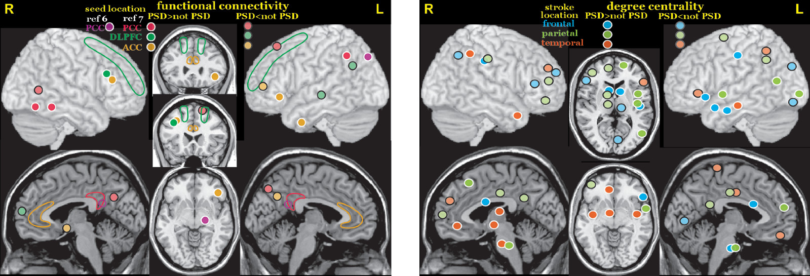
42 A rs fMRI study that utilized bilateral seeds (posterior cingulate/retrosplenial, anterior cingulate and dorsolateral prefrontal cortices) to assess functional connectivity in three networks (chosen because they have been implicated in MDD) in the early subacute (<2 weeks) period reported multiple areas in which stroke patients with and without PSD differed (Figure 2).7 Another rs fMRI study in the subacute (<1 month) stage also reported higher functional connectivity between posterior cingulate/retrosplenial cortex and inferior parietal cortex in stroke patients with PSD compared with stroke patients without PSD (Figure 2).6 A rs fMRI study in the early chronic (3 months) stage that utilized the amplitude of low frequency fluctuations (ALFF) to assess regional activity level reported higher fractional ALFF (fALFF) in the left dorsolateral prefrontal cortex and right precentral gyrus in patients with PSD compared with patients without PSD.31 One source of variance in these studies is that their groups encompassed all stroke locations. The other chronic stage (3–12 months) rs fMRI study compared patients with and without PSD grouped by stroke location (frontal, parietal, temporal).8 This study assessed whole brain functional connectivity at the voxel level (degree centrality, number of connections). Of note, stroke location affected where differences in degree centrality between patients with and without PSD were located and in some instances the direction of differences (Figure 2).8
FIGURE 2. Several studies have compared resting-state functional MRI (rs fMRI) functional connectivity between stroke patients with and without PSD to identify network changes that might be symptom related. Left. Two studies utilized bilateral seeds (outlined areas) to assess functional connectivity in the early subacute stage.6,7 Color-coded solid circles indicate areas in which connectivity differed between the groups. Both studies combined across stroke locations. Right. A study in the chronic stage poststroke (3–12 months) compared patients with and without PSD grouped by stroke location (frontal, parietal, temporal).8 This study assessed whole-brain functional connectivity at the voxel level (degree centrality, number of connections). Of note, stroke location affected where differences in degree centrality between patients with and without PSD were located and in some instances the direction of differences.
A few studies have assessed relationships between connectivity metrics and severity of depression symptoms. A DTI study compared structural connectivity (node degree) of 90 nodes (regions of interest) derived by using the automated anatomic labeling (AAL) atlas to parcellate the brain.27 Lower connectivity at one week correlated with higher severity of depression (HAM-D-24) at 1 month for 17 of the nodes (bilateral - superior frontal gyrus, posterior cingulate gyrus, fusiform gyrus, insula, caudate; left: inferior frontal gyrus, superior temporal gryus, olfactory cortex, precuneus; right: precentral gyrus, putamen).
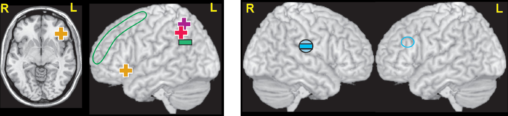
27 Both rs fMRI studies that utilized bilateral seeds reported that severity of depression (HAM-D-17; Beck Depression Inventory, BDI) in the PSD group was significantly positively correlated with increased functional connectivity between posterior cingulate/retrosplenial cortex (default mode network) and inferior parietal cortex (Figure 3 and cover).6,7 The study that assessed other seeds reported that the severity of depression (HAM-D-17) in the PSD group was also positively correlated with increased functional connectivity of the anterior cingulate cortex (affective network) to orbital inferior frontal gyrus, with a trend negative correlation with decreased functional connectivity of dorsolateral prefrontal cortex (cognitive control network) to angular gyrus (Figure 3 and cover).7 In a set of rs fMRI studies in the early chronic stage (3 months) after mostly minor strokes, severity of depression symptoms (Patient Health Questionnaire, PHQ-9) correlated with higher fALFF in the left insula/superior temporal gyrus and with lower seed-based functional connectivity of the left dorsolateral prefrontal cortex to right supramarginal gyrus (Figure 3).9,31 Another rs fMRI study that evaluated functional connectivity between all voxels and the default mode network (identified by independent component analysis) reported functional connectivity at ten days correlated with symptoms of depression (HAM-D-17) at 3 months (but not at ten days) for three areas.43 The correlation was positive for middle temporal cortex and precuneus, negative for neostriatum. As noted in many of these studies, these network changes are generally consistent with what has been reported for MDD.41,44,45
FIGURE 3 AND COVER. Left and Cover. Although there were many areas where seed based functional connectivity differed between patients with and without PSD in the rs fMRI studies detailed above, only a few correlated with severity of depressive symptoms. Both studies reported that increased functional connectivity between the seeds in the posterior cingulate cortex (default mode network) and left parietal cortex (pink plus sign, purple plus sign) positively correlated with severity of depression.6,7 One study also reported that severity of depression was positively correlated with increased functional connectivity between the anterior cingulate cortex seeds (affective network) and left orbital inferior frontal gyrus (gold plus signs), and a trend negative correlation with reduced connectivity between the dorsolateral prefrontal cortex seeds (cognitive control network) and left angular gyrus (green minus sign).7 Right: In contrast, a set of rs fMRI studies in the early chronic stage (3 months) reported that severity of depression correlated with lower functional connectivity between the seed in left dorsolateral prefrontal cortex (blue circle) and right supramarginal gyrus (blue filled circle and negative sign).9
Despite the consensus regarding the prevalence and burden of PSD, there is substantial disagreement regarding the pathophysiology. Proposed biological contributors to PSD include alterations in neurotrophic factors, hypothalamic-pituitary-adrenal axis dysfunction, the inflammatory cascade and cell-mediated immune activation that follows stroke.13,4648 A study that correlated the proteomic profile with depressive symptoms at 3 months poststroke supported the presence of peripheral immunodepression due to neuroinflammatory processes.49 The interaction of the inflammatory response with neurotrophic factors has become a promising avenue of study. The most important neurotrophin implicated thus far is brain-derived neurotrophic factor (BDNF), shown as early as 1995 to promote neuronal survival in vitro.50 Low circulating BDNF levels have been associated with an increased risk of stroke.51 Low levels of BDNF in the acute stage of stroke have been associated with poor functional outcomes at two-year and seven-year follow ups, independent of stroke severity.52 A recent meta-analysis found that peripheral BDNF levels in the acute stage were lower in stroke patients who went on to develop PSD, suggesting potential as a biomarker.53 Although a connection between polymorphisms in BDNF genotype and a propensity for PSD has been reported in some studies, a recent meta-analysis found a relationship to significantly lower risk of ischemic stroke, but no role in PSD.54 Many of these factors have also been implicated in MDD.46,5557 For example, meta-analyses indicate elevations of multiple peripheral biomarkers of inflammation (e.g., cytokines, interleukins, chemokines) in MDD, as well as decreased peripheral levels of some immune mediators following treatment with antidepressants.5862 A recent meta-analysis that focused on BDNF found support for a link between polymorphisms in BDNF genotype and risk of late life depression, decreased peripheral levels of BDNF in patients with MDD, and increases in BDNF following treatment with selective serotonin reuptake inhibitors (SSRIs) or electroconvulsive therapy.63 It is noteworthy serotonin-norepinephrine reuptake inhibitors did not increase BDNF levels.
Although multiple treatment modalities have been studied, there is considerable heterogeneity in key methodological aspects such as time from stroke and duration of treatment. Several meta-analyses of randomized control trials have determined that antidepressants were better than placebo for improving symptoms of depression in patients with PSD, although medications varied considerably in both efficacy and tolerability.6467 The potential of antidepressants to prevent development PSD in nondepressed stroke patients is also supported by several meta-analyses.64,68,69 In addition, there is growing evidence that treatment with SSRIs in particular can improve functional recovery (e.g., dependence, disability, neurological impairment) after stroke, although the meta-analysis did not address whether neurological recovery occurred independently of improvement in depression.65,70 A recent meta-analysis of randomized control trials in which SSRIs were initiated within the first month in stroke patients without PSD reported improved neurologic functioning (e.g., decreased disability, increased functional independence).71 Contrary to expectations, SSRIs did not decrease incidence of PSD. Animal studies indicate multiple possible mechanisms of action (e.g., promoting neurogenesis, increasing neurotrophins including BDNF, decreasing inflammation).70,72

Conclusions

Understanding of the prevalence, influence, and pathophysiology of PSD have advanced over the previous two decades. An important advance has been the shift from a primary focus on lesion location to study of brain functional and structural network changes after stroke. The study of PSD has also led to possible interventions that may facilitate motor and cognitive recovery. Emerging insights into the connectome, BDNF, and the role of the poststroke inflammatory cascade are leading to better understanding of PSD and may, in time, identify new prophylactic or therapeutic options.

References

1.
Ayerbe L, Ayis S, Crichton S, et al: The natural history of depression up to 15 years after stroke: the South London Stroke Register. Stroke 2013; 44:1105–1110
2.
Hackett ML, Pickles K: Part I: frequency of depression after stroke: an updated systematic review and meta-analysis of observational studies. Int J Stroke 2014; 9:1017–1025
3.
Shi YZ, Xiang YT, Wu SL, et al: The relationship between frontal lobe lesions, course of post-stroke depression, and 1-year prognosis in patients with first-ever ischemic stroke. PLoS One 2014; 9:e100456
4.
El Husseini N, Goldstein LB, Peterson ED, et al: Depression and antidepressant use after stroke and transient ischemic attack. Stroke 2012; 43:1609–1616
5.
Ouellet MC, Beaulieu-Bonneau S, Sirois MJ, et al: Depression in the first year after traumatic brain injury. J Neurotrauma 2018; 35:1620–1629
6.
Vicentini JE, Weiler M, Almeida SRM, et al: Depression and anxiety symptoms are associated to disruption of default mode network in subacute ischemic stroke. Brain Imaging Behav 2017; 11:1571–1580
7.
Zhang P, Wang J, Xu Q, et al: Altered functional connectivity in post-ischemic stroke depression: a resting-state functional magnetic resonance imaging study. Eur J Radiol 2018; 100:156–165
8.
Shi Y, Zeng Y, Wu L, et al: A study of the brain functional network of post-stroke depression in three different lesion locations. Sci Rep 2017; 7:14795
9.
Egorova N, Cumming T, Shirbin C, et al: Lower cognitive control network connectivity in stroke participants with depressive features. Transl Psychiatry 2018; 7:4
10.
Feigin VL, Norrving B, Mensah GA: Global burden of stroke. Circ Res 2017; 120:439–448
11.
Go AS, Mozaffarian D, Roger VL, et al: American Heart Association Statistics Committee and Stroke Statistics Subcommittee: Heart disease and stroke statistics—2014 update: a report from the American Heart Association. Circulation 2014; 129:e28–e292
12.
Feigin VL, Roth GA, Naghavi M, et al: Global Burden of Diseases, Injuries and Risk Factors Study 2013 and Stroke Experts Writing Group: Global burden of stroke and risk factors in 188 countries, during 1990–2013: a systematic analysis for the Global Burden of Disease Study 2013. Lancet Neurol 2016; 15:913–924
13.
Towfighi A, Ovbiagele B, El Husseini N, et al: American Heart Association Stroke Council; Council on Cardiovascular and Stroke Nursing; and Council on Quality of Care and Outcomes Research: Poststroke depression: a scientific statement for healthcare professionals from the American Heart Association/American Stroke Association. Stroke 2017; 48:e30–e43
14.
Das J, G K R: Post stroke depression: the sequelae of cerebral stroke. Neurosci Biobehav Rev 2018; 90:104–114
15.
Arwert HJ, Meesters JJL, Boiten J, et al: Poststroke depression: a long-term problem for stroke survivors. Am J Phys Med Rehabil 2018; 97:565–571
16.
Broomfield NM, Quinn TJ, Abdul-Rahim AH, et al: Depression and anxiety symptoms post-stroke/TIA: prevalence and associations in cross-sectional data from a regional stroke registry. BMC Neurol 2014; 14:198
17.
Maaijwee NA, Tendolkar I, Rutten-Jacobs LC, et al: Long-term depressive symptoms and anxiety after transient ischaemic attack or ischaemic stroke in young adults. Eur J Neurol 2016; 23:1262–1268
18.
Ponsford J, Alway Y, Gould KR: Epidemiology and natural history of psychiatric disorders after TBI. J Neuropsychiatry Clin Neurosci (Epub ahead of print, June 25, 2018)
19.
Kim ES, Kim JW, Kang HJ, et al: Longitudinal impact of depression on quality of life in stroke patients. Psychiatry Investig 2018; 15:141–146
20.
Carson AJ, MacHale S, Allen K, et al: Depression after stroke and lesion location: a systematic review. Lancet 2000; 356:122–126
21.
Wei N, Yong W, Li X, et al: Post-stroke depression and lesion location: a systematic review. J Neurol 2015; 262:81–90
22.
Yu L, Liu CK, Chen JW, et al: Relationship between post-stroke depression and lesion location: a meta-analysis. Kaohsiung J Med Sci 2004; 20:372–380
23.
Douven E, Köhler S, Rodriguez MMF, et al: Imaging markers of post-stroke depression and apathy: a systematic review and meta-analysis. Neuropsychol Rev 2017; 27:202–219
24.
Zhang Y, Zhao H, Fang Y, et al: The association between lesion location, sex and poststroke depression: meta-analysis. Brain Behav 2017; 7:e00788
25.
Narushima K, Kosier JT, Robinson RG: A reappraisal of poststroke depression, intra- and inter-hemispheric lesion location using meta-analysis. J Neuropsychiatry Clin Neurosci 2003; 15:422–430
26.
Nickel A, Thomalla G: Post-stroke depression: impact of lesion location and methodological limitations: a topical review. Front Neurol 2017; 8:498
27.
Yang S, Hua P, Shang X, et al: A significant risk factor for poststroke depression: the depression-related subnetwork. J Psychiatry Neurosci 2015; 40:259–268
28.
Murakami T, Hama S, Yamashita H, et al: Neuroanatomic pathways associated with poststroke affective and apathetic depression. Am J Geriatr Psychiatry 2013; 21:840–847
29.
Grajny K, Pyata H, Spiegel K, et al.: Depression symptoms in chronic left hemisphere stroke are related to dorsolateral prefrontal cortex damage. J Neuropsychiatry Clin Neurosci, 2016; 28:292–298
30.
Gozzi SA, Wood AG, Chen J, et al: Imaging predictors of poststroke depression: methodological factors in voxel-based analysis. BMJ Open 2014; 4:e004948
31.
Egorova N, Veldsman M, Cumming T, et al: Fractional amplitude of low-frequency fluctuations (fALFF) in post-stroke depression. Neuroimage Clin 2017; 16:116–124
32.
Carter AR, Shulman GL, Corbetta M: Why use a connectivity-based approach to study stroke and recovery of function? Neuroimage 2012; 62:2271–2280
33.
Fornito A, Zalesky A, Breakspear M: The connectomics of brain disorders. vol. 16. 2015, pp 159–172
34.
Siegel JS, Seitzman BA, Ramsey LE, et al: Re-emergence of modular brain networks in stroke recovery. Cortex 2018; 101:44–59
35.
Corbetta M, Siegel JS, Shulman GL: On the low dimensionality of behavioral deficits and alterations of brain network connectivity after focal injury. Cortex 2018; pii: S0010-9452(17)30425-2
36.
Achard S, Delon-Martin C, Vértes PE, et al: Hubs of brain functional networks are radically reorganized in comatose patients. Proc Natl Acad Sci USA 2012; 109:20608–20613
37.
Crossley NA, Mechelli A, Scott J, et al: The hubs of the human connectome are generally implicated in the anatomy of brain disorders. Brain 2014; 137:2382–2395
38.
Termenon M, Achard S, Jaillard A, et al: The “Hub Disruption Index”: a reliable index sensitive to the brain networks reorganization: a study of the contralesional hemisphere in stroke. Front Comput Neurosci 2016; 10:84
39.
Wang Z, Dai Z, Gong G, et al: Understanding structural-functional relationships in the human brain: a large-scale network perspective. Neuroscientist 2015; 21:290–305
40.
Batista-García-Ramó K, Fernández-Verdecia CI: What we know about the brain structure-function relationship. Behav Sci (Basel) 2018; 8:E39
41.
Li BJ, Friston K, Mody M, et al: A brain network model for depression: from symptom understanding to disease intervention. CNS Neurosci Ther 2018; Epub ahead of print
42.
Siegel JS, Shulman GL, Corbetta M: Measuring functional connectivity in stroke: Approaches and considerations. J Cereb Blood Flow Metab 2017; 37:2665–2678
43.
Lassalle-Lagadec S, Sibon I, Dilharreguy B, et al: Subacute default mode network dysfunction in the prediction of post-stroke depression severity. Radiology 2012; 264:218–224
44.
Brakowski J, Spinelli S, Dörig N, et al: Resting state brain network function in major depression - Depression symptomatology, antidepressant treatment effects, future research. J Psychiatr Res 2017; 92:147–159
45.
Williams LM: Defining biotypes for depression and anxiety based on large-scale circuit dysfunction: a theoretical review of the evidence and future directions for clinical translation. Depress Anxiety 2017; 34:9–24
46.
Pascoe MC, Crewther SG, Carey LM, et al: Inflammation and depression: why poststroke depression may be the norm and not the exception. Int J Stroke 2011; 6:128–135
47.
Noonan K, Carey LM, Crewther SG: Meta-analyses indicate associations between neuroendocrine activation, deactivation in neurotrophic and neuroimaging markers in depression after stroke. J Stroke Cerebrovasc Dis 2013; 22:e124–e135
48.
Villa RF, Ferrari F, Moretti A: Post-stroke depression: mechanisms and pharmacological treatment. Pharmacol Ther 2018; 184:131–144
49.
Nguyen VA, Carey LM, Giummarra L, et al: A pathway proteomic profile of ischemic stroke survivors reveals innate immune dysfunction in association with mild symptoms of depression: a pilot study. Front Neurol 2016; 7:85
50.
Acheson A, Conover JC, Fandl JP, et al: A BDNF autocrine loop in adult sensory neurons prevents cell death. Nature 1995; 374:450–453
51.
Pikula A, Beiser AS, Chen TC, et al: Serum brain-derived neurotrophic factor and vascular endothelial growth factor levels are associated with risk of stroke and vascular brain injury: Framingham Study. Stroke 2013; 44:2768–2775
52.
Stanne TM, Åberg ND, Nilsson S, et al: Low circulating acute brain-derived neurotrophic factor levels are associated with poor long-term functional outcome after ischemic stroke. Stroke 2016; 47:1943–1945
53.
Xu HB, Xu YH, He Y, et al: Decreased serum brain-derived neurotrophic factor may indicate the development of poststroke depression in patients with acute ischemic stroke: a meta-analysis. J Stroke Cerebrovasc Dis 2018; 27:709–715
54.
Bao MH, Zhu SZ, Gao XZ, et al: Meta-analysis on the association between brain-derived neurotrophic factor polymorphism rs6265 and ischemic stroke, poststroke depression. J Stroke Cerebrovasc Dis 2018; 27:1599–1608
55.
Kim YK, Na KS, Myint AM, et al: The role of pro-inflammatory cytokines in neuroinflammation, neurogenesis and the neuroendocrine system in major depression. Prog Neuropsychopharmacol Biol Psychiatry 2016; 64:277–284
56.
Farooq RK, Asghar K, Kanwal S, et al: Role of inflammatory cytokines in depression: focus on interleukin-1β. Biomed Rep 2017; 6:15–20
57.
Jeon SW, Kim YK: The role of neuroinflammation and neurovascular dysfunction in major depressive disorder. J Inflamm Res 2018; 11:179–192
58.
Dowlati Y, Herrmann N, Swardfager W, et al: A meta-analysis of cytokines in major depression. Biol Psychiatry 2010; 67:446–457
59.
Haapakoski R, Mathieu J, Ebmeier KP, et al: Cumulative meta-analysis of interleukins 6 and 1β, tumour necrosis factor α and C-reactive protein in patients with major depressive disorder. Brain Behav Immun 2015; 49:206–215
60.
Eyre HA, Air T, Pradhan A, et al: A meta-analysis of chemokines in major depression. Prog Neuropsychopharmacol Biol Psychiatry 2016; 68:1–8
61.
Goldsmith DR, Rapaport MH, Miller BJ: A meta-analysis of blood cytokine network alterations in psychiatric patients: comparisons between schizophrenia, bipolar disorder and depression. Mol Psychiatry 2016; 21:1696–1709
62.
Köhler CA, Freitas TH, Stubbs B, et al: Peripheral alterations in cytokine and chemokine levels after antidepressant drug treatment for major depressive disorder: systematic review and meta-analysis. Mol Neurobiol 2018; 55:4195–4206
63.
Kishi T, Yoshimura R, Ikuta T, et al: Brain-derived neurotrophic factor and major depressive disorder: evidence from meta-analyses. Front Psychiatry 2018; 8:308
64.
Hackett ML, Anderson CS, House A, et al: Interventions for preventing depression after stroke. Cochrane Database Syst Rev 2008; (3):CD003689
65.
Mead GE, Hsieh CF, Hackett M: Selective serotonin reuptake inhibitors for stroke recovery. JAMA 2013; 310:1066–1067
66.
Xu XM, Zou DZ, Shen LY, et al: Efficacy and feasibility of antidepressant treatment in patients with post-stroke depression. Medicine (Baltimore) 2016; 95:e5349
67.
Sun Y, Liang Y, Jiao Y, et al: Comparative efficacy and acceptability of antidepressant treatment in poststroke depression: a multiple-treatments meta-analysis. BMJ Open 2017; 7:e016499
68.
Yi ZM, Liu F, Zhai SD: Fluoxetine for the prophylaxis of poststroke depression in patients with stroke: a meta-analysis. Int J Clin Pract 2010; 64:1310–1317
69.
Salter KL, Foley NC, Zhu L, et al: Prevention of poststroke depression: does prophylactic pharmacotherapy work? J Stroke Cerebrovasc Dis 2013; 22:1243–1251
70.
Siepmann T, Penzlin AI, Kepplinger J, et al: Selective serotonin reuptake inhibitors to improve outcome in acute ischemic stroke: possible mechanisms and clinical evidence. Brain Behav 2015; 5:e00373
71.
Gu SC, Wang CD: Early selective serotonin reuptake inhibitors for recovery after stroke: a meta-analysis and trial sequential analysis. J Stroke Cerebrovasc Dis 2018; 27:1178–1189
72.
Jin HJ, Pei L, Li YN, et al: Alleviative effects of fluoxetine on depressive-like behaviors by epigenetic regulation of BDNF gene transcription in mouse model of post-stroke depression. Sci Rep 2017; 7:14926

Information & Authors

Information

Published In

Go to The Journal of Neuropsychiatry and Clinical Neurosciences
The Journal of Neuropsychiatry and Clinical Neurosciences
Pages: A4 - 261
PubMed: 30351997

History

Published in print: Fall 2018
Published online: 23 October 2018

Keywords

  1. Stroke and Other Cerebral Vascular Disease (Neuropsychiatric Aspects)
  2. Imaging Techniques
  3. Neuroanatomy

Authors

Affiliations

Benjamin N. Lisle, M.D.
Dr. Lisle is affiliated with the Department of Neurology at Wake Forest School of Medicine in Winston-Salem, North Carolina. Drs. Hurley and Taber are affiliated with the Veterans Affairs Mid Atlantic Mental Illness Research, Education, and Clinical Center, and the Research and Academic Affairs Service Line at the Salisbury VA Healthcare System in Salisbury, North Carolina. Dr. Hurley is affiliated with the Departments of Psychiatry and Radiology at Wake Forest School of Medicine in Winston-Salem, North Carolina, and the Menninger Department of Psychiatry and Behavioral Sciences at Baylor College of Medicine in Houston. Dr. Taber is affiliated with the Division of Biomedical Sciences at the Via College of Osteopathic Medicine in Blacksburg, Virginia, and the Department of Physical Medicine and Rehabilitation at Baylor College of Medicine in Houston,
Robin A. Hurley, M.D. [email protected]
Dr. Lisle is affiliated with the Department of Neurology at Wake Forest School of Medicine in Winston-Salem, North Carolina. Drs. Hurley and Taber are affiliated with the Veterans Affairs Mid Atlantic Mental Illness Research, Education, and Clinical Center, and the Research and Academic Affairs Service Line at the Salisbury VA Healthcare System in Salisbury, North Carolina. Dr. Hurley is affiliated with the Departments of Psychiatry and Radiology at Wake Forest School of Medicine in Winston-Salem, North Carolina, and the Menninger Department of Psychiatry and Behavioral Sciences at Baylor College of Medicine in Houston. Dr. Taber is affiliated with the Division of Biomedical Sciences at the Via College of Osteopathic Medicine in Blacksburg, Virginia, and the Department of Physical Medicine and Rehabilitation at Baylor College of Medicine in Houston,
Katherine H. Taber, Ph.D.
Dr. Lisle is affiliated with the Department of Neurology at Wake Forest School of Medicine in Winston-Salem, North Carolina. Drs. Hurley and Taber are affiliated with the Veterans Affairs Mid Atlantic Mental Illness Research, Education, and Clinical Center, and the Research and Academic Affairs Service Line at the Salisbury VA Healthcare System in Salisbury, North Carolina. Dr. Hurley is affiliated with the Departments of Psychiatry and Radiology at Wake Forest School of Medicine in Winston-Salem, North Carolina, and the Menninger Department of Psychiatry and Behavioral Sciences at Baylor College of Medicine in Houston. Dr. Taber is affiliated with the Division of Biomedical Sciences at the Via College of Osteopathic Medicine in Blacksburg, Virginia, and the Department of Physical Medicine and Rehabilitation at Baylor College of Medicine in Houston,

Notes

Corresponding author: Send correspondence to Dr. Hurley; e-mail: [email protected]

Metrics & Citations

Metrics

Citations

Export Citations

If you have the appropriate software installed, you can download article citation data to the citation manager of your choice. Simply select your manager software from the list below and click Download.

For more information or tips please see 'Downloading to a citation manager' in the Help menu.

Format
Citation style
Style
Copy to clipboard

There are no citations for this item

View Options

View options

PDF/ePub

View PDF/ePub

Get Access

Login options

Already a subscriber? Access your subscription through your login credentials or your institution for full access to this article.

Personal login Institutional Login Open Athens login
Purchase Options

Purchase this article to access the full text.

PPV Articles - Journal of Neuropsychiatry and Clinical Neurosciences

PPV Articles - Journal of Neuropsychiatry and Clinical Neurosciences

Not a subscriber?

Subscribe Now / Learn More

PsychiatryOnline subscription options offer access to the DSM-5-TR® library, books, journals, CME, and patient resources. This all-in-one virtual library provides psychiatrists and mental health professionals with key resources for diagnosis, treatment, research, and professional development.

Need more help? PsychiatryOnline Customer Service may be reached by emailing [email protected] or by calling 800-368-5777 (in the U.S.) or 703-907-7322 (outside the U.S.).

Media

Figures

Other

Tables

Share

Share

Share article link

Share