Full access
Windows to the Brain
Published Online: 28 January 2019

Neurodegenerative Dementias: Improving Brain Health to Decrease Risk

Publication: The Journal of Neuropsychiatry and Clinical Neurosciences
Major neurocognitive disorders (dementias) are expected to double worldwide over the next two decades (20). Alzheimer’s disease (AD) is the most commonly encountered in clinical practice, followed by vascular dementia (21). It is estimated that by 2050, 13.8 million people will be living with dementia due to AD (22). In the absence of disease-modifying therapies, the possibility of delaying or preventing dementia has become an important focus (16). There is a growing consensus that addressing potentially modifiable risk factors is essential to caring for the cognitive health of an aging population. The neurobiological underpinnings of “brain health” approaches will become increasingly important for clinicians to understand. Some of the potentially modifiable factors and their physiologic impact will be reviewed. These include sleep, physical activity, and diet.
Multiple groups now conceptualize the brain and body as a complex nonlinear system (ecosystem) optimized to respond effectively and appropriately (allostasis, adaptive homeostasis, adaptive calibration, resilience thinking framework) to internal and external challenges (stressors) (2, 2326). A stressor is not necessarily intrinsically good or bad, as many are beneficial at one dose level and detrimental at another. The physiologic changes associated with the stress-response may aid in survival when a potential predator is seen. In contrast, when that stress-response is chronic and excessive (e.g., anxiety disorders), there can be deleterious effects (e.g., allostatic load). In addition, individuals interface with stressors in different and distinct ways. Known influences include underlying genetic predisposition, temperament, and prior life experiences. Under normal circumstances, flexible adaptation to challenges maintains the internal environment in a healthy range (allostasis). Severe and/or chronic challenges (high allostatic load) may degrade capacity to cope effectively when a new challenge presents. If not corrected, this may lead to the development of disease (allostatic overload).
Neurodegenerative dementias typically afflict individuals later in life. However, epidemiological studies have identified multiple early, mid, and late-life factors that alter risk (811). Early-life exposure to education and enrichment appears to be a protective factor, independent of ultimate occupational attainment (27, 28). Treatment of acquired sensory impairments (e.g., hearing loss, cataracts) may slow the rate of cognitive decline (29, 30). Sensory loss may promote social isolation, which is independently associated with an increased risk of dementia (though cause and effect determinations cannot be drawn from epidemiological studies) (31, 32). Physical activity is associated with lower risks of dementia and depression (3335). Poor sleep quality, a common complaint across the lifespan, has multiple detrimental effects on brain health (36). Conditions such as metabolic syndrome and smoking are clearly associated with increasing risk of dementia and represent important and well-established targets for prevention (
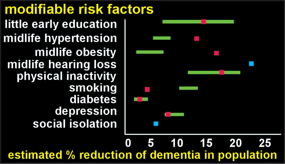
5). Several recent studies have estimated the population impact of eliminating specific risks (Figure 1) (79). Many of these modifiable factors are interdependent. For example, physical inactivity is also associated with increased risk of hypertension, obesity, depression, and diabetes (all risk factors for dementia) (33, 37, 38). Reported combined risk (after correction for nonindependence) ranged from 28%−48%, indicating substantial potential for reducing the population burden of dementia (79). Many of these factors have been incorporated into the various “brain health” approaches to these disorders (13, 5). While evidence in the form of randomized, placebo-controlled trials is rare, there is robust support in the preclinical and clinical spheres (6).
FIGURE 1. In the absence of disease modifying therapies for dementia, the possibility of delaying or preventing development has become an important focus (16). Epidemiological studies indicate that risk for developing dementia is modulated by a wide range of factors (e.g., genetics, medical conditions, education, and environmental exposures) (711). Several recent studies have estimated the population impact of eliminating specific risks (79). The range is indicated (green) for the study that included four different populations (worldwide, United States, Europe, United Kingdom) (7). The study assessing the same risk factors for Australia (pink) reported both similarities and differences (9). The third study evaluated two additional risk factors (blue) (8). Combined risk attributable to these modifiable factors (after correction for nonindependence) ranged from 28% to 48%, supporting the value of prevention.
The system that maintains the brain microenvironment in an optimal state by delivering needed substances (e.g., nutrients, gases, signaling molecules) and removing wastes is an important focus (3941). The cerebral vasculature is one important part of this system, as an intact blood-brain barrier is essential for tight control of movement of substances (e.g., carrier mediated transport, transcytosis) (42, 43). Glia also contribute to maintaining an optimal brain microenvironment both by secretion of neuroactive compounds and by removal of wastes (i.e., uptake and degradation of toxic substances and debris) (44, 45). Circulation of CSF via a perivascular route is also important for delivery and removal of substances, although many aspects continue to be actively debated (19, 42, 4650). One major area of contention is whether movement of fluid and solutes through the extracellular spaces is purely passive (diffusion) or involves an active process (convective flow from the arterial to the venous perivascular spaces). The participation of astroglial aquaporin-4 (AQP4) water channels in fluid flow is also contentious. Recently, identification of lymphatic vessels in the meninges has occurred. Preclinical studies suggest that these lymphatic vessels contribute to clearance of wastes, including amyloid beta (Aβ), from the brain to the cervical lymph nodes (16, 
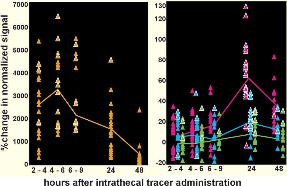
51). Although most studies are preclinical, several important aspects have now been demonstrated in human studies. The passage into the brain of a CSF-borne tracer and its clearance to cervical lymph nodes has been confirmed by sequential MRI (Figure 2) (12, 14, 15, 52, 
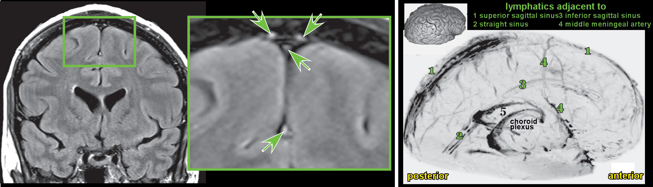
53). The presence of lymphatic vessels in human meninges have been confirmed by both in vivo MRI (Figure 3) and postmortem microscopy (16, 54). Modifiable factors that can influence some aspect of this system are potential targets for interventions, some of the more established of which are discussed below (39).
FIGURE 2. MRI-based measurement of the passage of contrast-enhanced CSF through the brain parenchyma and into the cervical lymph nodes has been performed in humans following intrathecal administration of contrast agent (1215). Plotted here are group averages (solid lines) and data for individual subjects (triangles) color coded by regions of interest placed within CSF (gold), the thalamus (blue), the inferior frontal gyrus (pink), and a cervical lymph node (green) (14). Each individual’s highest value is marked (white outline). Note that there is tremendous individual variability in signal change, indicating that the time course of gadolinium movement from the intrathecal space, into brain, and finally into cervical lymph nodes varies considerably between subjects. Intrathecal gadolinium administration is only performed when clinically necessary. Thus, the kinetics presented here may not reflect normal physiology.
FIGURE 3 and COVER. Recently, the existence of true lymphatic vessels in the dura has been confirmed in humans and nonhuman primates (1618). This adds dural lymphatics to the routes through which substances might travel to exit the brain. Others include resorption of CSF in the arachnoid villi, perivascular flow (i.e., glymphatic), and pericranial nerve flow (e.g., through cribriform plate) (19). Left: Dural lymphatic vessels (green arrows) are well visualized on contrast-enhanced (gadobutrol) fluid attenuated inversion recovery (FLAIR) MRI (16).*Right and Cover: A three-dimensional rendering (skull-stripped subtraction T1 black blood MRI) demonstrates that lymphatics are widespread throughout the brain adjacent to the sinuses (13) and middle meningeal artery (4) (16).* *Reproduced under the terms of the Creative Commons Attribution License.
Abnormal protein aggregation and deposition is a core feature of numerous degenerative illnesses in all organ systems, including many affecting the brain (39). Multiple protein aggregates are associated with neurodegenerative dementias (e.g., Aβ, microtubule associated protein tau, transactivation response-DNA binding protein 43 kDa, alpha-synuclein, prion protein, huntingtin protein) (55). The exact role of each of these proteins in neurodegeneration remains in question, as only some correlate highly with symptom presentation and cognitive decline (56). While a minority of these neurodegenerative proteinopathies have clear genetic mutations, the majority present sporadically. There may be shared physiologic alterations that lead to abnormal protein deposition (e.g., enhanced mis-folding/aggregation, reduced clearance). If impaired clearance is a central pathologic feature early in the development of dementia, then understanding the various factors that affect clearance is critical to understanding prevention strategies.
Sleep difficulties are present in multiple neurodegenerative dementias (5760). The majority of evidence for causal contributions to neuropathologic changes comes from AD (60). Impaired sleep has been implicated in both the pathophysiology of AD and as a symptom of AD (6164). There is emerging evidence that tau hyperphosphorylation appears very early in regions of the brain that are responsible for sleep-wake cycle regulation (57). Overnight Aβ production in cognitively healthy individuals is increased by sleep deprivation (62, 65). Two longitudinal studies of aging in community-dwelling participants reported that presence of excessive daytime sleepiness predicted increased amyloid accumulation (Pittsburgh compound B positron emission tomography imaging) (61). A cross sectional study in cognitively healthy older adults found that the relationship between self-reported impaired sleep and amyloid burden was mediated by AQP4 genetic variation (66). Certain AQP4 variants have been associated with more rapid decline in patients with AD, while others have been associated with a slower rate of decline (67).
In preclinical studies (mouse), locus coeruleus degeneration (an area important for wakefulness) appears to promote Aβ pathology (68). Preclinical models of AD also show a diurnal variation in Aβ levels, with higher levels present during wakefulness and lower levels during sleep (69, 70). Clearance of substances including Aβ is enhanced during states of decreased arousal (e.g., sleep, anesthetic-induced suppression of norepinephrine release) (7177). Thus, it is possible that sleep disruption may lead to impaired waste clearance of pathologically relevant proteins, and that deposition of those same proteins may lead to further sleep disruption. If so, the positive feedback loop set up may be difficult to interrupt after a critical threshold is crossed (71).
There is a clear association between physically active lifestyles and a reduced relative risk of cognitive decline (34). However, the evidence for exercise as an intervention in older adults has been mixed. A recent review noted that only some studies reported beneficial effects on sleep and depression (78). Two well-conducted randomized trials found no cognitive benefit in either healthy older adults (79) or patients with mild to moderate AD (80). In Parkinson’s disease the benefit of exercise in patients already experiencing symptoms is more promising (81). In mice, voluntary exercise has been shown to improve both influx and clearance of substances, resulting in less Aβ accumulation and reduced neuroinflammation (82, 83). In mouse models of AD, exercise was able to reduce pathology including preventing the deposition of Aβ triggered by a high fat diet (84, 85). Other beneficial effects include increases in neurotrophic factors and neurogenesis (78, 85). The differences in outcomes across clinical and preclinical studies suggest that the type, timing, and chronicity of exercise are all likely to be important.
The clinical evidence for dietary interventions in AD is reasonably robust. The Mediterranean-DASH Intervention for Neurodegenerative Delay (MIND) diet has been shown to reduce AD risk with only moderate adherence, whereas the Mediterranean diet (MeDi) appears to require high adherence (86, 87). In a 3-year study, the MeDi was associated with a better AD biomarker profile in those with higher dietary adherence (88). Another dietary approach to AD targets impaired brain glucose metabolism, which may occur decades before the disease is clinically evident (89). Induced hyperglycemia may improve memory in AD (likely mediated through effects on the insulin response), although maintaining a hyperglycemic state is an untenable treatment strategy (89, 90). Ketone bodies (produced either through a ketogenic diet or ingestion of medium-chain fatty acids) provide an alternative energy source for the brain and have some evidence of cognitive benefit (89, 9195). The initial enthusiasm for simply supplementing a “regular” diet with medium chain fatty acids has been tempered due to recent failure in a phase 3 trial (https://www.reuters.com/article/us-accera-study-idUSKBN1671LG). Dietary change aimed at increased ketosis may be more effective. The beneficial effects of carbohydrate-restriction on risk factors for dementia (e.g., metabolic syndrome, diabetes) are supported by multiple studies (96). Recently, a 12-week intervention that included a ketogenic diet, high intensity interval exercise, and memory training in a patient with comorbid mild cognitive impairment and metabolic syndrome resulted in improved cognition and metabolic profile (97). The potential mechanisms of a ketogenic diet include rescue from the hypometabolic state, reduced oxidative stress, and anti-inflammatory effects (91, 92, 96, 98). The role of diet is likely complex and intersects with other factors that are independently important in AD pathogenesis such as sleep and metabolic factors (99).
In conclusion, there is emerging evidence that deceasing the rate and/or increasing the age of onset for neurodegenerative dementias may be possible. A central concept is that an increased toxic load can occur within the brain due to increased production and/or insufficient clearance. Efforts to understand this production and clearance have led to a focus on possible modifiable risk factors and their contributions to this process. Some of those factors may include sleep, physical activity, and diet. A population health approach to preventive efforts, focusing on early-, mid, and late-life modifiable factors, offers a potential roadmap for risk reduction.

References

1.
Centers for Disease Control and Prevention: Association As: The Healthy Brain Initiative: A National Public Health Road Map to Maintaining Cognitive Health. Chicago, Alzheimer’s Association, 2007
2.
Pomorska G, Ockene JK: A general neurologist’s perspective on the urgent need to apply resilience thinking to the prevention and treatment of Alzheimer’s disease. Alzheimers Dement 2017; 3:498–506
3.
Sagner M, McNeil A, Puska P, et al: The P4 Health Spectrum: a Predictive, Preventive, Personalized and Participatory Continuum for Promoting Healthspan. Prog Cardiovasc Dis 2017; 59:506–521
4.
Kivipelto M, Mangialasche F, Ngandu T: Lifestyle interventions to prevent cognitive impairment, dementia and Alzheimer disease. Nat Rev Neurol 2018; 14:653–666
5.
Qiu C, Fratiglioni L: Aging without dementia is achievable: current evidence from epidemiological research. J Alzheimers Dis 2018; 62:933–942
6.
Yaffe K: Modifiable risk factors and prevention of dementia: what is the latest evidence? JAMA Intern Med 2018; 178:281–282
7.
Norton S, Matthews FE, Barnes DE, et al: Potential for primary prevention of Alzheimer’s disease: an analysis of population-based data. Lancet Neurol 2014; 13:788–794
8.
Livingston G, Sommerlad A, Orgeta V, et al: Dementia prevention, intervention, and care. Lancet 2017; 390:2673–2734
9.
Ashby-Mitchell K, Burns R, Shaw J, et al: Proportion of dementia in Australia explained by common modifiable risk factors. Alzheimers Res Ther 2017; 9:11
10.
Vemuri P, Knopman DS, Lesnick TG, et al: Evaluation of amyloid protective factors and Alzheimer disease neurodegeneration protective factors in elderly individuals. JAMA Neurol 2017; 74:718–726
11.
Wang HX, MacDonald SW, Dekhtyar S, et al: Association of lifelong exposure to cognitive reserve-enhancing factors with dementia risk: a community-based cohort study. PLoS Med 2017; 14:e1002251
12.
Eide PK, Ringstad G: MRI with intrathecal MRI gadolinium contrast medium administration: a possible method to assess glymphatic function in human brain. Acta Radiol Open 2015; 4:2058460115609635
13.
Eide PK, Ringstad G: Delayed clearance of cerebrospinal fluid tracer from entorhinal cortex in idiopathic normal pressure hydrocephalus: a glymphatic magnetic resonance imaging study. J Cereb Blood Flow Metab, 2018: 271678X18760974.
14.
Eide PK, Vatnehol SAS, Emblem KE, et al: Magnetic resonance imaging provides evidence of glymphatic drainage from human brain to cervical lymph nodes. Sci Rep 2018; 8:7194
15.
Ringstad G, Valnes LM, Dale AM, et al: Brain-wide glymphatic enhancement and clearance in humans assessed with MRI. JCI Insight 2018; 3:121537
16.
Absinta M, Ha SK, Nair G, et al: Human and nonhuman primate meninges harbor lymphatic vessels that can be visualized noninvasively by MRI. ELife 2017; pii: e29738. doi: 10.7554/eLife.29738
17.
Aspelund A, Antila S, Proulx ST, et al: A dural lymphatic vascular system that drains brain interstitial fluid and macromolecules. J Exp Med 2015; 212:991–999
18.
Louveau A, Smirnov I, Keyes TJ, et al: Structural and functional features of central nervous system lymphatic vessels. Nature 2015; 523:337–341
19.
Benveniste H, Lee H, Volkow ND: The glymphatic pathway: waste removal from the CNS via cerebrospinal fluid transport. Neuroscientist 2017; 23:454–465
20.
Rizzi L, Rosset I, Roriz-Cruz M: Global epidemiology of dementia: Alzheimer’s and vascular types. BioMed Res Int 2014; 2014:908915
21.
Brunnström H, Gustafson L, Passant U, et al: Prevalence of dementia subtypes: a 30-year retrospective survey of neuropathological reports. Arch Gerontol Geriatr 2009; 49:146–149
22.
Hebert LE, Weuve J, Scherr PA, et al: Alzheimer disease in the United States (2010–2050) estimated using the 2010 census. Neurology 2013; 80:1778–1783
23.
Lomeli N, Bota DA, Davies KJA: Diminished stress resistance and defective adaptive homeostasis in age-related diseases. Clin Sci 2017; 131:2573–2599
24.
McEwen BS: Neurobiological and systemic effects of chronic stress. chronic stress 2017; doi: 10.1177/2470547017692328
25.
Oken BS, Chamine I, Wakeland W: A systems approach to stress, stressors and resilience in humans. Behav Brain Res 2015; 282:144–154
26.
Tonhajzerova I, Mestanik M: New perspectives in the model of stress response. Physiol Res 2017; 66(Suppl 2):S173–S185
27.
Dekhtyar S, Wang HX, Fratiglioni L, et al: Childhood school performance, education and occupational complexity: a life-course study of dementia in the Kungsholmen Project. Int J Epidemiol 2016; 45:1207–1215
28.
Ko K, Byun MS, Yi D, et al: Early-life cognitive activity is related to reduced neurodegeneration in Alzheimer signature regions in late life. Front Aging Neurosci 2018; 10:70
29.
Maharani A, Dawes P, Nazroo J, et al: SENSE-Cog WP1 Group: Longitudinal relationship between hearing aid use and cognitive function in older Americans. J Am Geriatr Soc 2018; 66:1130–1136
30.
Maharani A, Dawes P, Nazroo J, et al: SENSE-Cog WP1 group: Cataract surgery and age-related cognitive decline: a 13-year follow-up of the English Longitudinal Study of Ageing. PLoS One 2018; 13:e0204833
31.
Marioni RE, Proust-Lima C, Amieva H, et al: Social activity, cognitive decline and dementia risk: a 20-year prospective cohort study. BMC Public Health 2015; 15:1089
32.
Zhou Z, Wang P, Fang Y: Social engagement and its change are associated with dementia risk among Chinese older adults: a longitudinal study. Sci Rep 2018; 8:1551
33.
Bishwajit G, O’Leary DP, Ghosh S, et al: Physical inactivity and self-reported depression among middle- and older-aged population in South Asia: World Health Survey. BMC Geriatr 2017; 17:100
34.
Blondell SJ, Hammersley-Mather R, Veerman JL: Does physical activity prevent cognitive decline and dementia?: a systematic review and meta-analysis of longitudinal studies. BMC Public Health 2014; 14:510
35.
Shakersain B, Rizzuto D, Wang HX, et al: An active lifestyle reinforces the effect of a healthy diet on cognitive function: a population-based longitudinal study. Nutrients 2018; 10:E1297
36.
Gadie A, Shafto M, Leng Y, et al: Cam-CAN: How are age-related differences in sleep quality associated with health outcomes? an epidemiological investigation in a UK cohort of 2406 adults. BMJ Open 2017; 7:e014920
37.
Hegde SM, Solomon SD: Influence of physical activity on hypertension and cardiac structure and function. Curr Hypertens Rep 2015; 17:77
38.
Pietiläinen KH, Kaprio J, Borg P, et al: Physical inactivity and obesity: a vicious circle. Obesity 2008; 16:409–414
39.
Boland B, Yu WH, Corti O, et al: Promoting the clearance of neurotoxic proteins in neurodegenerative disorders of ageing. Nat Rev Drug Discov 2018; 17:660–688
40.
Louveau A, Plog BA, Antila S, et al: Understanding the functions and relationships of the glymphatic system and meningeal lymphatics. J Clin Invest 2017; 127:3210–3219
41.
Sun BL, Wang LH, Yang T, et al: Lymphatic drainage system of the brain: a novel target for intervention of neurological diseases. Prog Neurobiol 2018; 163-164:118–143
42.
Smith AJ, Verkman AS: The “glymphatic” mechanism for solute clearance in Alzheimer’s disease: game changer or unproven speculation? FASEB J 2018; 32:543–551
43.
Verheggen ICM, Van Boxtel MPJ, Verhey FRJ, et al: Interaction between blood-brain barrier and glymphatic system in solute clearance. Neurosci Biobehav Rev 2018; 90:26–33
44.
Simon MJ, Wang MX, Murchison CF, et al: Transcriptional network analysis of human astrocytic endfoot genes reveals region-specific associations with dementia status and tau pathology. Sci Rep 2018; 8:12389
45.
Zorec R, Parpura V, Verkhratsky A: Astroglial vesicular network: evolutionary trends, physiology and pathophysiology. Acta Physiol 2018; 222(2)doi: 10.1111/apha.12915
46.
Abbott NJ, Pizzo ME, Preston JE, et al: The role of brain barriers in fluid movement in the CNS: is there a glymphatic system? Acta Neuropathol 2018; 135:387–407
47.
Bacyinski A, Xu M, Wang W, et al: The paravascular pathway for brain waste clearance: current understanding, significance and controversy. Front Neuroanat 2017; 11:101
48.
Plog BA, Nedergaard M: The glymphatic system in central nervous system health and disease: past, present, and future. Annu Rev Pathol 2018; 13:379–394
49.
Taber KH, Hurley RA: Volume transmission in the brain: beyond the synapse. J Neuropsychiatry Clin Neurosci 2014; 26:iv-4
50.
Nicholson C, Hrabětová S: Brain extracellular space: the final frontier of neuroscience. Biophys J 2017; 113:2133–2142
51.
Da Mesquita S, Louveau A, Vaccari A, et al: Functional aspects of meningeal lymphatics in ageing and Alzheimer’s disease. Nature 2018; 560:185–191
52.
Akbar JJ, Luetmer PH, Schwartz KM, et al: The role of MR myelography with intrathecal gadolinium in localization of spinal CSF leaks in patients with spontaneous intracranial hypotension. AJNR Am J Neuroradiol 2012; 33:535–540
53.
Aydin K, Terzibasioglu E, Sencer S, et al: Localization of cerebrospinal fluid leaks by gadolinium-enhanced magnetic resonance cisternography: a 5-year single-center experience. Neurosurgery 2008; 62:584–589, discussion 584–589
54.
Goodman JR, Adham ZO, Woltjer RL, et al: Characterization of dural sinus-associated lymphatic vasculature in human Alzheimer’s dementia subjects. Brain Behav Immun 2018; 73:34–40
55.
Macedo MN, Kim EJ, Seeley WW: Neuropathology of dementia, in The Behavioral Neurology of Dementia. Edited by Miller BL, Boeve BF. Cambridge, United Kingdom, Cambridge University Press, 2009, pp 142–160
56.
Nelson PT, Alafuzoff I, Bigio EH, et al: Correlation of Alzheimer disease neuropathologic changes with cognitive status: a review of the literature. J Neuropathol Exp Neurol 2012; 71:362–381
57.
Holth J, Patel T, Holtzman DM: Sleep in Alzheimer’s disease: beyond amyloid. Neurobiol Sleep Circadian Rhythms 2017; 2:4–14
58.
McCarter SJ, St Louis EK, Boeve BF: Sleep disturbances in frontotemporal dementia. Curr Neurol Neurosci Rep 2016; 16:85
59.
McKeith IG, Boeve BF, Dickson DW, et al: Diagnosis and management of dementia with Lewy bodies: fourth consensus report of the DLB Consortium. Neurology 2017; 89:88–100
60.
Wennberg AMV, Wu MN, Rosenberg PB, et al: Sleep disturbance, cognitive decline, and dementia: a review. Semin Neurol 2017; 37:395–406
61.
Carvalho DZ, St Louis EK, Knopman DS, et al: Association of excessive daytime sleepiness with longitudinal β-amyloid accumulation in elderly persons without dementia. JAMA Neurol 2018; 75:672–680
62.
Lucey BP, Hicks TJ, McLeland JS, et al: Effect of sleep on overnight cerebrospinal fluid amyloid β kinetics. Ann Neurol 2018; 83:197–204
63.
Spira AP, An Y, Wu MN, et al: Excessive daytime sleepiness and napping in cognitively normal adults: associations with subsequent amyloid deposition measured by PiB PET. Sleep (Epub ahead of print, September 25, 2018)
64.
Winer JR, Mander BA: Waking up to the importance of sleep in the pathogenesis of Alzheimer disease. JAMA Neurol 2018; 75:654–656
65.
Shokri-Kojori E, Wang GJ, Wiers CE, et al: β-Amyloid accumulation in the human brain after one night of sleep deprivation. Proc Natl Acad Sci USA 2018; 115:4483–4488
66.
Rainey-Smith SR, Mazzucchelli GN, Villemagne VL, et al: AIBL Research Group: Genetic variation in aquaporin-4 moderates the relationship between sleep and brain Aβ-amyloid burden. Transl Psychiatry 2018; 8:47
67.
Burfeind KG, Murchison CF, Westaway SK, et al: The effects of noncoding aquaporin-4 single-nucleotide polymorphisms on cognition and functional progression of Alzheimer’s disease. Alzheimers Dement (N Y) 2017; 3:348–359
68.
Heneka MT, Ramanathan M, Jacobs AH, et al: Locus ceruleus degeneration promotes Alzheimer pathogenesis in amyloid precursor protein 23 transgenic mice. J Neurosci 2006; 26:1343–1354
69.
Kang JE, Lim MM, Bateman RJ, et al: Amyloid-beta dynamics are regulated by orexin and the sleep-wake cycle. Science 2009; 326:1005–1007
70.
Roh JH, Huang Y, Bero AW, et al: Disruption of the sleep-wake cycle and diurnal fluctuation of β-amyloid in mice with Alzheimer’s disease pathology. Sci Transl Med 2012; 4:150ra122
71.
Cedernaes J, Osorio RS, Varga AW, et al: Candidate mechanisms underlying the association between sleep-wake disruptions and Alzheimer’s disease. Sleep Med Rev 2017; 31:102–111
72.
Xie L, Kang H, Xu Q, et al: Sleep drives metabolite clearance from the adult brain. Science 2013; 342:373–377
73.
Achariyar TM, Li B, Peng W, et al: Glymphatic distribution of CSF-derived APOE into brain is isoform specific and suppressed during sleep deprivation. Mol Neurodegener 2016; 11:74
74.
Taoka T, Jost G, Frenzel T, et al: Impact of the glymphatic system on the kinetic and distribution of gadodiamide in the rat brain: observations by dynamic MRI and effect of circadian rhythm on tissue gadolinium concentrations. Invest Radiol 2018; 53:529–534
75.
Lundgaard I, Lu ML, Yang E, et al: Glymphatic clearance controls state-dependent changes in brain lactate concentration. J Cereb Blood Flow Metab 2017; 37:2112–2124
76.
Benveniste H, Lee H, Ding F, et al: Anesthesia with dexmedetomidine and low-dose isoflurane increases solute transport via the glymphatic pathway in rat brain when compared with high-dose isoflurane. Anesthesiology 2017; 127:976–988
77.
Gakuba C, Gaberel T, Goursaud S, et al: General anesthesia inhibits the activity of the “glymphatic system.” Theranostics 2018; 8:710–722
78.
Veronese N, Solmi M, Basso C, et al: Role of physical activity in ameliorating neuropsychiatric symptoms in Alzheimer disease: a narrative review. Int J Geriatr Psychiatry (Epub ahead of print, August 29, 2018)
79.
Sink KM, Espeland MA, Castro CM, et al: LIFE Study Investigators: Effect of a 24-month physical activity intervention vs health education on cognitive outcomes in sedentary older adults: the LIFE Randomized Trial. JAMA 2015; 314:781–790
80.
Lamb SE, Sheehan B, Atherton N, et al: DAPA Trial Investigators: Dementia and Physical Activity (DAPA) trial of moderate to high intensity exercise training for people with dementia: randomised controlled trial. BMJ 2018; 361:k1675
81.
Ramaswamy B, Jones J, Carroll C: Exercise for people with Parkinson’s: a practical approach. Pract Neurol 2018; 18:399–406
82.
He XF, Liu DX, Zhang Q, et al: Voluntary exercise promotes glymphatic clearance of amyloid beta and reduces the activation of astrocytes and microglia in aged mice. Front Mol Neurosci 2017; 10:144
83.
von Holstein-Rathlou S, Petersen NC, Nedergaard M: Voluntary running enhances glymphatic influx in awake behaving, young mice. Neurosci Lett 2018; 662:253–258
84.
Maesako M, Uemura K, Iwata A, et al: Continuation of exercise is necessary to inhibit high fat diet-induced β-amyloid deposition and memory deficit in amyloid precursor protein transgenic mice. PLoS One 2013; 8:e72796
85.
Ryan SM, Kelly AM: Exercise as a pro-cognitive, pro-neurogenic and anti-inflammatory intervention in transgenic mouse models of Alzheimer’s disease. Ageing Res Rev 2016; 27:77–92
86.
Anastasiou CA, Yannakoulia M, Kosmidis MH, et al: Mediterranean diet and cognitive health: Initial results from the Hellenic Longitudinal Investigation of Ageing and Diet. PLoS One 2017; 12:e0182048
87.
Morris MC, Tangney CC, Wang Y, et al: MIND diet slows cognitive decline with aging. Alzheimers Dement 2015; 11:1015–1022
88.
Berti V, Walters M, Sterling J, et al: Mediterranean diet and 3-year Alzheimer brain biomarker changes in middle-aged adults. Neurology 2018; 90:e1789–e1798
89.
Costantini LC, Barr LJ, Vogel JL, et al: Hypometabolism as a therapeutic target in Alzheimer’s disease. BMC Neurosci 2008; 9(Suppl 2):S16
90.
Watson GS, Craft S: Modulation of memory by insulin and glucose: neuropsychological observations in Alzheimer’s disease. Eur J Pharmacol 2004; 490:97–113
91.
Cunnane SC, Courchesne-Loyer A, Vandenberghe C, et al: Can ketones help rescue brain fuel supply in later life? implications for cognitive health during aging and the treatment of Alzheimer’s disease. Front Mol Neurosci 2016; 9:53
92.
Pinto A, Bonucci A, Maggi E, et al: Anti-oxidant and anti-inflammatory activity of ketogenic diet: new perspectives for neuroprotection in Alzheimer’s disease. Antioxidants 2018; 7:E63
93.
Sharma A, Bemis M, Desilets AR: Role of medium chain triglycerides (Axona) in the treatment of mild to moderate Alzheimer’s disease. Am J Alzheimers Dis Other Demen 2014; 29:409–414
94.
Taylor MK, Sullivan DK, Mahnken JD, et al: Feasibility and efficacy data from a ketogenic diet intervention in Alzheimer’s disease. Alzheimers Dement (N Y) 2017; 4:28–36
95.
Ota M, Matsuo J, Ishida I, et al: Effects of a medium-chain triglyceride-based ketogenic formula on cognitive function in patients with mild-to-moderate Alzheimer’s disease. Neurosci Lett 2018; 690:232–236
96.
Ludwig DS, Willett WC, Volek JS, et al: Dietary fat: from foe to friend? Science 2018; 362:764–770
97.
Dahlgren K, Gibas KJ: Ketogenic diet, high intensity interval training (HIIT) and memory training in the treatment of mild cognitive impairment: A case study. Diabetes Metab Syndr 2018; 12:819–822
98.
Torosyan N, Sethanandha C, Grill JD, et al: Changes in regional cerebral blood flow associated with a 45 day course of the ketogenic agent, caprylidene, in patients with mild to moderate Alzheimer’s disease: Results of a randomized, double-blinded, pilot study. Exp Gerontol 2018; 111:118–121
99.
Pistollato F, Sumalla Cano S, Elio I, et al: Associations between sleep, cortisol regulation, and diet: possible implications for the risk of Alzheimer disease. Adv Nutr 2016; 7:679–689

Information & Authors

Information

Published In

Go to The Journal of Neuropsychiatry and Clinical Neurosciences
The Journal of Neuropsychiatry and Clinical Neurosciences
Pages: A4 - 5
PubMed: 30686211

History

Published in print: Winter 2019
Published online: 28 January 2019

Keywords

  1. Aging of the Brain
  2. Dementia

Authors

Affiliations

James R. Bateman, M.D., M.P.H.
From the Veterans Affairs Mid Atlantic Mental Illness Research, Education, and Clinical Center and the Research and Academic Affairs Service Line at the W.G. Hefner Veterans Affairs Medical Center in Salisbury, N.C. (JRB, RAH, KHT); the Department of Neurology at Wake Forest School of Medicine in Winston-Salem, N.C. (JRB); the Departments of Psychiatry and Radiology at Wake Forest School of Medicine in Winston-Salem, N.C., and the Menninger Department of Psychiatry and Behavioral Sciences at Baylor College of Medicine in Houston, Tex. (RAH); and the Division of Biomedical Sciences at the Via College of Osteopathic Medicine in Blacksburg, Va., and the Department of Physical Medicine and Rehabilitation at Baylor College of Medicine in Houston, Tex. (KHT).
Robin A. Hurley, M.D. [email protected]
From the Veterans Affairs Mid Atlantic Mental Illness Research, Education, and Clinical Center and the Research and Academic Affairs Service Line at the W.G. Hefner Veterans Affairs Medical Center in Salisbury, N.C. (JRB, RAH, KHT); the Department of Neurology at Wake Forest School of Medicine in Winston-Salem, N.C. (JRB); the Departments of Psychiatry and Radiology at Wake Forest School of Medicine in Winston-Salem, N.C., and the Menninger Department of Psychiatry and Behavioral Sciences at Baylor College of Medicine in Houston, Tex. (RAH); and the Division of Biomedical Sciences at the Via College of Osteopathic Medicine in Blacksburg, Va., and the Department of Physical Medicine and Rehabilitation at Baylor College of Medicine in Houston, Tex. (KHT).
Katherine H. Taber, Ph.D.
From the Veterans Affairs Mid Atlantic Mental Illness Research, Education, and Clinical Center and the Research and Academic Affairs Service Line at the W.G. Hefner Veterans Affairs Medical Center in Salisbury, N.C. (JRB, RAH, KHT); the Department of Neurology at Wake Forest School of Medicine in Winston-Salem, N.C. (JRB); the Departments of Psychiatry and Radiology at Wake Forest School of Medicine in Winston-Salem, N.C., and the Menninger Department of Psychiatry and Behavioral Sciences at Baylor College of Medicine in Houston, Tex. (RAH); and the Division of Biomedical Sciences at the Via College of Osteopathic Medicine in Blacksburg, Va., and the Department of Physical Medicine and Rehabilitation at Baylor College of Medicine in Houston, Tex. (KHT).

Notes

Send correspondence to Dr. Hurley; e-mail: [email protected]

Funding Information

Department of Veterans' Affairs, United States: OAA MIRECC fellowshi[
Supported by the Department of Veterans Affairs Office of Academic Affiliations Advanced Fellowship Program in Mental Illness, Research, and Treatment.

Metrics & Citations

Metrics

Citations

Export Citations

If you have the appropriate software installed, you can download article citation data to the citation manager of your choice. Simply select your manager software from the list below and click Download.

For more information or tips please see 'Downloading to a citation manager' in the Help menu.

Format
Citation style
Style
Copy to clipboard

There are no citations for this item

View Options

View options

PDF/ePub

View PDF/ePub

Get Access

Login options

Already a subscriber? Access your subscription through your login credentials or your institution for full access to this article.

Personal login Institutional Login Open Athens login
Purchase Options

Purchase this article to access the full text.

PPV Articles - Journal of Neuropsychiatry and Clinical Neurosciences

PPV Articles - Journal of Neuropsychiatry and Clinical Neurosciences

Not a subscriber?

Subscribe Now / Learn More

PsychiatryOnline subscription options offer access to the DSM-5-TR® library, books, journals, CME, and patient resources. This all-in-one virtual library provides psychiatrists and mental health professionals with key resources for diagnosis, treatment, research, and professional development.

Need more help? PsychiatryOnline Customer Service may be reached by emailing [email protected] or by calling 800-368-5777 (in the U.S.) or 703-907-7322 (outside the U.S.).

Media

Figures

Other

Tables

Share

Share

Share article link

Share