Full access
Regular Article
Published Online: 1 February 2005

Increasing Memory Load Modulates Regional Brain Activity in Older Adults as Measured by fMRI

Publication: The Journal of Neuropsychiatry and Clinical Neurosciences

Abstract

Several recent studies using functional magnetic resonance imaging (fMRI) during recognition memory tests have suggested that the ability to neuromodulate as a function of cognitive demand may be impaired in older adults due to age-related cell loss and neural volume reduction in memory specific regions. In the current study, older adults (ages 59–77) were tested with fMRI during a delayed-recognition task in which memory load for faces was varied across trials. Activity was greater in amplitude for three- versus one-face stimuli within the superior, middle, and inferior frontal gyri, intraparietal sulcus, and fusiform gyrus. It was concluded that the ability to modulate activity with increasing load is preserved in older adults despite reductions in neural volume.
A paradigm requiring the recognition of a recently learned face from a novel distractor is often used to examine the integrity of memory and the underlying neural substrates in both normal and clinical populations. Human lesion studies and functional imaging studies have shown that face recognition depends on the integrity of the right fusiform gyrus,15 medial temporal lobe structures,614 and frontal regions.1518
Reduced ability to recognize recently learned faces has been observed in healthy older adults.1923 Corresponding neural substrates have shown reduced activation in healthy older adults,24 particularly in the hippocampus,20 during face recognition tasks. Several studies, however, have reported increased activation in the prefrontal cortex and increased communication between the prefrontal and the occipital cortices in older adults during such tasks.25,26 This may indicate a compensatory mechanism so that behavioral performance is maintained at a high level.27,28
Recent reports using neuroimaging techniques have suggested that age-related reduction in neural volume in regions critical to memory, with cell loss in serotonergic and dopaminergic systems, may influence the brain’s ability to neuromodulate or change activity.29 Several studies using functional magnetic resonance imaging (fMRI) have suggested that the ability to neuromodulate as a function of cognitive demand may be impaired in older adults. For example, in a study of young and older subjects, Grady et al.30 assessed the effect of cognitive demand on modulation of cortical activity during the maintenance phase of a variable delay working memory test for faces. Lengthening the delay interval increased memory load demand. Young subjects were able to effectively increase activity, with increasing delay interval length within a number of areas, including the anterior prefrontal and ventral extrastriate cortex. On the other hand, activity among older subjects was invariant to changes in delay interval duration. Varied results demonstrating regionally-dependent increased or decreased load-related activation in older subjects have been reported using functional MRI during tasks requiring working memory for verbal stimuli.31
In previous studies of young subjects alone, increases in memory load have resulted in increases in activity within prefrontal cortex during continuous performance tasks3234 and delayed-recognition tasks.3537 In a recent study by Jha and McCarthy, the time-course of activity was investigated in high-resolution regions of interest analysis of the prefrontal, posterior parietal, and fusiform regions during a working memory task for faces in which the number of items to maintain was varied across trials.37 In addition to increases in prefrontal activity with higher load, increases were observed within intraparietal sulcus and fusiform gyrus. In the current study, the methodology of Jha and McCarthy was used to investigate the impact of memory load for faces during delayed-recognition performance in a group of older adults. The purpose of this study was to determine whether increasing memory load led to: 1) increased amplitude of activity within activated regions and 2) recruitment of additional activated neural volume.

METHODS

Subjects

The Duke University Medical Center Institutional Review Board approved the study, and each subject provided written informed consent after it had been fully explained. A total of 14 volunteer subjects (eight men and six women) participated (age range 59–77 years, mean 68 years). One subject was left-handed. All subjects were screened to rule out significant neuropsychiatric disorders, including dementia. The Mini-Mental State Examination38 scores ranged from 28 to 30, with a mean of 29.3.

Cognitive Activation Task

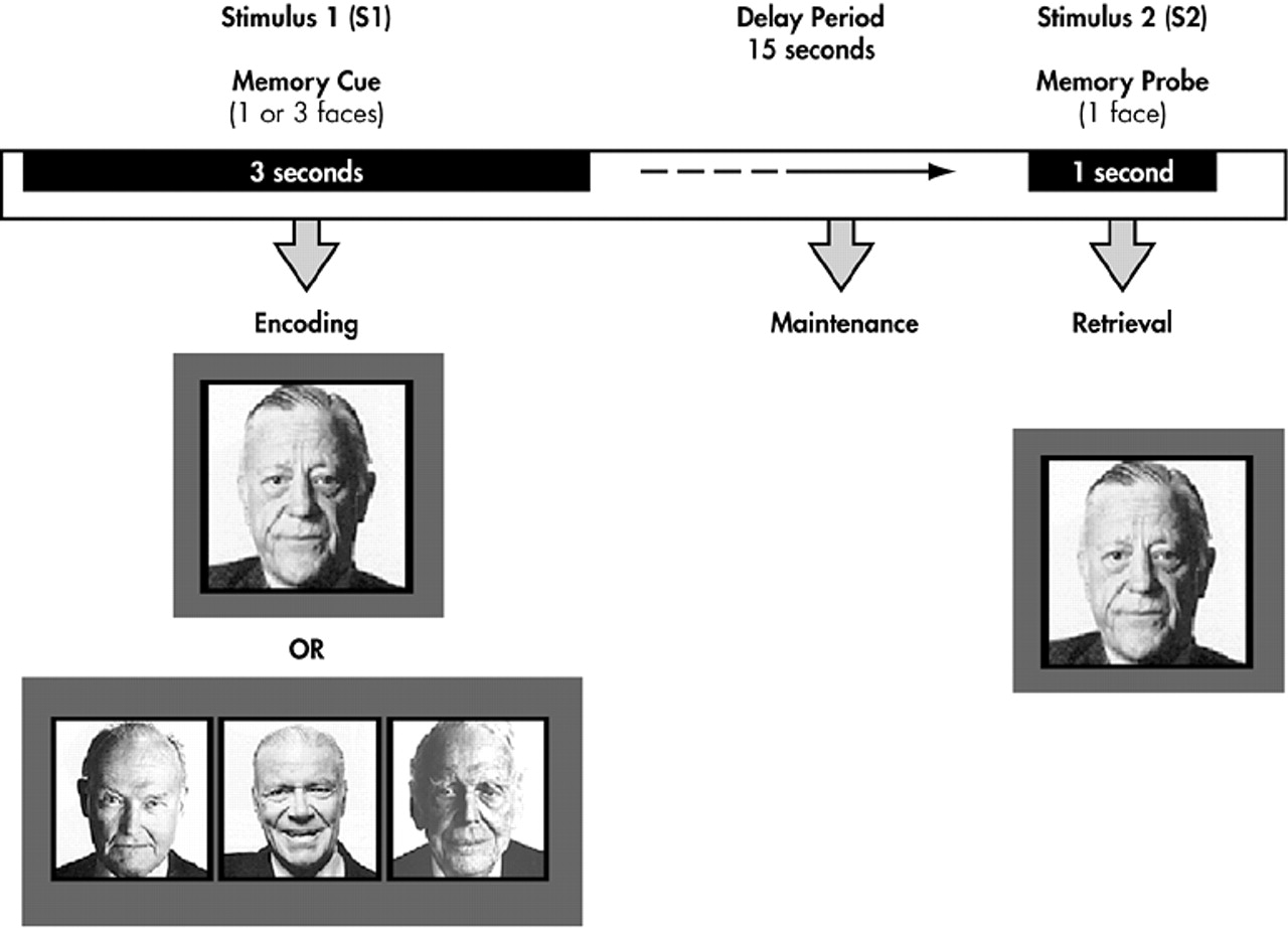
Each trial consisted of a memory array (S1) presented for 3 seconds, a 15-second-delay interval without any stimuli, and a single probe stimulus (S2) presented for 1.5 seconds that terminated the delay interval (Figure 1). The long-delay interval was employed to disentangle transient evoked responses to S1 and S2 stimuli from delay-interval activity.37 In one-half of the trials, the probe stimulus matched one of the items in the memory array, while in the remaining one-half the probe was a new stimulus. Subjects made a forced choice-button response with the index finger of their left or right hand to indicate whether the probe matched one of the memory items.
The memory array consisted of either one or three unfamiliar and moderately confusable faces. Faces presented in each S1-S2 trial were matched for general facial characteristics, ethnicity, gender, and luminescence. The one face and three face conditions were randomly yet equally distributed over all trials. The intertrial interval, measured from the offset of S2 to the onset of the next S1, was 29 seconds. All subjects performed six runs, with each run consisting of 6 trials (except one who only completed three runs due to an acquisition error).
All stimuli were presented using a liquid crystal display (LCD) projector (XGA resolution, 900 lumens) equipped with a specially designed Buhl lens. Stimuli were projected upon a 10-inch wide screen located within the magnet bore directly behind the subject’s head and subjects viewed the stimuli through mirrored glasses. Behavioral responses were acquired using a button box incorporating a fiber-optic loop connected to a TTL driver circuit located outside of the magnet room.

Image Acquisition

All scanning was performed on a 1.5-T NVi scanner (GE Medical Systems, Milwaukee), equipped with an Advanced Development Workstation for real time echoplanar imaging. A scout series of T1-weighted images were obtained in the sagittal plane (two-dimensional SE TR/TE=500/10, 256×192, field of view 24 cm, 5.0 mm thk/2.5 sp), followed by axial dual-echo FSE images (TR/TE 2700/20/80 256×192, field of view 24 cm, 5.0 mm thk/0.0 sp), obtained for screening purposes. A board-certified neuroradiologist (J.R.P.) evaluated all anatomical scans clinically, screening for mass lesions or other clinically significant findings. The anterior and posterior commissures were identified in the mid-sagittal slice of the initial scout series, and high-resolution coronal anatomical images were acquired from 60 contiguous 5-mm thick coronal slices using a gradient echo sequence (three-dimensional SPGR TR/TE=6/minute, 256×256, field of view 24 cm, FA=25, 5.0 mm/0.0 sp). A time series of contiguous coronal images, sensitive to blood-oxygenation-dependent contrast, were acquired from 32 contiguous coronal slice locations chosen from the three-dimensional spoiled gradient-echo (SPGR) series (GE EPI TR/TE=3000/40 ms, 24 cm field of view, 64×64 image matrix, 90° flip angle, slice thickness=5.0 mm, gap=0.0 mm), during performance of the cognitive activation task. A total of 98 brain volumes were obtained over each of the six runs described in the previous section.

Behavioral Performance

Behavioral performance results were obtained from 11 of the subjects (equipment failure precluded behavioral data collection in three subjects). Reaction time and percent correct measures were recorded. Percent correct was defined as the number of correct trials divided by the total number of trials.

Image Analysis

Preprocessing.

The center of mass for each echo planar imaging (EPI) volume within each time series was computed and plotted to determine head motion during scanning, revealing submillimeter motion in all subjects. The time series of EPI volumes was adjusted to compensate for the interleaved slice acquisition order within each repetition time (TR) interval. Fitting the time series of each voxel with a cubic spline and then resampling this function for all voxels at the onset of each TR interval accomplished this.

Processing.

Average time-epochs synchronized to S1 were computed for each memory load condition according to the methodology of Jha and McCarthy.37 In brief, 16 image time segments, from the five image volumes preceding to 10 following each S1 stimulus, were excised from the continuous time series of volumes for each run comprising each subject’s data. These single trial volume epochs were then grouped by stimulus type (1- or three-face trials) and averaged, with the temporal order of each volume relative to S1 maintained. The baseline was calculated as the mean intensity of the two time points prior to S1. The average MR signal values were then converted to percent signal change relative to this pre-S1 baseline. The averaged time-epochs were then subjected to the analytic procedures described below.

Regions of Interest

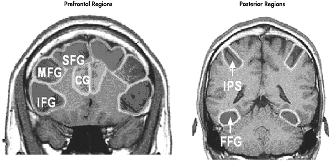
Anatomical regions of interest (ROIs) were drawn on each subject’s high-resolution coronal anatomical images. Regions of interest were drawn on the left and right superior frontal gyri, middle frontal gyri, inferior frontal gyri, anterior cingulate gyri, intraparietal sulci, and fusiform gyri. The ROIs for superior frontal gyrus, anterior cingulate gyrus, middle frontal gyrus, and inferior frontal gyrus were drawn on nine slices ranging from 35 mm anterior to 5 mm posterior to the anterior commissure. The fusiform gyrus was drawn on six slices ranging from 20 mm to 45 mm posterior to the anterior commissure. The intraparietal sulcus was drawn on eight slices ranging from 50 to 85 mm posterior to the anterior commissure (Figure 2).

Time Course Analysis

Since the activity measured within an ROI might represent the activity of only a few activated voxels diluted by surrounding unactivated space, a subset of activated voxels were defined, and time-activation waveforms were computed for only activated voxels located within each anatomical ROI. To identify a subset of activated voxels, a correlation analysis was performed using a reference waveform, based on empirical data obtained during a similar task.37 Active voxels were defined as those that had a waveform that significantly correlated with the reference waveform (t>1.96, p<0.05, uncorrected). By using a low statistical criterion for the significance of the correlation, we purposely defined “activated” as those voxels that had waveforms that could vary somewhat from the reference waveform. As there were differences in the number of activated voxels as a function of memory load, the time waveforms were computed for the union of active voxels for both load conditions for each ROI. Each subject contributed 24 waveforms (six anatomical regions by two hemispheres by two memory load conditions).

Activated Voxel Counts

Using the ROIs defined above, we tabulated the number of active voxels within each anatomical ROI for each load condition. These counts were converted to percentages relative to the number of voxels in each ROI to normalize for differences in brain volume across subjects.

Statistics

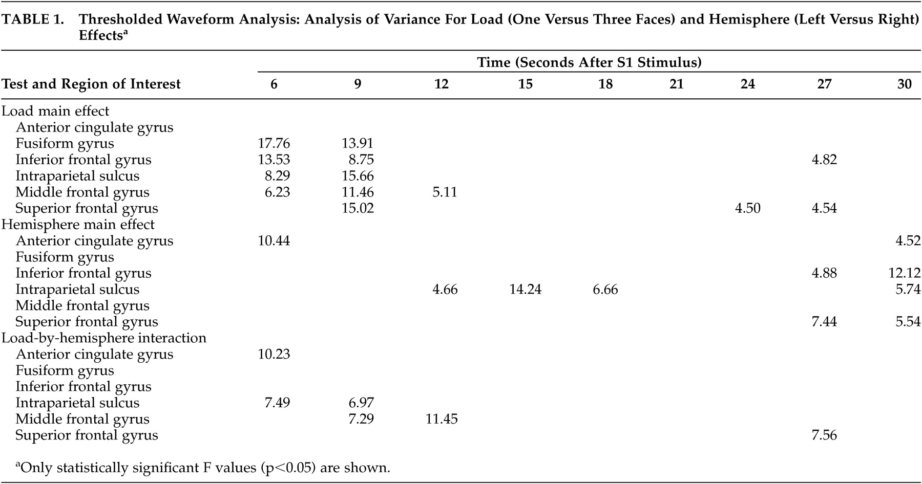
For comparison of behavioral responses between memory load conditions, reaction time and percent correct measures were entered into separate paired Student’s t tests. For comparison of the waveforms, repeated measures analyses of variance (ANOVA) were performed to determine if activity differed as a function of memory load at each time point in the activation time-waveforms for each ROI . Finally, for comparison of activated voxel counts, repeated measures ANOVA were performed to determine if the number of activated voxels differed across group, gyrus, hemisphere, and memory load condition. Our statistics did not include corrections for multiple tests.

RESULTS

Behavioral Performance

Memory load had a significant impact on behavioral performance and reaction times. Paired t tests revealed a significant difference (p<0.05) between memory load conditions for both performance accuracy and reaction time. Performance accuracy was significantly better for one-face (mean=96%, SD=5.83) versus three-face trials (mean=75%, SD=11.95) (p<0.0008). Mean reaction times were significantly faster in one-face (mean=1491 msec, SD=591.32) versus three-face trials (mean=2288 msec, SD=1018.59) (p<0.02).

Time Course Analysis

The mean activation time courses for activated voxels in all ROIs are depicted in Figure 3. Significant S1/S2-evoked hemodynamic response peaks were observed approximately 6 to 9 seconds following S1 onset and approximately 6 seconds following S2 onset. Activity during the S1-S2 delay interval was load invariant and varied widely between ROIs. All ROIs produced the same general time course response. The mean signal within the posterior ROIs (fusiform gyrus and intraparietal sulcus) was twice as great in amplitude as that within the anterior ROIs comprising the prefrontal cortex (superior frontal gyrus, middle frontal gyrus, inferior frontal gyrus).
Load sensitivity was assessed by testing the influence of load upon the mean amplitude values at each of the time points following S1 onset (i.e., 3, 6, 9, 12, 15, 18, 21, 24, 27, 30 seconds). The significance levels (F values) are summarized in Table 1. Amplitude differences were classified as significant for p values <0.05. During the encoding period, at 6 seconds, mean signal values were significantly greater for the three-face compared to one-face trials within fusiform gyrus, inferior frontal gyrus, intraparietal sulcus, and middle frontal gyrus. At 9 seconds, mean signal values were significantly greater within fusiform gyrus, inferior frontal gyrus, intraparietal sulcus, middle frontal gyrus, and superior frontal gyrus. At 12 seconds, the mean signal value was significantly greater only in the middle frontal gyrus. During the delay period (12–18 seconds), there were no statistically significant load sensitive differences. However, during the retrieval period, the superior frontal gyrus showed statistically significant load sensitive differences at 24 and 27 seconds. The inferior frontal gyrus showed a statistically significant difference at 27 seconds.
For all gyri, there was a general trend toward right hemispheric lateralization during encoding and retrieval but toward left lateralization during the delay period. However, a statistically significant difference in the hemispheric activity was observed only during the delay period within the intraparietal sulcus, encoding period in the anterior cingulate gyrus, and retrieval period in the superior and inferior frontal gyri.

Activated Voxel Counts

The percentage of activated voxels in each ROI varied from a low of 10% in the anterior cingulate gyrus to 28% in the intraparietal sulcus. Figure 4 presents the mean percentage of activated voxels (relative to the total number of voxels) in each ROI as a function of memory load. Between the two memory load conditions, there were no statistically significant differences for any of the gyri tested, either individually or as a whole. However, there was a distinct trend for a greater number of activated voxels in the three-face condition compared to the one-face condition across all gyri.

DISCUSSION

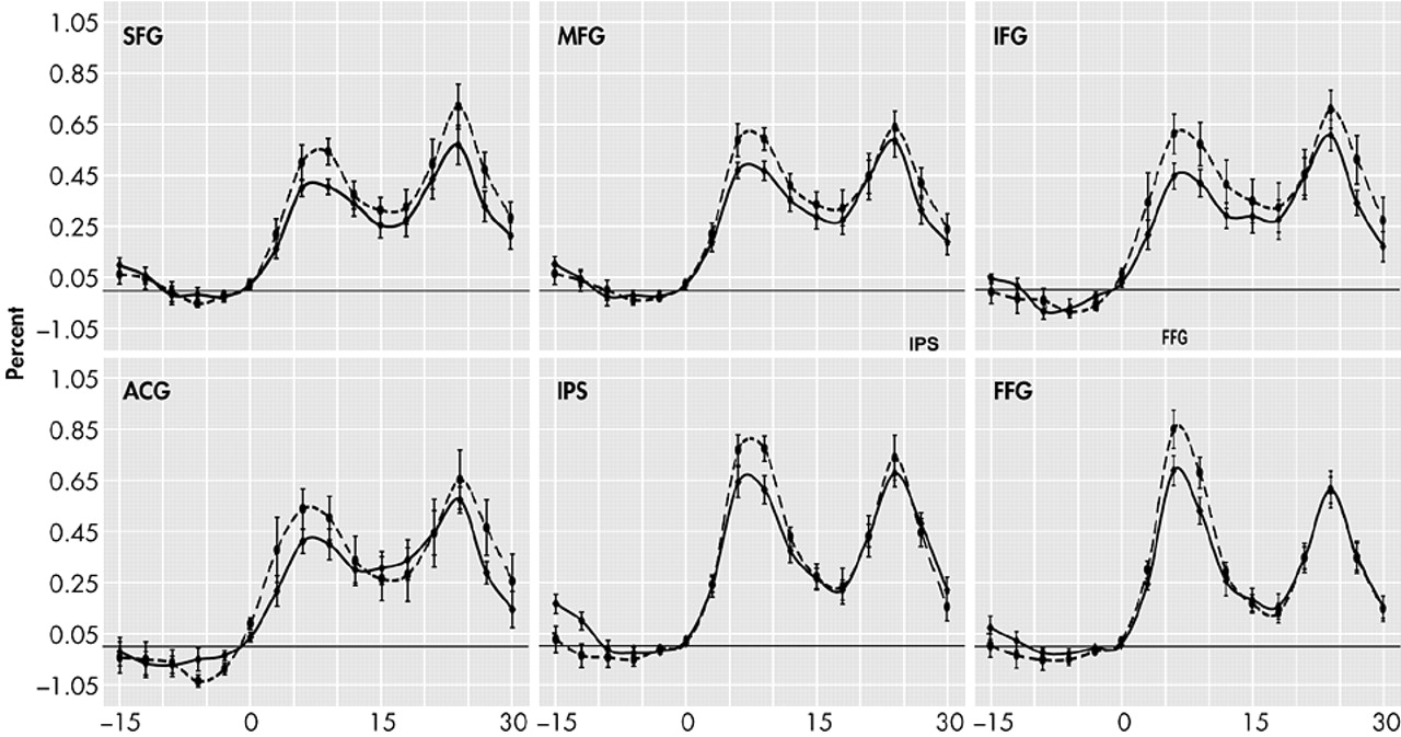
Our study demonstrates that there is an increase in magnitude of brain activation in response to increased memory load in healthy older subjects. These findings suggest that such individuals may have neurofunctional reserves that can be called upon to handle more challenging memory tasks. The pattern of increased activation is highly similar to that observed in younger individuals doing a similar task in which they demonstrated increased activation in both three-face and two-face tasks compared to a one-face task.37
Memory load only affected the waveform during the encoding and transition (early delay) period of our memory task. The prefrontal cortex, intraparietal sulcus, and fusiform gyrus all showed statistically significant activation differences due to memory load at the 6- and 9-second time points, but by 12 seconds, only the middle frontal gyrus still showed significant activity change. These findings are consistent with those of D’Esposito et al.,39 who found the encoding process in older subjects was significantly affected by memory load.
None of our subjects showed load-related differences in the delay interval. These results are consistent with the patterns found in young individuals by Jha and McCarthy, who observed that while phasic processes such as encoding were affected by memory load, maintenance processes were not.37 During the retrieval period, our results show statistically significant load-related activation differences in the superior frontal gyrus at two time points and in the inferior frontal gyrus at one time point. Prior studies have shown load sensitive retrieval effects only in young subjects.40
Our observation that the intraparietal sulcus responded similarly to the prefrontal cortex when the memory load increased replicates findings of Braver et al.,34 who observed increased activation bilaterally in the prefrontal cortex and the parietal regions with increased memory load in a young population. Manoach et al,41 however, found that while both dorsolateral prefrontal cortex and intraparietal sulcus were activated by memory tasks, only the dorsolateral prefrontal cortex was affected by changes in memory load.
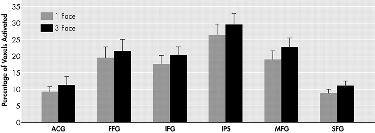
Our findings demonstrate a significant increase in the magnitude of activation in the prefrontal cortex region (inferior frontal gyrus, middle frontal gyrus, superior frontal gyrus) in the three-face condition compared to the one-face condition. However, we were unable to demonstrate a statistically significant increase in extent of activation measured by voxel counts. Rypma et al.31 suggested that older individuals might be limited in their ability to recruit additional dorsolateral prefrontal cortex regions for more challenging memory loads. Alhough it is possible that new regions of the prefrontal cortex cannot be recruited, and only regions already activated in the lower memory load condition may have the ability to raise their level of activation when presented with a memory load challenge, the current study suggests that this recruitment ability is indeed preserved. A clear trend toward a positive load affect on voxel count was present in every ROI and approached statistical significance when all regions were combined (Figure 4).
Using a very similar paradigm with younger subjects, Jha and McCarthy37 noted a statistically greater number of activated voxels in their three-face and two-face trials compared to their one-face trial. This was found in a number of the same regions tested in this study: the middle frontal gyrus, inferior frontal gyrus, anterior cingulate gyrus and intraparietal sulcus. Therefore, a functional explanation needs to be considered for the ability to increase the magnitude, but to a lesser degree, the extent of activation in response to increasing memory load in older adults. Age-related neuronal loss has been well documented (see reference 42). Though vascular reserve capacity may be relatively preserved in older subjects,
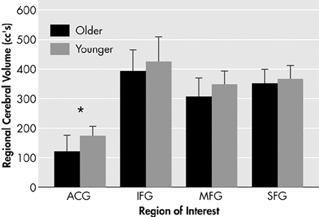
43 it is possible that there is less capacity to recruit additional neurons to deal with increased task load due to age related neuronal loss. A comparison of regional brain volumes was performed between this study and that of Jha and McCarthy,37 in regions of interest defined by identical anatomical criteria, namely the superior frontal gyrus, middle frontal gyrus, inferior frontal gyrus and anterior cingulate gyrus. The total number of voxels in each region was tabulated for each subject in both studies and multiplied by the appropriate voxel size. Results show a trend toward decreased volume in older subjects in the prefrontal cortex, with a statistically significant decrease in the anterior cingulate gyrus (Figure 5).
In conclusion, this study in healthy older adult subjects demonstrates similar changes to those described in young subjects, namely that increasing memory load increases the magnitude of signal change in activated voxels in memory-specific brain regions in the frontal, temporal, and parietal lobes. Load-related increases in the extent of activation are less robust, which may be a consequence of age-related volume loss with decreased capacity for neuronal recruitment.

ACKNOWLEDGMENTS

Dr. Petrella’s research is supported by grants from the Radiological Society of North America Research and Education Foundation and National Institute of Aging, 1R01 AG-019728-01. The authors thank Gregory McCarthy, Ph.D. for his advice on experimental design and analysis techniques, and Haris Sair, B.S., and Lakshmi Murty, B.S. for assistance with analysis and proofreading. Aspects of this study were presented at the Ninth Annual Meeting of the Cognitive Neuroscience Society, April 14–16, 2002.
FIGURE 1.  Schematic of Cognitive Activation Task
One or Three faces (cue) were displayed during the encoding period. Following a 15-second delay, a test face (probe) was shown for one second during the retrieval period. Subjects responded as to whether or not the test face matched the face or faces shown during the encoding period.
FIGURE 2.  Prefrontal and Posterior Regions of Interest
ACG=anterior cingulate gyrus, FFG=fusiform gyrus, IFG=inferior frontal gyrus, IPS=intraparietal sulcus, MFG=middle frontal gyrus, SFG=superior frontal gyrus
FIGURE 3.  Thresholded Waveform Analysis: Mean Activation Time Course for Activated Voxels in Each Region as a Function of Memory Load
(solid curve=1 face condition, dashed curve=3 face condition); ACG=anterior cingulate gyrus, FFG=fusiform gyrus, IFG=inferior frontal gyrus, IPS=intraparietal sulcus, MFG=middle frontal gyrus, SFG=superior frontal gyrus. Error bars denote standard deviation.
FIGURE 4.  Voxel Count Analysis: Mean Percentage of Activated Voxels Within Each Region as a Function of Memory Load
ACG=anterior cingulate gyrus, FFG=fusiform gyrus, IFG=inferior frontal gyrus, IPS=intraparietal sulcus, MFG=middle frontal gyrus, SFG=superior frontal gyrus. Error bars denote standard deviation.
FIGURE 5.  Comparison of Regional Brain Volumes Between the Current Study in Older Subjects and That of Jha and McCarthy37 in Young Subjects
ACG=anterior cingulate gyrus, IFG=inferior frontal gyrus, MFG=middle frontal gyrus, SFG=superior frontal gyrus. Error bars denote standard deviation. Asterisk deontes statistically significant difference.
TABLE 1. Thresholded Waveform Analysis: Analysis of Variance For Load (One Versus Three Faces) and Hemisphere (Left Versus Right) Effectsa

References

1.
Damasio AR, Tranel D, Damasio H: Face agnosia and the neural substrates of memory. Annu Rev Neurosci 1990; 13:89–109
2.
McCarthy G, Puce A, Gore JC, et al: Face-specific processing in the human fusiform gyrus. J Cogn Neurosci 1997; 9:604–609
3.
Haxby JV, Ungerleider LG, Horwitz B, et al: Face encoding and recognition in the human brain. Proc Natl Acad Sci USA 1996; 93:922–927
4.
Kanwisher N, McDermott J, Chun MM: The fusiform face area: a module in human extrastriate cortex specialized for face perception. J Neurosci 1997; 17:4302–4311
5.
Puce A, Allison T, Gore JC, et al: Face-sensitive regions in human extrastriate cortex studied by functional MRI. J Neurophysiol 1995; 74:1192–1199
6.
Bookheimer SY, Strojwas MH, Cohen MS, et al: Patterns of brain activation in people at risk for Alzheimer’s disease. N Engl J Med 2000; 343:450–456
7.
Brewer JB, Zhao Z, Desmond JE, et al: Making memories: brain activity that predicts how well visual experience will be remembered. Science 1998; 281:1185–1187
8.
Dolan RJ, Fletcher PC: Dissociating prefrontal and hippocampal function in episodic memory encoding. Nature 1997; 388:582–585
9.
Gabrieli JDE, Brewer JB, Desmond JE, et al: Separate neural bases of two fundamental memory processes in the human medial temporal lobe. Science 1997; 276:264–266
10.
Martin A, Wiggs CL, Weisberg J: Modulation of human medial temporal lobe activity by form, meaning, and experience. Hippocampus 1997; 7:587–593
11.
Schacter DL, Alpert NM, Savage CR, et al: Conscious recollection and the human hippocampal formation: evidence from positron emission tomography. Proc Natl Acad Sci USA 1996; 93:321–325
12.
Stark CE, Squire LR: Functional magnetic resonance imaging (fMRI) activity in the hippocampal region during recognition memory. J Neurosci 2000; 20:7776–7781
13.
Stern CE, Corkin S, Gonzalez RG, et al: The hippocampal formation participates in novel picture encoding: evidence from functional magnetic resonance imaging. Proc Natl Acad Sci USA 1996; 93:8660–8665
14.
Strange BA, Fletcher PC, Henson RN, et al: Segregating the functions of human hippocampus. Proc Natl Acad Sci USA 1999; 96:4034–4039
15.
D’Esposito M, Detre JA, Alsop DC, et al: The neural basis of the central executive system of working memory. Nature 1995; 378:279–281
16.
McCarthy G, Puce A, Constable RT, et al: Activation of human prefrontal cortex during spatial and nonspatial working memory tasks measured by functional MRI. Cereb Cortex 1996; 6:600–611
17.
Rypma B, D’Esposito M: The roles of prefrontal brain regions in components of working memory: effects of memory load and individual differences. Proc Natl Acad Sci USA 1999; 96:6558–6563
18.
Wagner AD, Poldrack RA, Eldridge LL, et al: Material-specific lateralization of prefrontal activation during episodic encoding and retrieval. Neuroreport 1998; 9:3711–3717
19.
Bartlett JC, Leslie JE: Aging and memory for faces versus single views of faces. Mem Cognit 1986; 14:371–381
20.
Grady CL, Bernstein LJ, Beig S, et al: The effects of encoding task on age-related differences in the functional neuroanatomy of face memory. Psychol Aging 2002; 17:7–23
21.
Bartlett JC, Leslie JE, Tubbs A, et al: Aging and memory for pictures of faces. Psychol Aging 1989; 4:276–283
22.
Crook T, Larrabee G: Changes in facial recognition memory across the adult life span. J Gerontol 1992; 47:138–141
23.
Smith AD, Winograd E: Adult age differences in remembering faces. Dev Psychol 1978; 14:443–444
24.
Grady CL, McIntosh AR, Horwitz B, et al: Age-related reductions in human recognition memory due to impaired encoding. Science 1995; 269:218–221
25.
Horwitz B, McIntosh AR, Haxby JV, et al: Network analysis of PET-mapped visual pathways in Alzheimer type dementia. Neuroreport 1995; 6:2287–2292
26.
McIntosh AR, Grady CL, Ungerleider LG, et al: Network analysis of cortical visual pathways mapped with PET. J Neurosci 1994; 14:655–666
27.
Cabeza R, Grady CL, Nyberg L, et al: Age-related differences in neural activity during memory encoding and retrieval: a positron emission tomography study. J Neurosci 1997; 17:391–400
28.
Grady CL, Maisog JM, Horwitz B, et al: Age-related changes in cortical blood flow activation during visual processing of faces and location. J Neurosci 1994; 14(3, part 2):1450–1462
29.
Li S, Lindenberger U, Sikstrom S: Aging cognition: from neuromodulation to representation. Trends Cogn Sci 2001; 5:479–486
30.
Grady C, McIntosh A, Bookstein F, et al: Age-related changes in regional cerebral blood flow during working memory for faces. Neuroimage 1998; 8:409–425
31.
Rypma B, Prabhakaran V, Desmond JE, et al: Age differences in prefrontal cortical activity in working memory. Psychol Aging 2001; 16:371–384
32.
Barch DM, Braver TS, Nystrom LE, et al: Dissociating working memory from task difficulty in human prefrontal cortex. Neuropsychologia 1997; 35:1373–1380
33.
Callicott JH, Ramsey NF, Tallent K, et al: Functional magnetic resonance imaging brain mapping in psychiatry: methodological issues illustrated in a study of working memory in schizophrenia. Neuropsychopharmacology 1998; 18:186–196
34.
Braver TS, Cohen JD, Nystrom LE, et al: A parametric study of prefrontal cortex involvement in human working memory. Neuroimage 1997; 5:49–62
35.
Rypma B, Prabhakaran V, Desmond JE, et al: Load-dependent roles of frontal brain regions in the maintenance of working memory. Neuroimage 1999; 9:216–226
36.
Postle BR, Berger JS, D’Esposito M: Functional neuroanatomical double dissociation of mnemonic and executive control processes contributing to working memory performance. Proc Natl Acad Sci USA 1999; 96:12959–12964
37.
Jha AP, McCarthy G: The influence of memory load upon delay interval activity in a working memory task: an event-related functional MRI study. J Cogn Neurosci 2000; 12:90–105
38.
Folstein MF, Folstein SE, McHugh PR: “Mini-Mental State”: a practical method for grading the cognitive state of patients for the clinician. J Psychiatr Res 1975; 12:189–198
39.
D’Esposito M, Zarahn E, Aguirre GK, et al: The effect of normal aging on the coupling of neural activity to the bold hemodynamic response. Neuroimage 1999; 10:6–14
40.
Rypma B, D’Esposito M: Isolating the neural mechanisms of age-related changes in human working memory. Nat Neurosci 2000; 3:509–515
41.
Manoach DS, Schlaug G, Siewert B, et al: Prefrontal cortex fMRI signal changes are correlated with working memory load. Neuroreport 1997; 8:545–549
42.
Morrison JH, Hof PR: Life and death of neurons in the aging brain. Science 1997; 278:412–419
43.
Petrella JR, DeCarli C, Dagli M, et al: Age-related vasodilatory response to acetazolamide challenge in healthy adults: a dynamic contrast-enhanced MR study. AJNR Am J Neuroradiol 1998; 19:39–44

Information & Authors

Information

Published In

Go to The Journal of Neuropsychiatry and Clinical Neurosciences
The Journal of Neuropsychiatry and Clinical Neurosciences
Pages: 75 - 83
PubMed: 15746486

History

Published online: 1 February 2005
Published in print: February 2005

Authors

Affiliations

Jeffrey R. Petrella, M.D.
Received March 13, 2003; revised September 23, 2003; accepted February 10, 2004. From the Department of Radiology and Brain Imaging and Analysis Center and the Department of Psychiatry, Duke University Medical Center, Durham, North Carolina; the Department of Psychology, University of Pennsylvania, Philadelphia; the Department of Psychiatry, Naval Medical Center, San Diego, California; the Department of Psychology, Washington University, St. Louis, Missouri.; and the Department of Psychology, University of North Carolina, Chapel Hill, North Carolina. Address correspondence to Dr. Petrella, Department of Radiology, DUMC, Box 3808, Durham NC 27710-3808; [email protected] (E-mail).
Brent A. Townsend, B.S.
Received March 13, 2003; revised September 23, 2003; accepted February 10, 2004. From the Department of Radiology and Brain Imaging and Analysis Center and the Department of Psychiatry, Duke University Medical Center, Durham, North Carolina; the Department of Psychology, University of Pennsylvania, Philadelphia; the Department of Psychiatry, Naval Medical Center, San Diego, California; the Department of Psychology, Washington University, St. Louis, Missouri.; and the Department of Psychology, University of North Carolina, Chapel Hill, North Carolina. Address correspondence to Dr. Petrella, Department of Radiology, DUMC, Box 3808, Durham NC 27710-3808; [email protected] (E-mail).
Amishi P. Jha, Ph.D.
Received March 13, 2003; revised September 23, 2003; accepted February 10, 2004. From the Department of Radiology and Brain Imaging and Analysis Center and the Department of Psychiatry, Duke University Medical Center, Durham, North Carolina; the Department of Psychology, University of Pennsylvania, Philadelphia; the Department of Psychiatry, Naval Medical Center, San Diego, California; the Department of Psychology, Washington University, St. Louis, Missouri.; and the Department of Psychology, University of North Carolina, Chapel Hill, North Carolina. Address correspondence to Dr. Petrella, Department of Radiology, DUMC, Box 3808, Durham NC 27710-3808; [email protected] (E-mail).
Lauretta A. Ziajko, M.D.
Received March 13, 2003; revised September 23, 2003; accepted February 10, 2004. From the Department of Radiology and Brain Imaging and Analysis Center and the Department of Psychiatry, Duke University Medical Center, Durham, North Carolina; the Department of Psychology, University of Pennsylvania, Philadelphia; the Department of Psychiatry, Naval Medical Center, San Diego, California; the Department of Psychology, Washington University, St. Louis, Missouri.; and the Department of Psychology, University of North Carolina, Chapel Hill, North Carolina. Address correspondence to Dr. Petrella, Department of Radiology, DUMC, Box 3808, Durham NC 27710-3808; [email protected] (E-mail).
Melissa J. Slavin, Ph.D.
Received March 13, 2003; revised September 23, 2003; accepted February 10, 2004. From the Department of Radiology and Brain Imaging and Analysis Center and the Department of Psychiatry, Duke University Medical Center, Durham, North Carolina; the Department of Psychology, University of Pennsylvania, Philadelphia; the Department of Psychiatry, Naval Medical Center, San Diego, California; the Department of Psychology, Washington University, St. Louis, Missouri.; and the Department of Psychology, University of North Carolina, Chapel Hill, North Carolina. Address correspondence to Dr. Petrella, Department of Radiology, DUMC, Box 3808, Durham NC 27710-3808; [email protected] (E-mail).
Cindy Lustig, Ph.D.
Received March 13, 2003; revised September 23, 2003; accepted February 10, 2004. From the Department of Radiology and Brain Imaging and Analysis Center and the Department of Psychiatry, Duke University Medical Center, Durham, North Carolina; the Department of Psychology, University of Pennsylvania, Philadelphia; the Department of Psychiatry, Naval Medical Center, San Diego, California; the Department of Psychology, Washington University, St. Louis, Missouri.; and the Department of Psychology, University of North Carolina, Chapel Hill, North Carolina. Address correspondence to Dr. Petrella, Department of Radiology, DUMC, Box 3808, Durham NC 27710-3808; [email protected] (E-mail).
Sarah J. Hart, B.S.
Received March 13, 2003; revised September 23, 2003; accepted February 10, 2004. From the Department of Radiology and Brain Imaging and Analysis Center and the Department of Psychiatry, Duke University Medical Center, Durham, North Carolina; the Department of Psychology, University of Pennsylvania, Philadelphia; the Department of Psychiatry, Naval Medical Center, San Diego, California; the Department of Psychology, Washington University, St. Louis, Missouri.; and the Department of Psychology, University of North Carolina, Chapel Hill, North Carolina. Address correspondence to Dr. Petrella, Department of Radiology, DUMC, Box 3808, Durham NC 27710-3808; [email protected] (E-mail).
P. Murali Doraiswamy, M.D.
Received March 13, 2003; revised September 23, 2003; accepted February 10, 2004. From the Department of Radiology and Brain Imaging and Analysis Center and the Department of Psychiatry, Duke University Medical Center, Durham, North Carolina; the Department of Psychology, University of Pennsylvania, Philadelphia; the Department of Psychiatry, Naval Medical Center, San Diego, California; the Department of Psychology, Washington University, St. Louis, Missouri.; and the Department of Psychology, University of North Carolina, Chapel Hill, North Carolina. Address correspondence to Dr. Petrella, Department of Radiology, DUMC, Box 3808, Durham NC 27710-3808; [email protected] (E-mail).

Metrics & Citations

Metrics

Citations

Export Citations

If you have the appropriate software installed, you can download article citation data to the citation manager of your choice. Simply select your manager software from the list below and click Download.

For more information or tips please see 'Downloading to a citation manager' in the Help menu.

Format
Citation style
Style
Copy to clipboard

There are no citations for this item

View Options

View options

PDF/ePub

View PDF/ePub

Get Access

Login options

Already a subscriber? Access your subscription through your login credentials or your institution for full access to this article.

Personal login Institutional Login Open Athens login
Purchase Options

Purchase this article to access the full text.

PPV Articles - Journal of Neuropsychiatry and Clinical Neurosciences

PPV Articles - Journal of Neuropsychiatry and Clinical Neurosciences

Not a subscriber?

Subscribe Now / Learn More

PsychiatryOnline subscription options offer access to the DSM-5-TR® library, books, journals, CME, and patient resources. This all-in-one virtual library provides psychiatrists and mental health professionals with key resources for diagnosis, treatment, research, and professional development.

Need more help? PsychiatryOnline Customer Service may be reached by emailing [email protected] or by calling 800-368-5777 (in the U.S.) or 703-907-7322 (outside the U.S.).

Media

Figures

Other

Tables

Share

Share

Share article link

Share