T he human brain is anatomically, neurochemically, and functionally asymmetric.
1,
2 Development of lateralization and hemispheric asymmetry is a result of interactions of endogenous influences of genes, hormones, and the environment.
3 Normal anatomical left-right fetal brain asymmetry begins at 20–22 weeks of gestation.
4 Disrupted development of lateralization and brain asymmetry is an important cause in the genesis of mental disorders,
5 in particular schizophrenia.
6 Mental retardation, microcephalus, seizures, and cognitive impairment have been found in prenatally irradiated survivors of atomic bombing, especially after exposure at weeks 8–15 and weeks 16–25 of gestation.
7 –
9 The fetal dose threshold for the development of mental retardation after in utero irradiation at weeks 8–15 of gestation is 0.06–0.31 Gy. At weeks 16–25, it is 0.28–0.87 Gy.
10 The lifetime prevalence of schizophrenia in people prenatally exposed to atomic bomb radiation in Japan is 0.96%,
11 which is higher than an established rate of 0.33% in the Japanese population.
12 However, data are not available for brain laterality patterns following prenatal exposure as a result of atomic bombing. Moreover, it is difficult to compare the Japanese circumstances to those of the Chernobyl nuclear power plant accident; the latter incident caused significantly lower fetal radiation doses, but higher exposure to the fetal thyroid with the incorporation of radioactive iodine. The Japanese atom bomb survivors were acutely externally irradiated with gamma rays and neutrons without thyroid exposure to radioactive iodine.
The World Health Organization’s pilot project “Brain Damage in Utero,” conducted within the framework of the International Program on the Health Effects of the Chernobyl Accident, found an increased prevalence of mild mental retardation and emotional-behavioral disorders in the prenatally exposed children.
13 –
15It has been reported that the prenatally exposed children in Belarus manifest a relative increase in psychological impairment and a lower IQ relative to the comparison group children, but these effects were not attributed to radiation.
16 –
18 It was also claimed that the exposed Ukrainian children showed only nonsignificant differences in the applied tests relative to the comparison group.
19 –
20 In these studies it was concluded that unfavorable psychosocial factors, such as relocation, broken social contacts, and adaptation difficulties, explained the differences between the exposed and nonexposed groups.
In contrast, the French-German Initiative for Chernobyl subproject 3.4.1, “Potential Effects of Prenatal Brain Irradiation as a Result of the Chernobyl Accident (Ukraine),” revealed significantly more neuropsychiatric disorders, lower full-scale IQ and verbal IQ, and an increased frequency of verbal/performance IQ discrepancy in the prenatally exposed children. When IQ discrepancies of the exposed children exceeded 25 points, there appeared to be a correlation with the amount of fetal exposure. The mothers of the children in the exposed and comparison groups did not show differences in verbal abilities, but mothers evacuated from Prypiat (adjacent to the site of the Chernobyl plant) had more psychological disorders.
21 –
23The UN Chernobyl Forum Expert Group “Health” has concurred that the effects on the developing brain must be the focus of particular attention.
24 Although we do know that the developing brain is extremely radiosensitive, information is not available on the effects of exposure to ionizing radiation in utero in relation to cerebral asymmetry in humans.
Whether one cerebral hemisphere is more vulnerable than the other, with respect to different exogenous agents in utero, is still undetermined. It has been reported that diagnostic ultrasound exposure in the fetal stage of life increases the rate of left-handedness.
25 Prenatal malnutrition results in a significant reduction of the normal interhemispheric asymmetry of visual evoked responses.
26 Children prenatally exposed to cocaine had greater right frontal EEG asymmetry.
27 Prenatal stress resulted in elevated dopaminergic activity lateralized to the right hemisphere
28 together with increased anxiety behavior.
29 At the same time, thalidomide embryopathy asymmetry in EEG was found to be infrequent, in spite of mental retardation or epilepsy.
30 In children with heavy prenatal alcohol exposure, cerebral laterality effects were not found,
31 nor was there a significant reduction in normal anatomical cerebral asymmetry.
32The pattern of brain laterality following exposure to different exogenous prenatal factors is quite different. It has been reported that there is prominent impairment of the left, dominant, cerebral hemisphere functions in children irradiated in utero as a result of the Chernobyl accident.
33 –
37 The objective of this study was to assess the brain laterality pattern of cognitive and neurophysiological measurements in children irradiated in utero as a result of the Chernobyl accident and to evaluate whether this asymmetry could be attributed to prenatal exposure to ionizing radiation.
METHOD
Participants
We examined 100 children (ages 11–13 years) born between April 26, 1986, and February 26, 1987 (the dates of the Chernobyl accident), to mothers evacuated from Pripyat to Kiev (exposed group). At the same time, 50 nonexposed classmates from Kiev along with their parents were also examined. The comparison group was not truly “unexposed” as they did experience stress and prenatal radiation, but at much lower levels than those evacuated from Pripyat. The urban, cultural, and social characteristics of those evacuated to Kiev and the comparison group who had always lived in Kiev were very similar and among the highest in the country. Informed consent was obtained from the mother or legal guardian and child after the procedures had been fully explained.
Prenatal Age at Time of Exposure
Modified formulas
38 were used for the estimation of prenatal age: days of pregnancy (Y)=280−(date of birth−April 26, 1986). The date of birth was obtained during the mother’s interview; the mean duration of pregnancy was taken to be 280 days. The days from birth were counted back until the date of the Chernobyl accident and subtracted from the gestation period (280 days). Taking into account the last menstrual cycle, an additional 14 days were deducted. Gestational weeks after fertilization at the time of the accident were calculated using the following equation: gestational weeks (G)=(Y−14 days)/7 days; G was taken to be zero if it was less than zero.
Dosimetry
Individual dose reconstruction for internal and external exposure was undertaken. Using the models from International Commission on Radiological Protection’s (ICRP) Publication 88,
39 we calculated the effective fetal, brain, and thyroid internal doses for children in both groups.
The external dose received by a pregnant woman included doses received at her place of permanent residence from the time of the accident until evacuation, the dose during evacuation, and any additional doses received in transit.
40,
41 Mothers were interviewed using a questionnaire.
41 The behavioral factor (the part of a day time spent in the open air, out of an average 9.6 hours) for a pregnant woman was taken to be 0.4. The concrete/brick buildings are sufficient radioprotective shelters, and they are characteristic for urban settlements. For rural settlements the typical wood/clay buildings have lower radioprotective characteristics. Taking into account the actual measurements, it was found that the protection factor of the urban settlements (a reduction of the dose exposure outside comparison to the inside of a concrete/brick building) was 10, while for a rural wood/clay building it was only 3.
41,
42 We obtained fetal brain
external exposure dose calculations using the results from tissue-equivalent human dosimetric phantoms. For calculation of the fetal brain doses due to the
internal exposure in the pregnant body, we used the data of the ICRP Publication 88.
39The basis for fetal thyroid dose calculation were the results of direct measurements of radioiodine content in the thyroids of adults in Pripyat and the 30-km exclusion zone. According to the ICRP Publication 88
39 model, the in utero thyroid dose is calculated taking into account iodine-131 internal intake by a pregnant woman. The inhalation model assumes that iodine-131 intake before evacuation is in proportion to average standardized thyroid doses for Pripyat inhabitants prior to evacuation (605 mGy) without a stable iodine prophylactic.
41 There were no iodine deficiencies in the examined groups, as they resided in areas where iodine supplements are available.
Confounding Factors
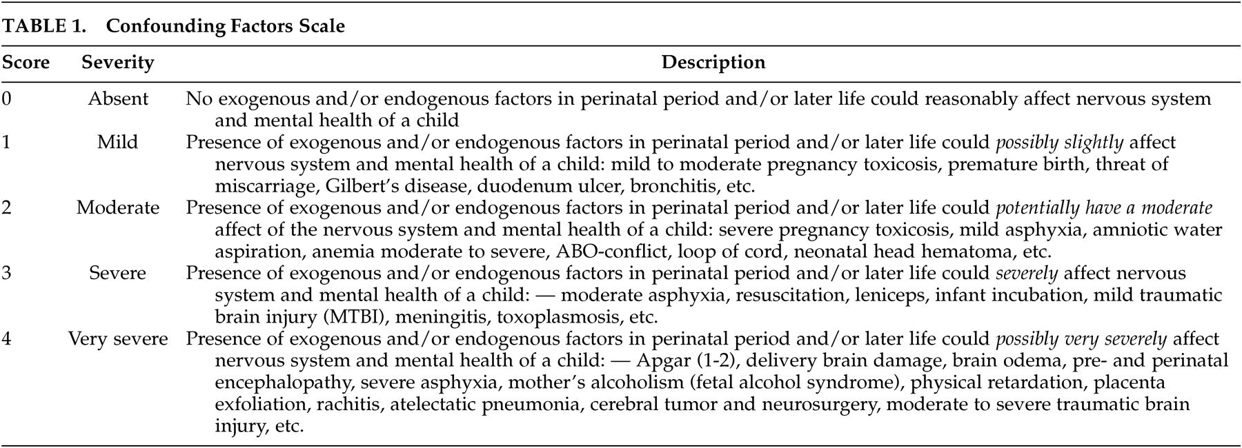
The severity of confounding factors, which could influence the mental health of children—traditional risk factors, such as antenatal and postnatal complications, physical diseases, traumatic brain injuries, infections—was quantified with the elaborated 5-point scale (scores of 0–4), as shown in
Table 1 . This scale was devised and validated within the framework of Subproject 3.4.1, “Potential Effects of Prenatal Irradiation on the Brain as a Result of the Chernobyl Accident (Ukraine),” and Project 3, “Health Effects of the Chernobyl Accident” from the French-German Initiative for Chernobyl.
Assessment of Children
Children in the exposed and comparison groups were examined using a clinical psychiatric interview and a clinical neurological examination and were assessed according to ICD-10 criteria.
Scales and Measurements for Children
The Minor Physical Anomalies Scale was used for the measurement of stigmas of dysembryogenesis.
43 –
44 A special assessment of movement for the quantitative evaluation of fine motor functions
45 –
47 was used, as was the Autonomic Index for assessment of parasympathetic and sympathetic system tone.
48A children’s WISC
49 version normalized for the Ukrainian population was used.
50 Full-scale IQ, verbal IQ, and performance IQ values were obtained. Verbal IQ assessed mainly the functions of the left hemisphere, while performance IQ assessed the right (nondominant) hemisphere.
51 The direction and amount of discrepancy in the verbal/performance scores of the WISC are lateralizing neuropsychological indices.
1 Where children had IQ discrepancies greater than 25 points, brain damage was suspected.
52We used the Rutter Scale A
2 for the assessment of a child’s problems associated with health, hyperactivity, and behavioral and emotional disorders.
53 Conventional and quantitative electroencephalography (qEEG) and checkerboard visual evoked potentials were obtained using a 19-channel brain biopotentials analyzer (Brain Surveyor, SAICO, Italy). Spectral analysis was carried out using classical Fast Fourier Transformation methods for the 1–32 Hz band. Visual evoked potentials were registered binocularly on 50 chess-pattern reversals with a frequency of 1 Hz. The 50 epochs selected for analysis were 400 msec in duration.
Laterality (asymmetry) was assessed using the following equation: Lat=(L−R)/(L+R)×100%, where Lat=laterality coefficient, L=parameter of symmetrical area of the left side, R=parameter of symmetrical area of the right side. Laterality was considered to be significant at Lat>5%.
The parasympathetic and sympathetic system tone was assessed using the Kerdo Autonomous Index (AI),
48 and it was calculated with the following equation: AI=(1−BP
D ×HR
–1 )×100, where BP
D =diastolic blood pressure in left and right upper extremities (mm Hg), HR=heart rate per minute. AI=0 is at autonomic balance (eutonia). Sympathetic tone corresponds to AI>0. Parasympathetic tone prevails when AI<0. Laterality of AI was assessed using the following equation: Lat(AI)=(L(AI)−R(AI)) / (L(AI)+R(AI))×100%, where Lat(AI)=AI laterality coefficient, L(AI)=AI at the left hand, R(AI)=AI at the right hand.
Assessment of Mothers
Scales and Questionnaires for Mothers
On the basis of the DSM-IV scale of stress factors, a 10-item stress-event scale was administered to expectant mothers in relation to the Chernobyl accident. The scale was designed for assessment of the level of real stress factors (but not their perception) from the time of the Chernobyl accident until the birth of their child. The possible scores for each item ranged from 1 (no event) to 5 (the greatest event). Factors included evacuation, lack of information about relatives, migration, and difficulties of medical care.
54The questionnaires for assessment of posttraumatic stress disorder (PTSD) include the Impact of Event Scale
55 and a clinical scale for self-assessment of irritability, depression, and anxiety.
56 The Zung Self-Rating Depression Scale was designed for self-estimation of depression.
57 The General Health Questionnaire (GHQ-28) was used for the self-estimation of psychopathology.
58 The vocabulary subtest of the WAIS
59 was used to determine the mothers’ verbal abilities. All evaluations as well as all scales and measurements for children were blind to the prenatal radiation doses. However, exposure history was a part of the psychiatric interview.
Data Analysis
Data analysis was performed using STATISTICA 5.0 and 6.0 software (Statsoft, Tulsa, Okla.). Statistical processing included descriptive statistics, analysis of variance, Student’s t test, the chi-square test (criterion χ 2 ), Pearson’s product-moment correlation, and regression analysis. The paired t test was used to analyze data when a pair of measurements was obtained for each individual. The differences between groups were considered to be significant at alpha <0.05, and the Bonferroni correction was used when multiple statistical tests were performed to reduce the probability of type I errors. If the expected frequencies of some of the cells of the 2×2 tables were too small (<5), the two-tailed Fisher’s exact test was used instead of the chi-square test.
In the multiple regression analysis, the independent or predictor variables included doses in utero; the week of gestation at the time of the Chernobyl accident; the level of experienced stress events measured by the stress-event scale; the level of confounding (traditional, nonradiation, factors); the mother’s intelligence; and the mother’s level of stress perception (PTSD), anxiety, and depression. The dependent or criterion variables included intelligence measurements and qEEG parameters. Following the multiple regression and correlation analyses, the two dimensional or variable space regression models were defined by the equation Y=a+b×X, where Y =the dependent variable, a =constant (intercept), b =slope (regression coefficient), and X =independent variable, and were calculated for dose-effect relationships for the most informative dependent variables, using the data from all children in the exposed group if none other were specified.
RESULTS
General and Exposure Data
There were 51 (51%) boys and 49 (49%) girls in the exposed group, and 27 (54%) boys and 23 (46%) girls in the comparison group. The children’s mean age was 136 months (11.3 years; SD=5.1 months) in the exposed group and 138.4 months (11.5 years; SD=8.1 months) in the comparison group. Within the exposed group, there were fewer children who were in the earliest stages of prenatal development (0–7 weeks of gestation), relative to the comparison group (12 [12%] versus 14 [28%]; χ 2 =5.96, df=1; p<0.05).
The in utero doses to the brain and thyroid of the embryo and fetus in the exposed group were significantly higher than in the comparison group (
Table 2 ). According to the model of ICRP Publication 88,
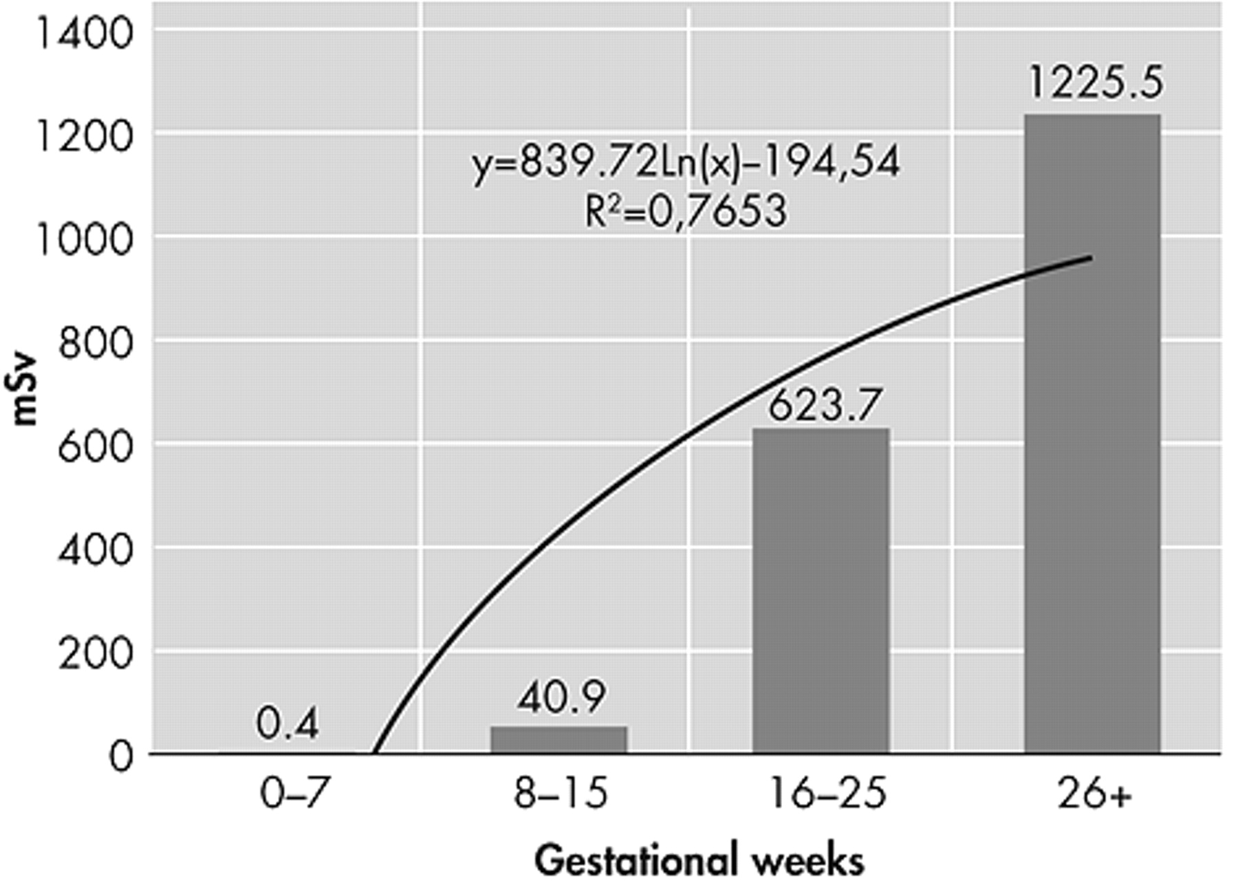
39 there was a strong influence of gestational age on the dose received by the thyroid in utero. The later the intrauterine period at the time of exposure, the higher the in utero thyroid dose absorbed (
Figure 1 ). The permitted limit for thyroid doses in utero, 0.3 Sv, was exceeded in 71% of exposed children, and in 35% of these children thyroid doses in utero exceeded 1 Sv.
Social and Demographic Characteristics of Families
There were no social or demographic differences between the groups concerning divorces, recurrent marriages, incomplete families, average age of parents at the time of the child’s birth, family economic level, and alcohol, smoking, and drug consumption (these additional hazards occurred at low levels in both groups).
Perinatal History
Pregnancy toxemia was more frequent in the exposed group than in the comparison group (63 [63%] versus 16 [32%]; χ 2 =12.85, df=1, p<0.001). There were more pregnancy complications in the exposed group than in the comparison group (42 [42%] versus 12 [24%]; χ 2 = 4.69, df=1, p<0.05). In the exposed group, there were more second-born children, which was not the case in the comparison group (43 [43%] versus 12 [24%]; χ 2 =5.18, df=1, p<0.05). Between the exposed and comparison groups, there were no anthropometric differences in newborns in body weight, length, or Apgar scores.
Psychomotor Development and Medical History
There were no differences between the groups with regard to feeding; mixed feeding (breast-feeding together with formula feeding) prevailed. Paroxysmal states, including epileptic syndromes during the child’s lifetime, were more frequent in the exposed group than in the comparison group (72 [72%] versus 6 [12%]; χ 2 =48.1, df=1, p<0.001).
Physical Health of Children and Confounding Factors
There were no anthropometric differences between the groups on body weight, height, and head circumference at the time of examination. Mean head circumference for exposed children was 53.9 cm (SD=0.9); for the comparison group it was 54 cm (SD=0.7). Children with mild to moderate health problems were more frequently found in the exposed group (87%) than in the comparison group (32 [64%]; χ 2 =10.7, df=1, p=0.001) as a result of pathology of the ear, nose, and throat organs; cardiovascular and blood systems; eyes; and skin. There were no significant differences between the groups in terms of the severity of the confounding factors. There was no difference in handedness between the groups: three children (3%) in the exposed group were left-handed and one child (2%) in the comparison group.
Neuropsychiatric Data
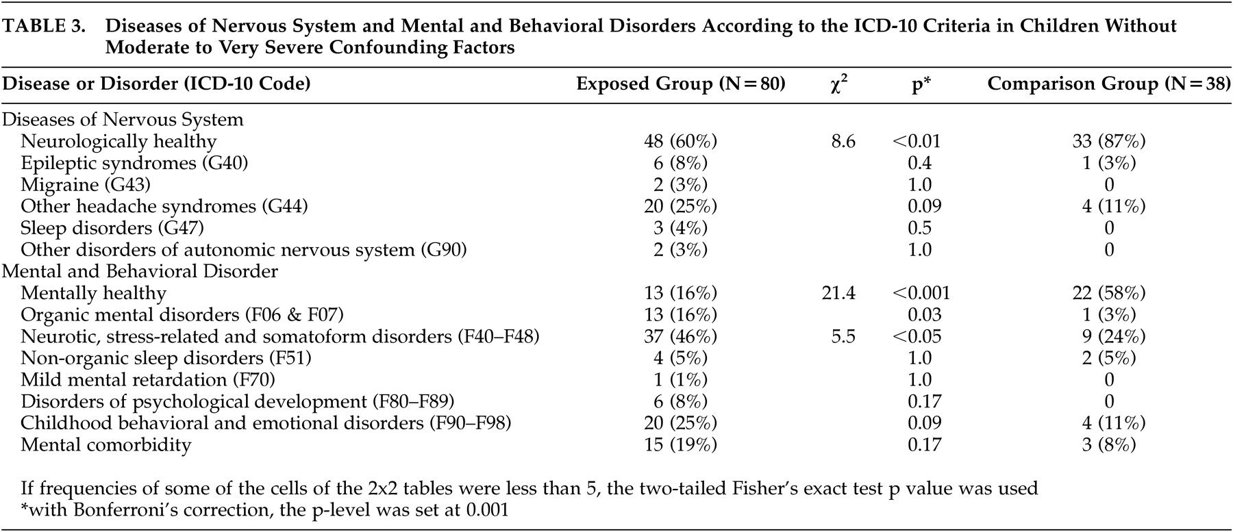
The exposed children were found to have more neuropsychiatric disorders than those in the comparison group. After we excluded children with moderate to very severe confounding factors using Bonferroni adjusted p level (p<0.001), children irradiated in utero had only a tendency to an increased frequency of neurological disorders but had more psychiatric disorders than those in the comparison group (
Table 3 ). In both groups mental and behavioral disorders included predominantly neurotic, stress-related, and somatoform disorders (ICD-10 F40–F48), as well as childhood behavioral and emotional disorders (F90–F98). Together, these disorders were revealed more frequently in the exposed group than in the comparison group (71 [71%] versus 13 [34%]; χ
2 =14.65, df=1, p<0.001). There was a tendency toward an increased frequency of organic mental disorders (organic emotionally labile [asthenic] disorder [F06.6]; organic anxiety disorder [F06.4]; and organic personality disorder [F07.0]) in the exposed group. Severe mental retardation, microcephalus, and epilepsy were not present among the children examined.
Minor Physical Anomalies
Relative to the comparison group, exposed children were more likely to have minor physical anomalies, such as malformed or asymmetrical ears (10 [10%] versus 0, two-tailed Fisher’s exact test, p=0.03) and a high palate (14 [14%] versus 1 [2%], two-tailed Fisher’s exact test, p=0.02; using Bonferroni adjusted p level (p<0.001).
Special Assessment of Movements
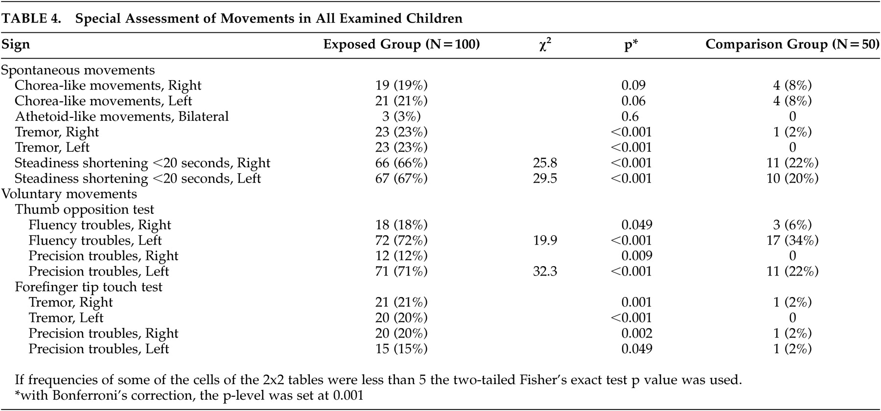
Exposed children were found to have more spontaneous movements (tremor and steadiness shortening less than 20 seconds) and malfunctioning in voluntary movements than those in the comparison group (
Table 4 ). In exposed children, left-sided extrapyramidal dysfunction (fluency and precision troubles, tremor) was more evident than in right-sided extrapyramidal dysfunction relative to those in the comparison group.
Autonomic Dysfunction
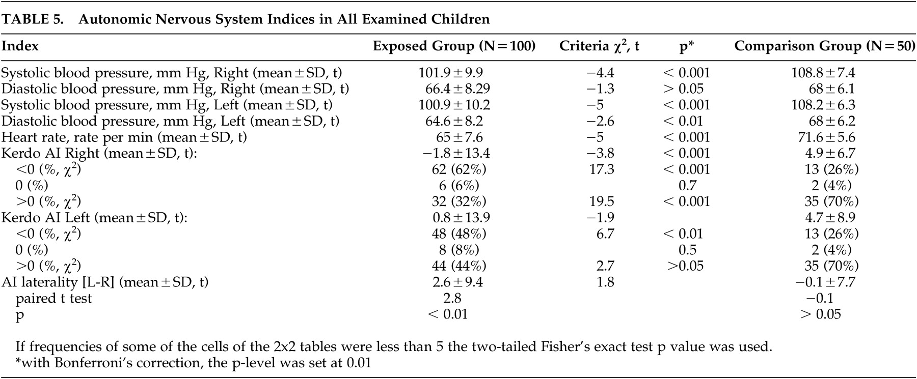
Exposed children had significantly more dysfunction with autonomic control of the cardiovascular system, as shown in
Table 5 . Thus, left and right systolic blood pressure, left diastolic blood pressure, and heart rate were lower in the exposed group than in the comparison group. Exposed children had lateralized Kerdo Autonomic Index (AI) to the left and parasympathetic tone prevalence.
WISC
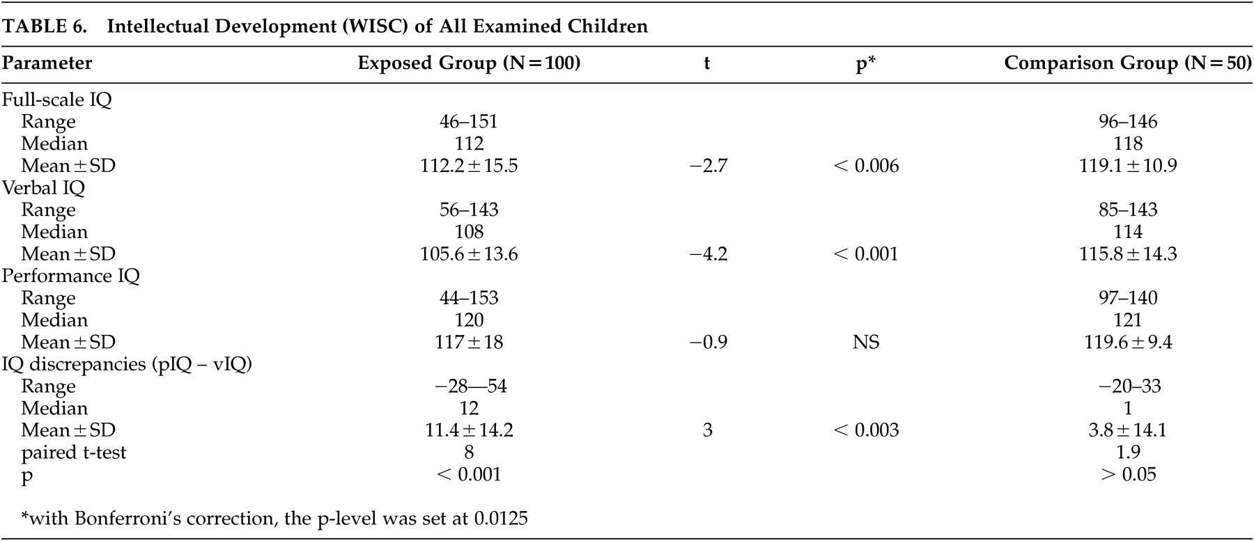
Exposed children had decreased full-scale and verbal IQ as well as increased IQ discrepancy due to verbal decrement compared with comparison group children (
Table 6 ). The intelligence of exposed children was lower on the similarities and vocabulary verbal subtests (
Table 7 ). The exposed children still showed lower verbal IQ and greater IQ discrepancy than those in the comparison group (
Table 8 ).
In the multiple regression model, the significant predictors of full-scale IQ in the exposed group were the mother’s vocabulary subtest score (df=5, 94, p=0.0003) and the confounding factors score (df=5, 94, p=0.003); the significant predictors of verbal IQ was the mother’s vocabulary subtest score (df=5, 94, p=0.0005); for the performance IQ predictors, they were the mother’s vocabulary subtest score (df=5, 94, p=0.004) and the confounding factors score (df=5, 94, p=0.002).
There were no significant predictors for IQ discrepancy due to verbal decrement in the exposed children. In the exposed children who had an IQ discrepancy of more than 25 points, suggesting the presence of brain damage,
52 the IQ discrepancy predictors (R
2 =0.48, F=6.1, df=2, 13, p<0.01) were the fetal radiation dose and the level of stress factors. Moreover, the significance of the multiple regression model increased in proportion to the discrepancy; at an IQ discrepancy of more than 29 points, the R
2 reaches 0.96 (F=24.99, df=2, 4, p<0.01), where the only significant predictor is the fetal dose (p=0.0059).
The full-scale IQ, verbal IQ, and performance IQ in the exposed children were correlated with the mother’s vocabulary subtest score (r=0.32, df=1, 98, p=0.002; r=0.32, df=1, 98, p=0.002; r=0.25, df=1, 98, p=0.013, respectively). The full-scale IQ and performance IQ in the exposed children fell in proportion to the mother’s mental health problems as assessed by the GHQ-28 (r=0.26, df=1, 98, p=0.013; r=0.27, df=1, 98, p=0.008, respectively). At the same time, PTSD in mothers as measured by the Impact of Events Scale was inversely correlated with IQ discrepancies (r=0.22, df=1, 98, p=0.034). There was no significant correlation between intelligence in exposed children and dose received in utero.
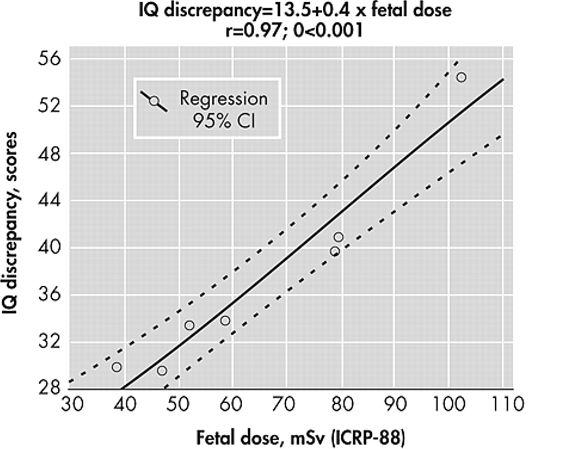
Among all exposed children, plotting the cases of IQ discrepancies greater than 25 points against prenatal fetal dose gives a positive correlation (r=0.5, df=1, 14, p=0.029). The power of this correlation between the discrepancy and fetal doses grows with increased discrepancy: at a discrepancy ≥30 (
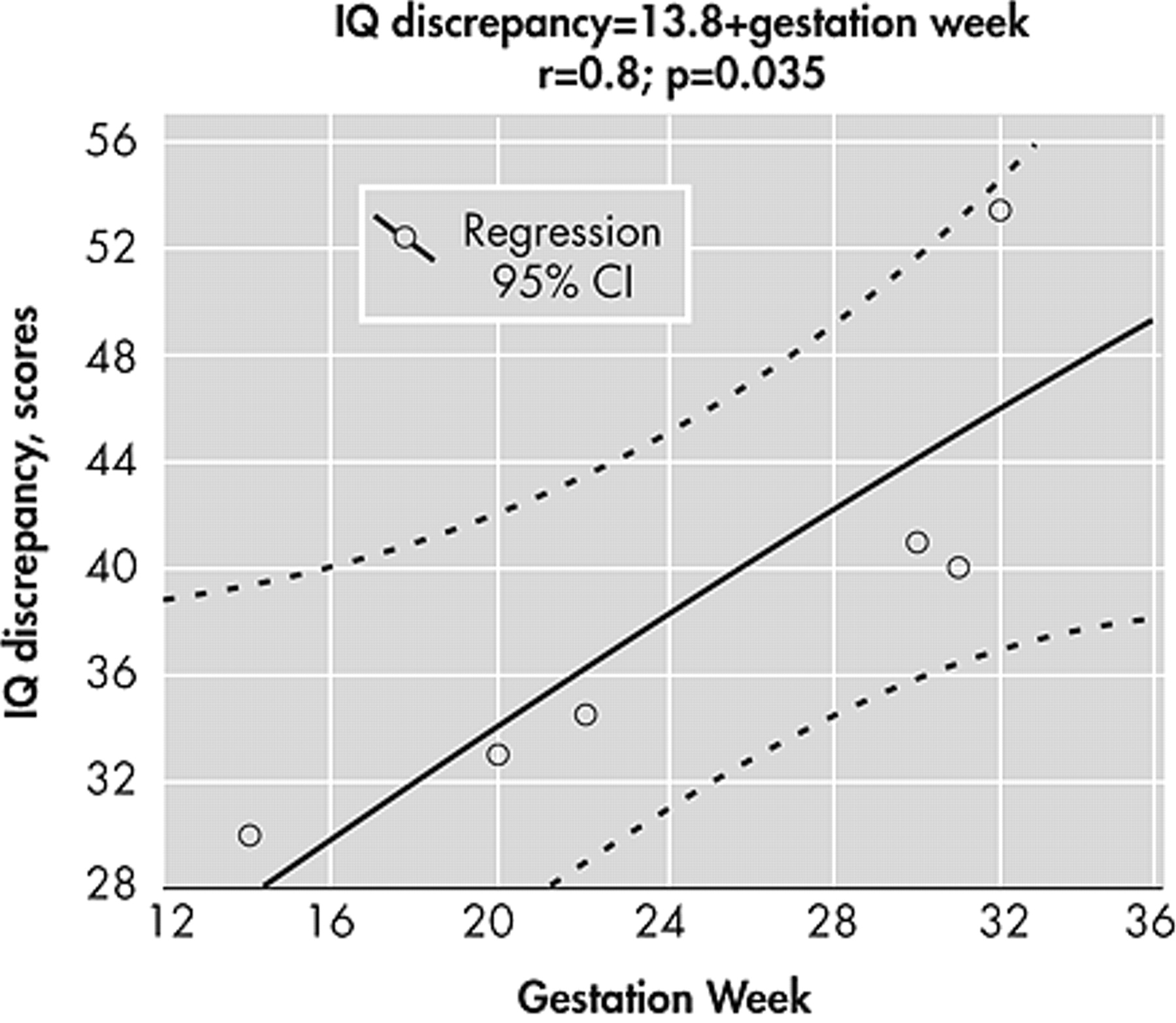
Figure 2 ), the correlation with fetal dose increased (r=0.97, df=1, 5, p<0.001), and correlation with the in utero thyroid dose according to ICRP Publication 88 is r=0.7 (df=1, 5, p≈0.05). IQ discrepancy greater than 25 points increased in proportion to the gestation week at the time of exposure (r=0.6, df=1, 14, p=0.027). The power of this correlation also grows with increased verbal/performance IQ discrepancy: at a discrepancy ≥30 (
Figure 3 ), there is a strong correlation with the gestation week (r=0.8, df=1, 5, p=0.035). It should be noted that IQ discrepancy greater than 25 points also increased in proportion to the level of stress events (r=0.6, df=1, 14, p=0.027), although this correlation disappears if the IQ discrepancy exceeds 30 points. There were no other significant correlations between IQ discrepancy and the tested independent variables.
Rutter Scale A
According to mothers interviewed, the severity of emotional and behavioral problems was similar in the exposed and the comparison groups (14.2±7.6 versus 13.2±7.2 scores; t=0.7, df=1, p>0.05). The number of children without emotional and behavioral disorders (scores <13 on the Rutter Scale A) was also similar: 42 (42%) in the exposed group versus 20 (40%) in the comparison group (χ 2 =0.05, df=1, p>0.05).
Conventional EEG
EEG patterns of the comparison group corresponded to norms for 10–12-year-old children. Children exposed in utero had more abnormal EEG patterns than did those in the comparison group: 29 exposed children (29%) exhibited low-voltage (flat) EEG versus four from the comparison group (8%) (two-tailed Fisher’s exact test, p=0.003 [Bonferroni adjusted p level p=0.01]); 18 exposed children (18%) showed disorganized slow EEG type versus seven comparison children (14%) (χ 2 =0.4, df=1, p>0.05); 27 exposed children (27%) showed disorganized EEG type with paroxysmal activity versus 11 comparison children (22%) (χ 2 =0.4, df=1, p>0.05); and 10 exposed children (10%) versus one comparison child (2%) showed epileptiform EEG pattern (two-tailed Fisher’s exact test, p=0.1).
Interhemispheric asymmetry of EEG (laterality) was more typical for the exposed group than the comparison group (73 exposed children [73%] versus 19 comparison children [38%]; χ 2 =17.2, df=1, p<0.001), according to Lat <5%: left hemisphere dysfunction (37 exposed children [37%] versus six comparison children [12%]; χ 2 =10.2, df=1, p<0.001); right hemisphere dysfunction (15 exposed children [15%] versus 10 comparison children [20%]; χ 2 =0.6, df=1, p>0.05); cross-hemisphere dysfunction (21 exposed children [21%] versus three comparison children [6%]; two-tailed Fisher’s exact test, p=0.02).
Quantified EEG
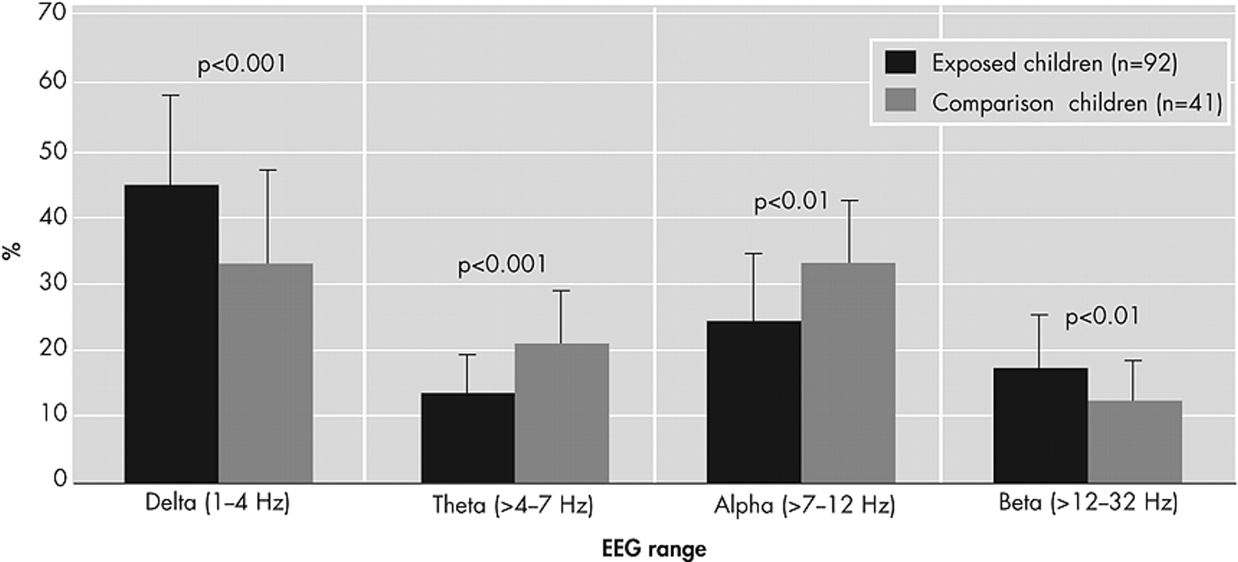
Exposed children were found to have increased delta and beta power and decreased alpha and theta power. After we excluded children with severe to very severe confounding factors, exposed children again were distinguishable from comparison group children, as shown in
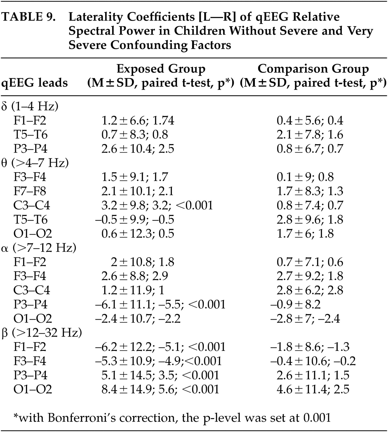
Figure 4 . Using laterality coefficients for qEEG (
Table 9 ), it was revealed in exposed children a tendency for delta power lateralization and shift of theta power to the left hemisphere, depression of alpha power in the left parietal area, depression of beta power in the left frontal area, and excess of beta power in the left parieto-occipital region.
In all exposed children, lateralization of theta power extension to the left temporal and occipital areas was correlated with the fetal dose (r=0.2–0.3, df=1, 98, p<0.01) and thyroid dose in utero (r=0.2, df=1, 98, p<0.05). At the same time, lateralization of alpha power depression to the left frontal area increased in proportion to fetal and thyroid doses in utero (r=0.3, df=1, 98, p<0.05). Lateralization of beta power extension to the left central area increased in proportion to fetal doses (r=0.2, df=1, 98, p<0.05). The dominant frequency of theta range in frontotemporal areas also increased in proportion to doses in utero (r=0.2–0.3, df=1, 98, p<0.05).
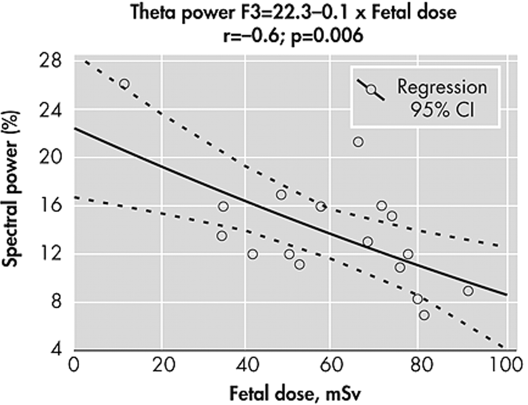
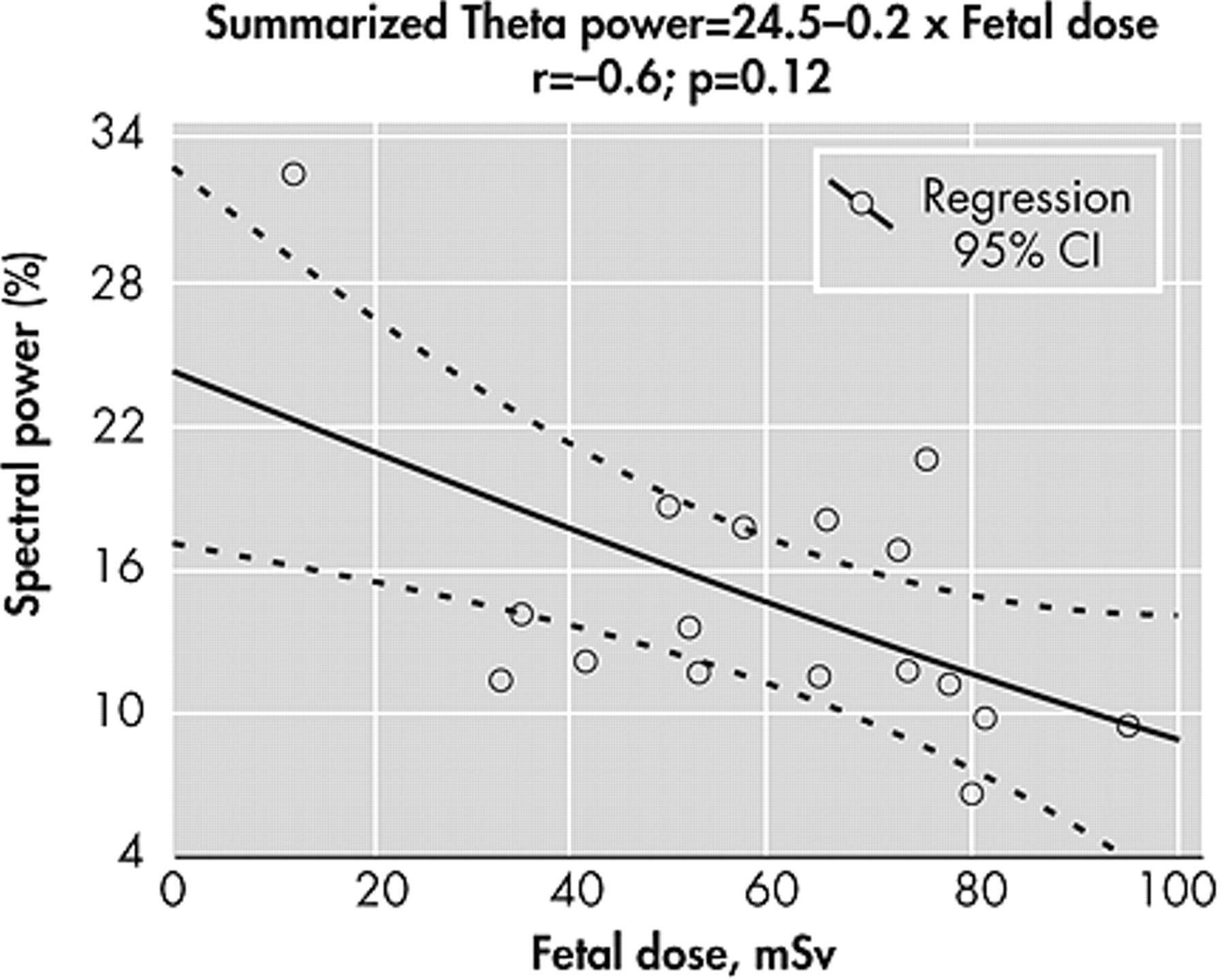
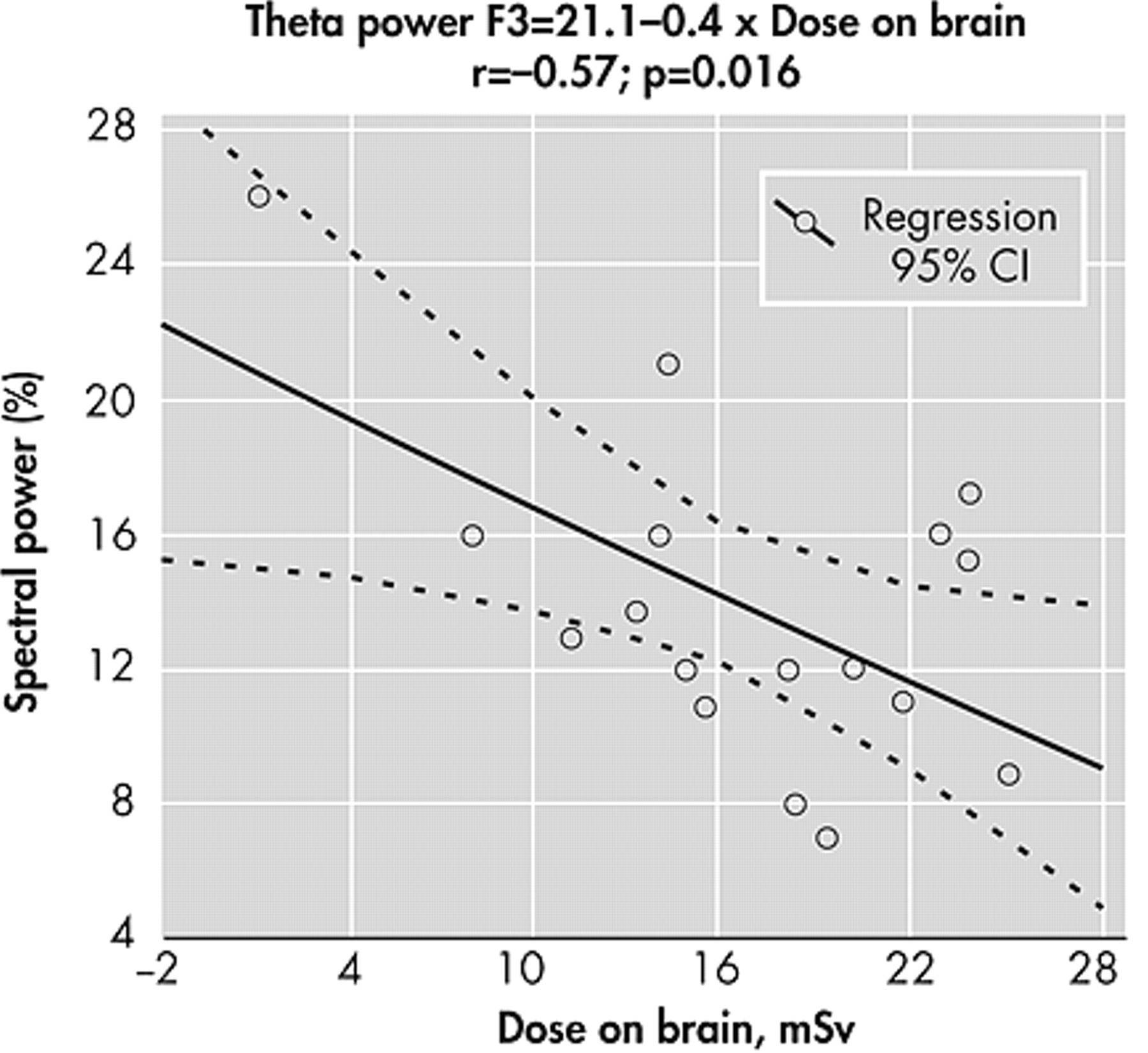
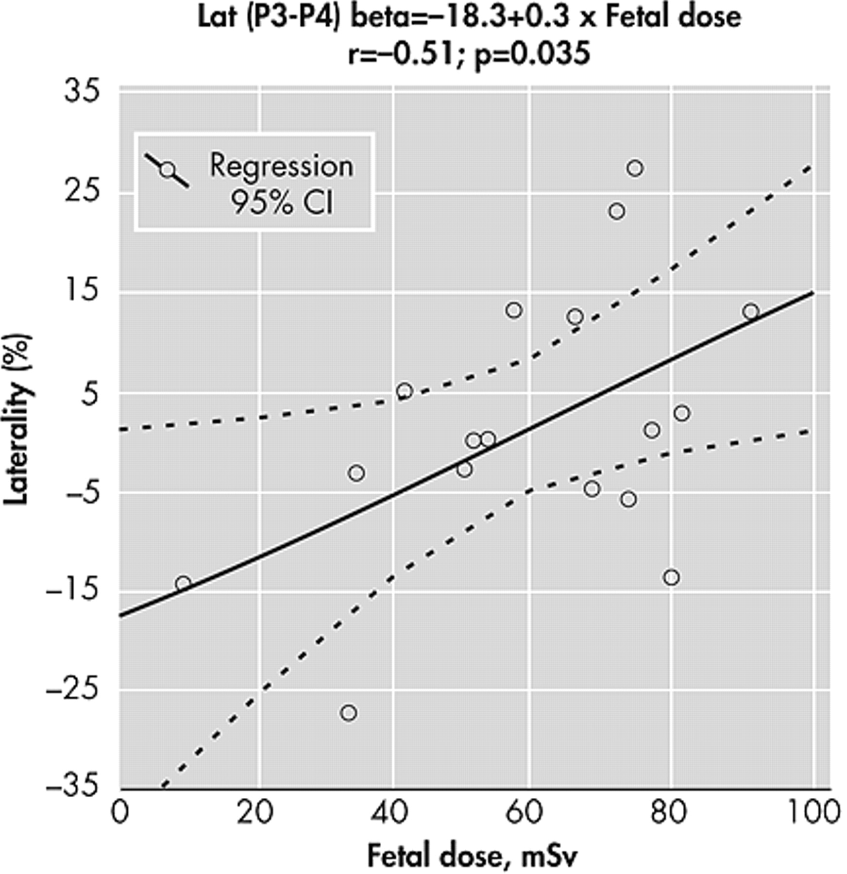
In exposed children without any moderate to very severe confounding factors who were at 16–25 weeks of gestation at the time of the Chernobyl accident, total relative theta power and left frontotemporal theta power decreased in proportion to the fetal dose received in utero (r=0.5–0.6, df=1, 11, p<0.05), as presented in
Figure 5,
Figure 6, and
Figure 7 . Lateralization of beta power to the left parietal area increases in proportion to the fetal dose (r=0.51, df=1, 11, p=0.035) (
Figure 8 ). The dominant frequency of the delta range decreased in relation with fetal dose in the temporal areas (r=0.5–0.6, p<0.05).
Visual Evoked Potentials
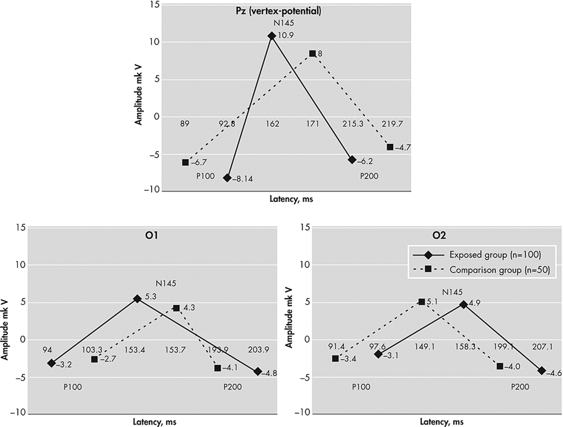
The characteristic features of visual evoked potentials in exposed children are shown in
Figure 9 . The characteristic pattern of visual evoked potentials in exposed children is a vertex potential, high amplitude (up 30.7 μV) double-phase potential with a component P100 latency of 42–152 msec, N145 75–245 msec and P200 115–302 msec registered in the central parietal lead (Pz). Vertex-potential was detected according to the amplitude of N145 components, and if its amplitude was >20 μV (the mean +2 standard deviations in the comparison group), this potential was considered to be abnormal. Among exposed children without severe and very severe confounding factors, 10 (10%) had abnormal vertex potential (>20 μV), while such potentials were absent in the comparison group children (two-tailed Fisher’s exact test, p=0.03). Paroxysmal (epileptiform) disorders were clinical equivalents of abnormal vertex potential.
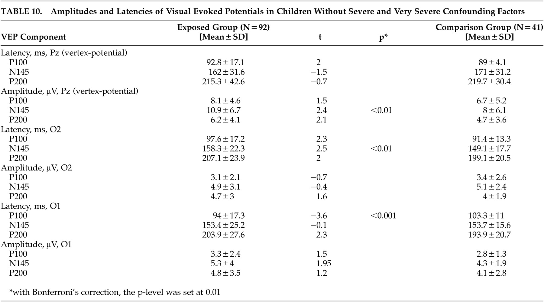
In children without severe to very severe confounding factors, the averaged visual evoked potential characteristics were different from those of exposed children (
Table 10 ), with increased latencies of N145 in the right occipital area O2 and decreased latency of P100 in the left occipital area O1. A positive laterality coefficient indicated an increased parameter at the left occipital area, while a negative laterality coefficient indicated a decreased parameter at the left occipital area. As is shown in
Table 11, in comparison group children P100 latency was larger at the occipital lead of the left (dominant) hemisphere O1 than at the occipital lead of the right (nondominant) hemisphere O2. This indicated more rapid visual information processing in the nondominant (right) hemisphere, as is typically found. In contrast, exposed children not only lost normal asymmetry in visual information processing but exhibited opposite lateralization predominantly in the dominant hemisphere, as followed by negative laterality coefficients of P200 latency. Taken together, these findings indicate the presence of abnormalities of visual information processing in prenatally irradiated children, where information processing is mainly in the left brain hemisphere rather than the right.
Mothers’ Mental Health
Mothers evacuated from Pripyat experienced many real stress events and, as shown in
Table 12, significant mental health problems, including depression, PTSD, and somatoform disorders. A correlation was found between the mothers’ mental health deterioration and the full-scale IQ in their children. Verbal IQ, however, did not correlate with the mothers’ mental health. There were statistically significant relationships between the mental health deterioration of the mothers and neuropsychiatric disorders in the children. However, there were no differences in the mothers’ verbal IQ between the groups, as assessed by the vocabulary subtest of the WAIS (
Table 12 ). Thus, the deterioration of the verbal IQ of the exposed children cannot be explained by the influence of the verbal IQ of their mothers, although there was a natural tendency of the verbal IQ of the children in both groups to increase with the mothers’ WAIS verbal subtest score.
DISCUSSION
The comparable clinical neuropsychiatric, neuropsychological, and neurophysiological findings suggest abnormal development of the dominant brain hemisphere, particularly in the frontal and temporal lobes, after prenatal exposure to ionizing radiation as a result of the Chernobyl accident. There is a dysfunction of the corticolimbic system, mainly in the dominant (left) hemisphere, with interhemispheric inversion of the maximal visual information processing from the nondominant hemisphere toward the dominant (left). The abnormal vertex potential verified limbic system irritation, which is considered to be the neurophysiological marker of epileptic spectrum disorders in children.
60The IQ of both exposed and nonexposed children was quite high despite a significant number of neuropsychiatric problems in both groups. At the same time, few severe neurological or psychiatric disorders were found among these children. The high IQ could be reasonably explained as an “urbanization effect,” where childhood development and learning occur in a large city (Kiev) with its rich cultural environment.
Dose-related cognitive and neurophysiological abnormalities in prenatally exposed children were observed following exposure to fetal doses >20 mSv and thyroid doses in utero >300 mSv in the 8th and later weeks of gestation, as well as fetal doses >10 mSv and thyroid doses in utero >200 mSv at 16–25 weeks of gestation.
54 These effects are attributed to irradiation in utero and are based on the “dose-effects” relationships and gestational stage.
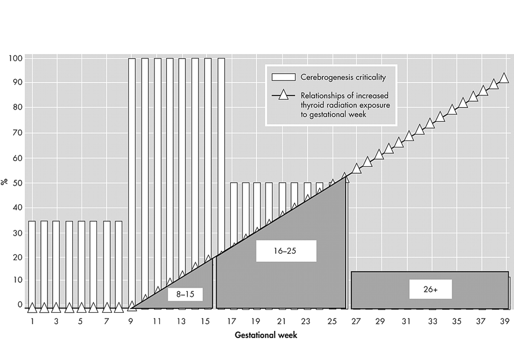
The most critical periods of cerebrogenesis are during weeks 8–15 and, to a lesser extent, weeks 16–25 of gestation and correspond to the most sensitive radioneuroembryological effects following external prenatal exposure during atomic bombing or radiological medical procedures. However, low doses of external fetal exposure as a result of the Chernobyl accident did not disrupt neuronal migration at the most critical stage of cerebrogenesis (8–15 weeks of gestation). At that time, the fetal thyroid is very small, and its metabolism is minimal. So thyroid doses in utero and, consequently, doses to the embryo and the fetus are also low. At the next critical stage of cerebrogenesis (16–25 weeks of gestation), neuronal differentiation and synaptogenesis increase, as does brain cytoarchitecture; the limbic system and its connections are developing, and brain asymmetry and hemisphere dominance are forming.
7,
61 –
63 Thyroid weight and metabolism rate also increase, with increased thyroid exposure in utero and, consequently, doses received by the embryo and the fetus. During this second critical period of cerebrogenesis, there is a certain intersection of cerebrogenetic vulnerability and increased prenatal susceptibility, as shown in
Figure 10 . These effects (neurological asymmetry, IQ discrepancy with verbal IQ decrement, EEG lateralization to the left, and visual information processing inversion to the left hemisphere) are caused by disrupted neuroembryological development at 16–25 weeks of gestation. Despite the reduced radiovulnerability of the developing brain, prenatal exposure of thyroid and fetus continues to increase. As a result, cognitive and neurophysiological abnormalities are still present through the gestational period (+26 weeks).
54,
64Left brain damage in utero caused by radiation and its relevance to schizophrenia should be discussed within the framework of the neurodevelopmental hypothesis of schizophrenia.
65 Schizophrenia is generally considered to be a disease of the left (dominant) brain.
1,
66 Neuropsychiatric, neurophysiological, neuropsychological, and neuroimaging studies of Chernobyl accident survivors have revealed dose-related left cortical-limbic brain damage.
67 –
74 Specific damage to the left hemisphere has been revealed in long-term studies of the aftermath of radiotherapy.
75 –
78 It is suggested that ionizing radiation is a risk factor for schizophrenia spectrum disorders.
79 –
80 Recently, important experimental radioneuroembryological research on nonhuman models of schizophrenia has also supported the hypothesis that prenatal exposure of brain tissue to ionizing (X-ray) radiation may be associated with an increased risk of schizophrenia later in life.
81 –
86 Consequently, persons exposed in utero after the Chernobyl accident may also have an increased risk of schizophrenia. Thus, whether prenatal irradiation is a risk factor for schizophrenia and other neuropsychiatric disorders should be verified in international follow-up epidemiological studies.
Acknowledgments
Supported in part by a grant from the Subproject 3.4.1 “Potential Effects of Prenatal Irradiation on the Brain as a Result of the Chernobyl Accident (Ukraine),” Project No. 3 “Health Effects of the Chernobyl Accident” of the French-German Initiative for Chernobyl from the Bavarian State Ministry of Environmental and Developmental Affairs. The authors thank our colleagues from Alberta Hospital Edmonton (Canada) - Professor Pierre Flor-Henry for his creative advice and kind support during the study; Dr. John Lind for help with statistical analysis; and Mrs. Kimberly Robbins-Bron for her editing services. We also thank Dr. Konstantin Yuryev (Kiev) for his significant help in the statistical analysis of the data.
