Full access
Letters
Published Online: 1 April 2011

EMDR Treatment for Posttraumatic Stress Disorder, With Focus on Hippocampal Volumes: A Pilot Study

Publication: The Journal of Neuropsychiatry and Clinical Neurosciences
To the Editor: Recent studies have suggested that hippocampal neurogenesis may be stimulated by successful treatment of posttraumatic stress disorder (PTSD), either by drugs14 or by behavioral interventions.5 Three investigations have evaluated the effects of pharmacotherapies and have suggested that antidepressant treatments may increase hippocampal volume.13 Lindauer and colleagues6 examined the effects of a psychotherapy intervention, the Brief Eclectic Psychotherapy, on hippocampal volumes and observed no changes on hippocampal volumes upon amelioration of PTSD symptoms. However, the authors recently reported
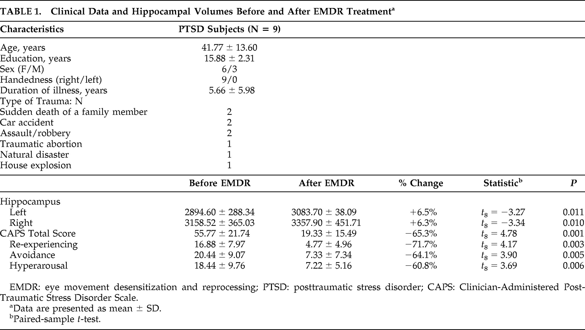
5 the case of a patient treated with Eye Movement Desensitization and Reprocessing (EMDR) who showed an increase in both left- and right-hippocampus upon improvement in PTSD symptoms. The aim of this study was to further investigate the relationship between EMDR treatment and hippocampal volumes in PTSD subjects. The study was conducted at the Department of Psychiatry, University of Siena Medical Center. Inclusion criteria were current PTSD and age 18–65 years. Exclusion criteria were current and/or lifetime comorbid psychiatric diagnoses as ascertained via the SCID; previous or current use of any psychotropic medication; history of head trauma; and presence of neurological, endocrine, or degenerative disorders. The authors recruited 10 outpatients, but one patient was excluded from the study because of MRI signal abnormalities. The remaining nine subjects were three men and six women, mean age: 41.77 (SD: 13.60) years. Subjects were assessed before and after EMDR treatment through the Clinician Administered Posttraumatic Stress Disorder Scale (CAPS) Italian Version,7 which is known to be a reliable measure of PTSD symptom severity. All volumetric MRI scans were performed with a 1.5 Tesla Philips Intera Machine (Philips Medical Systems; Best, the Netherlands). The imaging protocol included a T1-weighted Fast Field Echo (FFE) 1-mm-thick coronal images (matrix 512 x 512, slice thickness: 1 mm, slice number: 75). Hippocampal volumes were calculated by manually tracing the structure separately for the left and right hemispheres on each coronal slice and then summing these areas of successive coronal slices with the ANALYZE Software Package, Version 5.0 (Biomedical Imaging Resource, Mayo Clinic Foundation, Rochester, MN). The boundaries of the hippocampus were drawn following the guidelines described by Watson Laboratories, Inc.,8 as described in a previous study.9 The measurements were performed by a single rater blind to subjects' identity and treatment status. The treatment followed the guidelines by Shapiro.8 the therapy was given in a 90-minute session and was randomly assigned to three trained psychotherapists. Duration of the treatment was 3 months (12 ± 1 EMDR sessions). The bilateral sensory stimulation included both eye movements while following the finger of the therapist and tapping the hand of the patient. All sessions were videotaped; the fidelity to the treatment protocol was assessed by an independent evaluator, a psychologist and licensed psychotherapist who is an EMDR European-approved consultant and supervisor and who was not involved in the present study. Three months after the start of EMDR treatment, all subjects were reevaluated with MRI and CAPS.
Statistical analyses were performed with SPSS Version 13.0 for Windows (SPSS Inc, Chicago, IL). Hippocampal volumes were analyzed with a repeated-measures analysis of variance. Also, two-tailed, paired-sample t-tests were used to compare hippocampi, clinical assessments on CAPS total score, and each subscale of the CAPS before and after treatment. An alpha level of 0.05 was used for all statistical tests. The analysis showed that there was a significant difference in hippocampal volumes after the treatment (F=11.70; df=1,8; p=0.009) and a significant difference between the two sides (F=18.95; df=1,8; p=0.002), but no interaction between treatment phase and side (F=0.11; df=1,8; p=0.742). Detailed analyses indicated that there was an increase in both the left (+6.5%; t = –3.27; df=8; p=0.011) and the right side (+6.3%; t = –3.34; df=8, p=0.010; Table 1). After treatment, we also observed an improvement in PTSD symptoms as measured with CAPS total score (−65.3%; t=4.78; df=8; p=0.001), and on the three CAPS subscales (Table 1). After the treatment, all subjects no longer met the SCID criteria for PTSD. Despite these promising results, we acknowledge that the present study is only a pilot study because it has several limitations. First, the sample size was small. Second, there was no control group (this choice was motivated by ethical reasons). Third, we do not know whether the increase in hippocampal volume was caused by a promotion of neurogenesis or other reasons, such as, for example, a change in water content in the hippocampus. Despite these limitations, the results of this preliminary study suggest further consideration of the potential effects of psychotherapy on the neurobiology of psychiatric disorders, and not dismissing the question of whether psychotherapy may have beneficial effects on hippocampal structure. Further studies should be carried out in order to validate the present results, possibly with a larger sample of PTSD subjects, a control group, and different types of psychotherapy.
TABLE 1. Clinical Data and Hippocampal Volumes Before and After EMDR Treatmenta
EMDR: eye movement desensitization and reprocessing; PTSD: posttraumatic stress disorder; CAPS: Clinician-Administered Post-Traumatic Stress Disorder Scale.
a
Data are presented as mean ± SD.
bPaired-sample t-test.

References

1.
Vermetten E, Vythilingam M, Southwick SM, et al.: Long-term treatment with paroxetine increases verbal declarative memory and hippocampal volume in posttraumatic stress disorder. Biol Psychiatry 2003; 54:693–702
2.
Bremner JD, Mletzko T, Welter S, et al.: Effects of phenytoin on memory, cognition, and brain structure in post-traumatic stress disorder: a pilot study. J Psychopharmacol 2005; 19:159–165
3.
Bossini L, Tavanti M, Lombardelli A, et al.: Changes in hippocampal volume in patients with post-traumatic stress disorder after sertraline treatment: J Clin Psychopharmacol 2007; 27:233–235
4.
Bremner JD, Vermetten E: Neuroanatomical changes associated with pharmacotherapy in posttraumatic stress disorder. Ann N Y Acad Sci 2004; 1032:154–157
5.
Bossini L, Fagiolini A, Castrogiovanni P: Neuroanatomical changes after eye movement desensitization and reprocessing (EMDR) treatment in posttraumatic stress disorder. J Neuropsychiatry Clin Neurosci 2007; 19:475–476
6.
Lindauer RJ, Vlieger EJ, Jalink M, et al.: Effects of psychotherapy on hippocampal volume in outpatients with post-traumatic stress disorder: an MRI investigation. Psychol Med 2005; 35:1421–1431
7.
Blake DD, Weathers FW, Nagy LM, et al.: The development of a clinician-administered PTSD Scale. J Trauma Stress 1995; 8:75–90 in Repertorio delle Scale di Valutazione in Psichiatria. Firenze, S.E.E. (Società Editrice Europea), 1999. Edited by Conti L. pp 1239–1290
8.
Watson C, Andermann F, Gloor P, et al.: Anatomic basis of amygdaloid and hippocampal volume measurement by magnetic resonance imaging. Neurology 1992; 42:1743–1750
9.
Bossini L, Tavanti M, Calossi S, et al.: Magnetic resonance imaging volumes of the hippocampus in drug-naïve patients with post-traumatic stress disorder without comorbidity conditions. J Psychiatr Res 2008; 42:752–762
10.
Shapiro F: Eye Movement Desensitization and Reprocessing: Basic Principles, Protocols, and Procedures. New York, Guilford, 1995

Information & Authors

Information

Published In

Go to The Journal of Neuropsychiatry and Clinical Neurosciences
The Journal of Neuropsychiatry and Clinical Neurosciences
Pages: E1 - E2
PubMed: 21677204

History

Published online: 1 April 2011
Published in print: Spring 2011

Authors

Affiliations

Letizia Bossini, M.D.
Dept. of Neuroscience, Psychiatry Division Dept. of Neurological and Behavioral Sciences University of Siena School of Medicine University of Siena School of Medicine Siena, Italy
Maricla Tavanti, M.D.
Dept. of Neuroscience, Psychiatry Division Dept. of Neurological and Behavioral Sciences University of Siena School of Medicine University of Siena School of Medicine Siena, Italy
Sara Calossi, M.D.
Dept. of Neuroscience, Psychiatry Division Dept. of Neurological and Behavioral Sciences University of Siena School of Medicine University of Siena School of Medicine Siena, Italy
Nicola R. Polizzotto, M.D.
Dept. of Neuroscience, Psychiatry Division Dept. of Neurological and Behavioral Sciences University of Siena School of Medicine University of Siena School of Medicine Siena, Italy
Gianpaolo Vatti, M.D.
Dept. of Neuroscience, Psychiatry Division Dept. of Neurological and Behavioral Sciences University of Siena School of Medicine University of Siena School of Medicine Siena, Italy
Daniela Marino, M.D.
Dept. of Neuroscience, Psychiatry Division Dept. of Neurological and Behavioral Sciences University of Siena School of Medicine University of Siena School of Medicine Siena, Italy
Paolo Castrogiovanni, M.D.
Dept. of Neuroscience, Psychiatry Division Dept. of Neurological and Behavioral Sciences University of Siena School of Medicine University of Siena School of Medicine Siena, Italy

Metrics & Citations

Metrics

Citations

Export Citations

If you have the appropriate software installed, you can download article citation data to the citation manager of your choice. Simply select your manager software from the list below and click Download.

For more information or tips please see 'Downloading to a citation manager' in the Help menu.

Format
Citation style
Style
Copy to clipboard

There are no citations for this item

View Options

View options

PDF/ePub

View PDF/ePub

Get Access

Login options

Already a subscriber? Access your subscription through your login credentials or your institution for full access to this article.

Personal login Institutional Login Open Athens login
Purchase Options

Purchase this article to access the full text.

PPV Articles - Journal of Neuropsychiatry and Clinical Neurosciences

PPV Articles - Journal of Neuropsychiatry and Clinical Neurosciences

Not a subscriber?

Subscribe Now / Learn More

PsychiatryOnline subscription options offer access to the DSM-5-TR® library, books, journals, CME, and patient resources. This all-in-one virtual library provides psychiatrists and mental health professionals with key resources for diagnosis, treatment, research, and professional development.

Need more help? PsychiatryOnline Customer Service may be reached by emailing [email protected] or by calling 800-368-5777 (in the U.S.) or 703-907-7322 (outside the U.S.).

Media

Figures

Other

Tables

Share

Share

Share article link

Share