Full access
Clinical Research Reports
Published Online: 1 July 2011

Influence of Premorbid Psychopathology and Lesion Location on Affective and Behavioral Disorders After Ischemic Stroke

Publication: The Journal of Neuropsychiatry and Clinical Neurosciences

Abstract

Early recognition of psychopathological symptoms (PSs) after stroke is important because they greatly influence the recovery of patients. The aim of this study was to investigate the predictive factors of PSs occurring in patients with ischemic stroke. Eighty-nine patients were prospectively evaluated upon admission and 4, 12, and 26 weeks later with the Neuropsychiatric Inventory, Hamilton's Rating Scales for Depression and Anxiety, and a battery of neuropsychological and functional scales. Depression and apathy were the most frequent PSs detected after stroke. Premorbid psychopathologies and right-hemisphere location were the main predictive indicators of early and long-term PSs.
Assessment of behavioral and affective disorders in patients who have suffered an ischemic stroke is essential because these patients commonly suffer from psychopathological symptoms (PSs) that can greatly influence their recovery and quality of life.1,2
Several studies have drawn attention to the correlation between PSs and stroke. Depression and apathy have been reported as the most frequent disorders.36 Few studies have focused on the occurrence of depression or other psychiatric disorders in the acute and early post-acute phase of stroke, which is a crucial period for patients' rehabilitative treatment.7 Some authors have investigated the influence of premorbid personality traits on the occurrence of PSs after stroke,4,8 but psychopathological status before the event and its predictive potential for poststroke PSs have not been studied.
Some factors occurring in the acute phase of stroke, such as low consciousness level, aphasia, or comorbid pathologies, can limit patients' abilities to manifest and communicate their PSs.7 The Neuropsychiatric Inventory (NPI) uses a reliable informant to quantitatively assess a patient's behavioral symptoms over the past 4 weeks.9 The NPI was designed for patients with dementia, but it has also been used in such neurological disorders as multiple sclerosis10 and stroke.5,6 Given that this test does not require the patient's collaboration, the NPI can be used to provide information about the patient's psychopathological status before, and in the weeks immediately after, the occurrence of the stroke.
The aim of this study was to identify predictive factors of PSs associated with ischemic stroke that could be detected at the onset of the stroke, with a special focus on premorbid psychopathology, in order to identify a patient profile at risk of developing poststroke PSs. We also evaluated the frequency and evolution of these PSs as well as their correlation with the patient's cognitive and functional status.

METHOD

Participants

We prospectively studied a population of patients with ischemic stroke who were admitted to the Neurology Unit in the Hospital Virgen del Puerto. They were evaluated upon admission and again at 4, 12, and 26 weeks after their strokes. The recruitment period was from May 2007 to December 2008.

Inclusion/Exclusion Criteria

Patients admitted to the hospital with an acute cerebral infarct (CI) confirmed by neuroimaging techniques, and who had a responsible caregiver, qualified for the study. The exclusion criteria were the following: knowledge or suspicion of previous dementia or cognitive decline, as assessed by clinical record or by means of the Jorm's Informant Questionnaire on Cognitive Decline in the Elderly (IQCODE);11 cerebral hemorrhage or another suspected etiology of brain injury (tumor or others); transient ischemic attack; persistent coma or severe alteration of consciousness 4 weeks after the stroke; and death or appearance of a new brain lesion before 4 weeks had passed (stroke recurrence, cerebral hemorrhage, or others).

Instruments

We used the following instruments for patient evaluation: 1) A clinical record form for clinical, demographic, and social variables: age, gender, years of formal education, documented previous clinical diagnosis of hypertension, diabetes, or hypercholesterolemia; current smoking habit or hazardous/harmful alcohol consumption (defined as current intake of >280 g per week in men and >168g per week in women, according to the recommended criteria of the World Health Organization12); and documented before the diagnosis of depression, anxiety disorder, or psychosis; 2) The Spanish versions of the NPI,13 the 17-item Hamilton Rating Scale for Depression (Ham-D),14 and the Hamilton Rating Scale for Anxiety (Ham-A)15 were used for psychopathological assessment. The Ham-D has been identified as a reliable instrument for the diagnosis of poststroke depression in previous studies.7,16 The NPI is composed of 12 subscales that measure different behavioral symptoms (delusions, hallucination, agitation, dysphoria, anxiety, euphoria, apathy, disinhibition, irritability, aberrant motor behavior, nighttime behavioral disturbances, and eating abnormalities). Each subscale rates the frequency and severity of the behavior as well as the caregiver's resulting distress. The frequency rating multiplied by the severity rating gives a composite score for each behavior. A global score for the NPI is generated by summing up the total scores of the individual subscales.9 3) The Spanish versions of the Folstein's Mini-Mental State Exam (MMSE),17 the short form of the IQCODE,18 the Frontal Assessment Battery (FAB),19 and the Clock-Drawing Test20 for cognitive evaluation; 4) the Modified Rankin Scale,21 Barthel Index,22 Lawton Index,23 and the Canadian Neurological Scale24 for functional and neurological assessment.

Procedures

All patients were evaluated for inclusion within 48 hours after occurrence of the stroke. After checking inclusion and exclusion criteria, we obtained informed consent from the patient and/or caregiver. The clinical record form, NPI, IQCODE, Barthel Index, and Lawton Index were then administered to the caregiver. Patients with data from the IQCODE that suggested previous cognitive decline or dementia were excluded in order to avoid the inclusion of patients with previous psychopathology associated with neurodegenerative disease, which could bias the study. For those patients who qualified to participate in the study, all of the psychopathological, cognitive, and functional scales described in the instruments section were applied at 4, 12, and 26 weeks after the event.
CI locations were clinically diagnosed according to the Oxfordshire Community Stroke Project criteria25 and were confirmed by computerized tomography or brain MRI. For the purpose of this study, CIs were classified as right hemispheric, left hemispheric, or brainstem/cerebellum lesions, distinguishing whether or not they were lacunar infarcts.

Statistical Analysis

Statistical analyses were performed with the SPSS program (Version 11.0). We used descriptive statistics to analyze frequency distributions and summarize data. The Student's t-test was used for comparison of means, and Pearson's coefficient was used to examine correlations. Multivariate analysis was also performed, with a backward, stepwise linear-regression analysis. A p value of<0.05 was regarded as statistically significant.

RESULTS

Subject Characteristics and Clinical Features

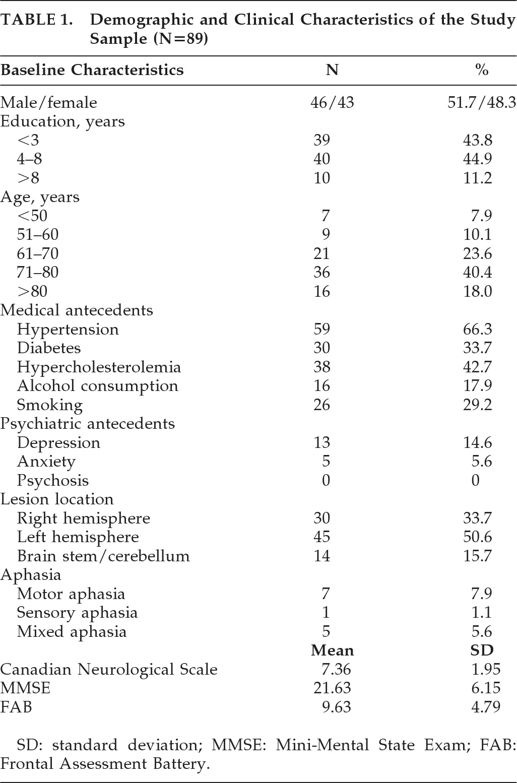
Eighty-nine patients fulfilled the inclusion criteria and met none of the exclusion criteria. Table 1 details their baseline demographic data, medical and psychiatric antecedents, and clinical characteristics.
TABLE 1. Demographic and Clinical Characteristics of the Study Sample (N=89)
SD: standard deviation; MMSE: Mini-Mental State Exam; FAB: Frontal Assessment Battery.
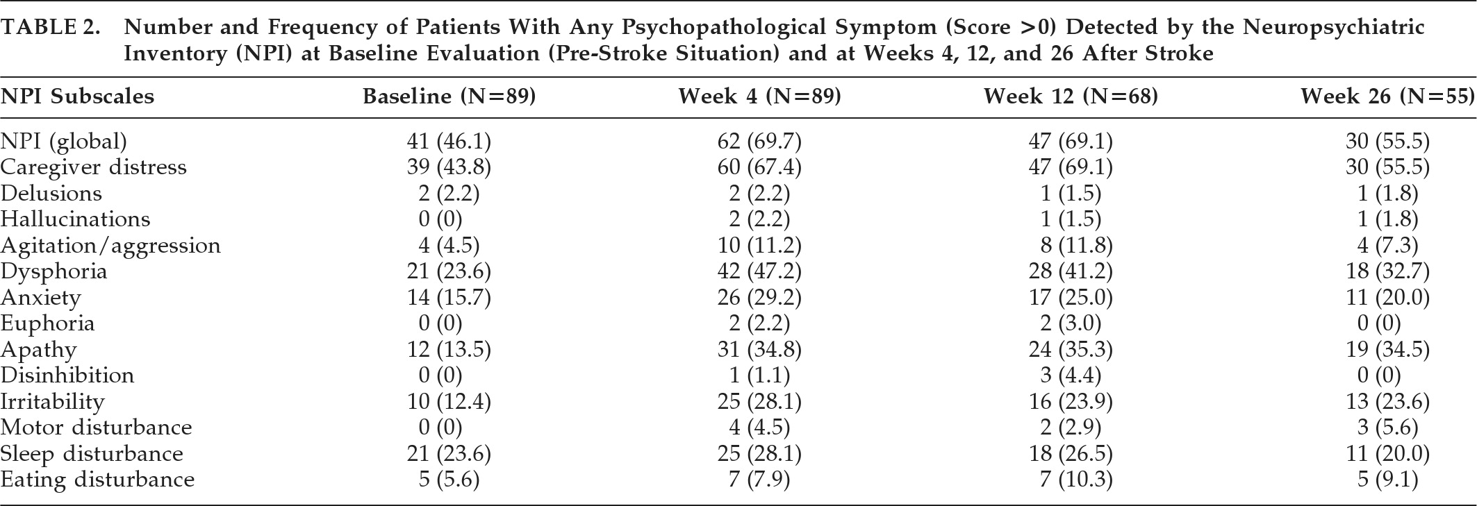
The number and percentage of patients who presented at least one PS, defined as a score >0 in any NPI subscale, at baseline (before stroke) and 4, 12, and 26 weeks after the acute event, are represented in Table 2.
TABLE 2. Number and Frequency of Patients With Any Psychopathological Symptom (Score >0) Detected by the Neuropsychiatric Inventory (NPI) at Baseline Evaluation (Pre-Stroke Situation) and at Weeks 4, 12, and 26 After Stroke
Eighty-three patients were able to accomplish an assessment with Hamilton's scales (Ham-D and Ham-A). Among the other six patients, five had a severe aphasic disorder, and the remaining patient refused to perform the test; 38% of patients showed detectable depression on the Ham-D (defined as a score >7). Moderate-to-severe depression (defined as a score >14) was seen in 19.9% of patients, whereas anxiety was detected with the Ham-A (defined as a score >5) in 33.7% of patients. Moderate-to-severe anxiety (defined as a score >14) was detected in 6% of patients. Pearson′s correlation coefficient between the NPI Dysphoria subscale and Ham-D scores at Week 4 was 0.837 (p<0.01), and the correlation coefficient between the NPI Anxiety subscale and Ham-A scores was 0.763 (p<0.01).
A comparison of the mean scores in the NPI-4W according to the presence or absence of vascular risk factors (HTA, diabetes, hypercholesterolemia, smoking), alcohol consumption, and psychiatric antecedents (depression, anxiety, or psychotic disorders) did not reveal any statistically significant differences. There were also no statistically significant differences on any of the NPI subscales regarding stroke location or size (lacunar versus nonlacunar infarcts).
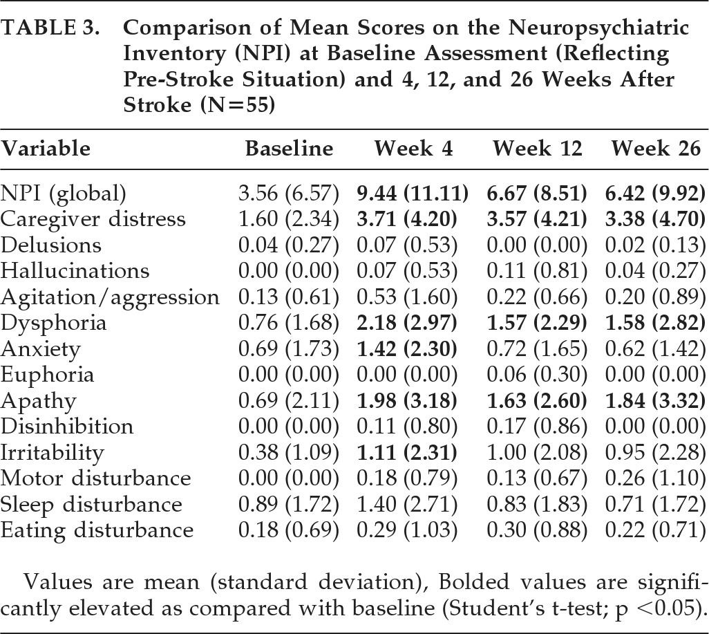
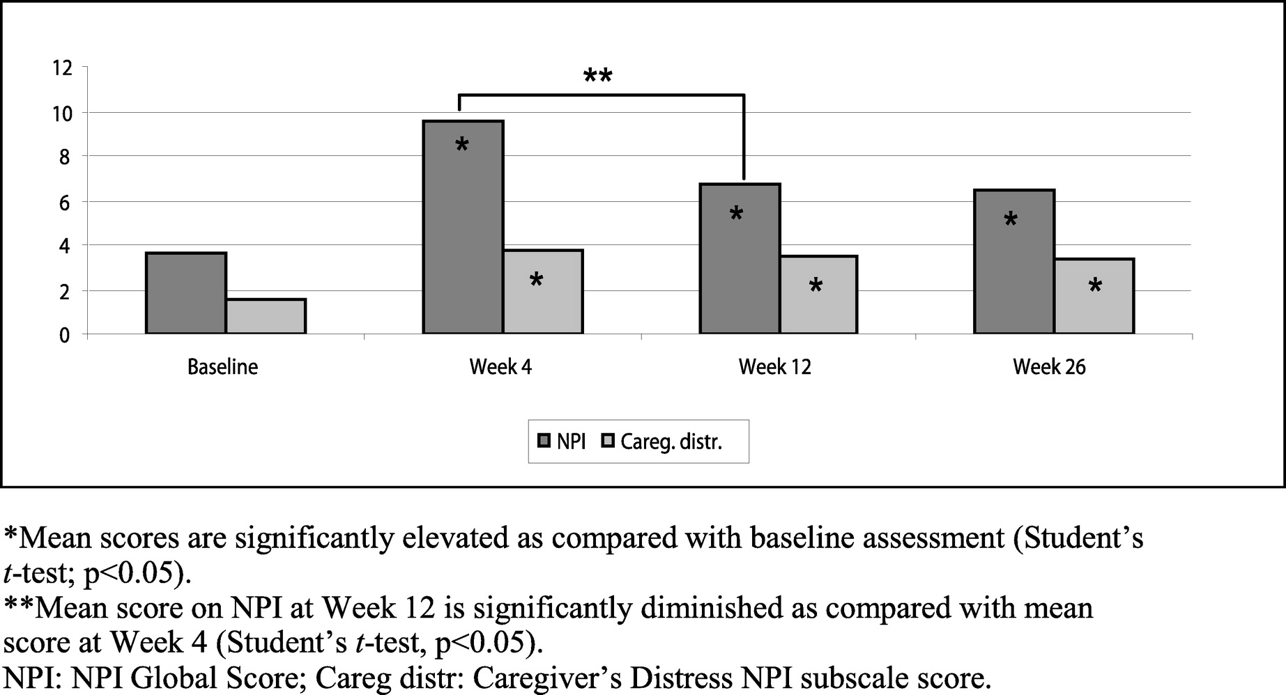
Fifty-five patients completed the study up to Week 26 (Table 3, Figure 1). In this population, the mean NPI Global score and the mean scores for the Caregiver's Distress, Dysphoria, and Apathy scales were significantly increased (p<0.05) at Weeks 4, 12, and 26, as compared with the baseline NPI. However, the mean NPI Global score and Anxiety score significantly decreased between Weeks 4 and 12. There were no statistically significant differences in the mean NPI Global score or in any NPI subscale between Weeks 12 and 26.
TABLE 3. Comparison of Mean Scores on the Neuropsychiatric Inventory (NPI) at Baseline Assessment (Reflecting Pre-Stroke Situation) and 4, 12, and 26 Weeks After Stroke (N=55)
Values are mean (standard deviation), Bolded values are significantly elevated as compared with baseline (Student's t-test; p <0.05).
FIGURE 1. Mean Neuropsychiatric Inventory (NPI) Global Score and Caregiver's Distress Score at Baseline and 4, 12, and 26 Weeks Post-Stroke (N=55)

Correlation With Other Variables

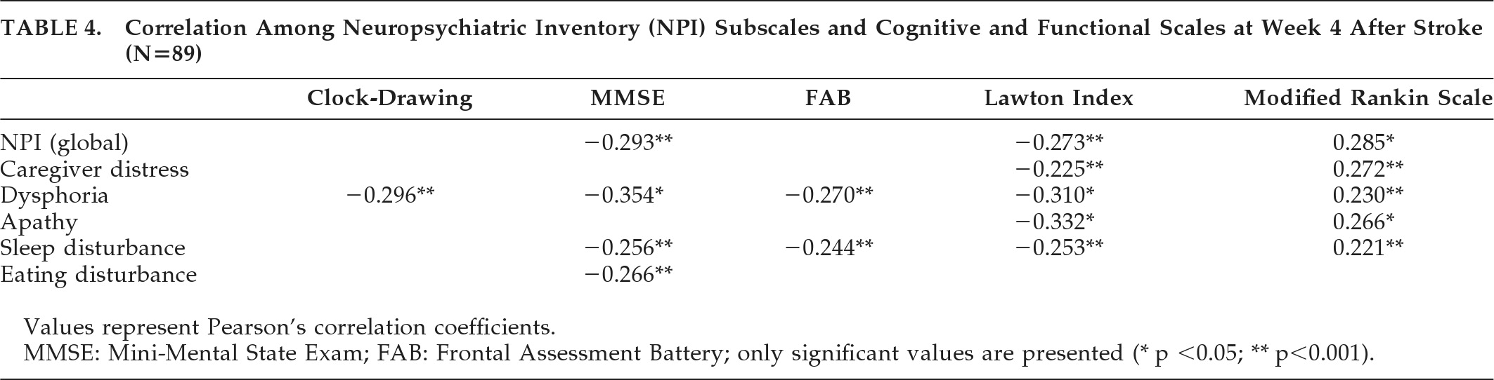
Table 4 shows the NPI-4W subscales that correlated significantly with cognitive and functional variables at Week 4. The Dysphoria score correlated negatively with more cognitive (Clock-Drawing Test, MMSE, and FAB) and functional (Lawton Index and Rankin Scale) scales. On the other hand, functional variables were negatively correlated with more psychopathological variables, including Caregiver's Distress.
TABLE 4. Correlation Among Neuropsychiatric Inventory (NPI) Subscales and Cognitive and Functional Scales at Week 4 After Stroke (N=89)
Values represent Pearson's correlation coefficients.
MMSE: Mini-Mental State Exam; FAB: Frontal Assessment Battery; only significant values are presented (* p <0.05; ** p<0.001).
In the group of patients who completed the follow-up (N=55), apathy was found to be significantly correlated with the Lawton Index (p<0.001) and Rankin score (p<0.05) at Week 12. At Week 26, the Lawton Index was significantly correlated with Dysphoria (p<0.05) and Apathy (p<0.001) scores, but there were no other correlations between psychopathological variables and the rest of the cognitive and functional scales.

Predictive Factors

Predictive factors for the occurrence of early and long-term PSs after stroke were determined by means of a backward, stepwise linear-regression analysis, which produced explanatory models for some of the psychopathological variables. The NPI Global score and NPI subscales scores at 4 (NPI-4W) and 26 weeks (NPI-26W) after stroke were introduced as dependent variables. The following variables, which could be assessed at the moment of admission, were included as independent variables: baseline NPI Global score and baseline NPI subscales (reflecting previous psychopathological status), psychiatric antecedents (depression, anxiety, or psychotic disorder), demographic characteristics (age, gender, and years of formal education), medical antecedents (vascular risk factors, smoking, and alcohol consumption) and stroke location (right hemispheric, left hemispheric, and brainstem/cerebellum). Qualitative variables were recodified as dummy variables to be introduced in the linear-regression models.
The following NPI-4W subscales produced valid regression models: Agitation, Dysphoria, Apathy, Disinhibition, Irritability, Motor Disturbance, Sleep Disturbance, and Eating Disturbance (Table 5). Predictive factors for PSs at 4 weeks were younger age, male gender, right-hemispheric location of the CI, and baseline NPI scores. Younger age was a predictive factor for Agitation and Disinhibition, whereas male gender was a predictive factor for Agitation and Irritability. Right-hemispheric location of the CI predicted the development of Dysphoria, Apathy, Disinhibition, and Sleep Disturbance, although its coefficient value was only significant for Sleep Disturbance.
TABLE 5. Linear-Regression Models for Neuropsychiatric Inventory (NPI) Variables at Week 4 and Week 26 After Stroke
-B: NPI subscale score at baseline evaluation; -Ant: antecedents in medical records.
The related baseline NPI subscale score was a predictive factor for the behavior studied at 4 weeks after stroke in every model except in Disinhibition and Sleep Disturbance (e.g., a high baseline NPI score for Dysphoria was a risk factor for having a high score for Dysphoria in the NPI-4W). Apathy at Week 4 was also predicted by a higher score in other baseline NPI subscales (Agitation and Dysphoria). Moreover, a history of hazardous/harmful alcohol consumption was a risk factor for Eating Disturbance, whereas antecedents of depression predicted the occurrence of Sleep Disturbance.
At 26 weeks after stroke, we found valid regression models for Agitation, Dysphoria, Anxiety, Apathy, Irritability, and Sleep Disturbance (Table 5). Antecedent depression, right-hemispheric location, and scores in the baseline NPI subscales of Agitation, Anxiety, Irritability, and Dysphoria emerged as predictive variables in these models. Right-hemispheric location persisted as a predictive factor for Dysphoria and Apathy, with a significant coefficient value for Dysphoria (p=0.03). Previous history of depression was a risk factor for Dysphoria and Agitation on the NPI-26W, but it made Irritability less probable.
The percentage of variance explained was higher at 26 weeks than at 4 weeks in the models for Agitation, Dysphoria, Apathy, and Irritability, with more than 50% of the variance explained by the models of agitation and dysphoria.

DISCUSSION

Nearly 70% of patients with CI presented some grade of psychopathology in the 4 weeks after stroke onset, as assessed by means of the NPI through a reliable informant. This percentage fell to 55.5% 6 months later. Dysphoria and apathy were the most frequent and persistent symptoms, followed by anxiety, irritability, sleep disturbance, and agitation. These results are consistent with previous reports,6,26,27 although some differences were present in the frequency of specific symptoms, probably due to methodological factors.
The same percentage of patients had detectable PSs at 4 and 12 weeks after stroke, but the intensity of these symptoms, determined by the mean NPI global score, showed a significant decrease by Week 12 and afterward remained almost unchanged.
In this study, the NPI was used not only for evaluating poststroke affective and behavioral disorders, but also to assess previous psychopathology, in order to evaluate its influence on the occurrence of PSs after stroke. Linear-regression analysis showed that the corresponding subscale score in the baseline NPI was a predictive factor for the most frequent symptoms after stroke, including dysphoria and apathy. Therefore, the development of some specific PSs probably depends, to some extent, on patients' previous affective and behavioral status.
Correlation analysis showed that PSs were related to patients' cognitive and functional status in the early poststroke period. However, in the follow-up, we observed that the only correlations remaining at Week 26 were those between dysphoria and apathy with one of the functional scales (the Lawton Index).
Using linear-regression analysis, we attempted to find out which factors of those that were assessable at stroke onset could be used to predict the occurrence of poststroke PSs. Younger age and male gender were predictive factors for disruptive behaviors (agitation, irritability, and disinhibition) during the first month after stroke. Right-hemispheric location and related baseline NPI subscale scores were the most consistently encountered factors across different models. It must be noted that right-hemisphere location and baseline dysphoria predicted the occurrence of dysphoria both at 4 and 26 weeks after stroke; therefore, these factors can be considered reliable predictors of the development of depressive symptomatology after a CI. Furthermore, right-hemisphere location also predicted the occurrence of apathy both in the early and delayed period after stroke.
In the first month poststroke, right-hemisphere location of CI was also a predictive factor for disinhibition and sleep disturbance. It has been reported that changes in sleep structure, with a reduced REM-to-NREM ratio, are more frequent in patients with right-sided CI;27 such alterations could contribute to the occurrence of nocturnal behavioral symptoms.
Other reports have also described right-hemisphere location of CI as a risk factor for some PSs, including depression, agitation, and apathy.2830 However, in contrast to our findings, most studies have reported an association between left-hemisphere lesions and depression in the early poststroke period, whereas poststroke depression in the chronic phase seems to be more frequent in patients with right-sided lesions.31 Several reasons have been postulated to explain this discrepancy, including different pathogenetic mechanisms for depression in the acute and chronic phases of stroke, but the measures used to assess depressive symptoms may also have an influence. Patients with right-hemisphere lesions may have indifference to their own symptoms, particularly in the acute phase, a fact that can result in underreporting of depressive symptoms in self-report measures.31 On the other hand, patients with left-hemisphere lesions frequently present with aphasia, which may be a limitation for the psychopathological evaluation. The NPI, which has been the main tool for PSs assessment in our study, may be adequate for this type of patient, as it obtains information through the caregiver, and the questionnaire refers to both verbal and nonverbal patients' expressions of a depressed mood. In our study, the assessment with the NPI could be completed in patients with aphasia, but we were unable to administer Hamilton's tests (Ham-D and Ham-A, which are patient-referred) to five individuals with a severe language disorder.
It is also important to note that the NPI is intended to evaluate the presence of PSs, but not to diagnose clinical syndromes, as depression. Thus, it cannot be assumed that patients with detected dysphoria in our study actually have poststroke depression. Nevertheless, the Ham-D, which has been previously validated in stroke patients,16 was used in our patients, in parallel with the NPI, in poststroke assessments, showing a high correlation with the NPI subscale for dysphoria, which indicates that there was good concurrent validity.
Regarding to the practical consequences of our study, it is clear that previous psychopathological status and psychiatric antecedents are important to predict the development of PSs after stroke, but the adequate instruments for psychopathological evaluation must be defined. NPI could be valid for this purpose, but some clinicians could find it time-consuming. However, some of its subscales did not provide any useful information (on hallucinations, delusions, euphoria, motor disturbances, sleep disturbances) in the population studied and could be avoided in these patients. Moreover, a brief questionnaire form of the NPI, the NPI–Q, has been developed as a more appropriate tool for neuropsychiatric assessment in clinical practice.32
In summary, this study has shown a high prevalence of PSs, mainly depressive symptoms and apathy. These disorders were associated with worse cognitive and functional conditions in the post-acute period, but only with functional status in the long term. PSs were more severe in the first month after stroke, but tended to improve with time until reaching a plateau around the third month. Younger age and male gender are risk factors for disruptive behaviors in the early poststroke period, whereas right-hemisphere location of CI and previous related psychopathology were the best predictive indicators for the occurrence of dysphoria/depression and apathy.

Acknowledgments

This work was previously presented at the 61st Meeting of the Spanish Society of Neurology (Barcelona, November 17–21, 2009).

References

1.
Kauhanen M, Korpelainen JT, Hiltunen P, et al.: Poststroke depression correlates with cognitive impairment and neurological deficits. Stroke 1999; 30:1875–1880
2.
Pohjasvaara T, Leppävuori A, Siira I, et al.: Frequency and clinical determinants of poststroke depression. Stroke 1998; 29:2311–2317
3.
Singh A, Herrmann N, Black SE: The importance of lesion location in poststroke depression: a critical review. Can J Psychiatry 1998; 43:921–927
4.
Greenop KR, Almeida OP, Hankey GJ, et al.: Premorbid personality traits are associated with post-stroke behavioral and psychological symptoms: a three-month follow-up study in Perth, Western Australia. Int Psychogeriatr 2009; 9:1–9
5.
Hama S, Yamashita H, Shigenobu M, et al.: Depression or apathy and functional recovery after stroke. Int J Geriatr Psychiatry 2007; 22:1046–1051
6.
Angelelli P, Paolucci S, Bivona U, et al.: Development of neuropsychiatric symptoms in post-stroke patients: a cross-sectional study. Acta Psychiatr Scand 2004; 110:55–63
7.
Fuentes B, Ortiz X, San Jose B, et al.: Post-stroke depression: can we predict its development from the acute stroke phase? Acta Neurol Scand 2009; 120:150–156
8.
Storor DL, Byrne GJ: Pre-morbid personality and depression following stroke. Int Psychogeriatr 2006; 18:457–469
9.
Cummings JL, Mega M, Gray K, et al.: The Neuropsychiatric Inventory: comprehensive assessment of psychopathology in dementia. Neurology 1994; 44:2308–2314
10.
Diaz-Olavarrieta C, Cummings JL, Velazquez J, et al.: Neuropsychiatric manifestations of multiple sclerosis. J Neuropsychiatry Clin Neurosci 1999; 11:51–57
11.
Jorm AF, Jacomb PA: The informant questionnaire on cognitive decline in the elderly (IQCODE): sociodemographic correlates, reliability, validity, and some norms. Psychol Med 1989; 19:1015–1022
12.
World Health Organization Global Status Report on Alcohol, 2004. World Health Organization: Geneva, 2004
13.
Vilalta-Franch J, Lozano-Gallego M, Hernández-Ferrándiz M, et al.: The Neuropsychiatric Inventory: psychometric properties of its adaptation into Spanish. Rev Neurol 1999; 29:15–19
14.
Ramos-Brieva JA, Cordero Villafáfila A: Validación de la versión castellana de la Escala de Hamilton Para la Depresión. Actas Luso-Esp Neurol Psiquiatr 1986; 14:324–334
15.
Lobo A, Chamorro L, Luque A, et al.: Validation of the Spanish versions of the Montgomery-Asberg Depression and Hamilton Anxiety Rating Scales. Med Clin (Barcelona) 2002; 118:493–499
16.
Aben I, Verhey F, Lousberg R, et al.: Validity of the Beck Depression Inventory, the Hospital Anxiety and Depression Scale, SCL–90, and Hamilton Depression Scales as screening instruments for depression in stroke patients. Psychosomatics 2002; 43:386–393
17.
Blesa R, Pujol M, Aguilar M, et al.: Clinical validity of the Mini-Mental state for Spanish-speaking communities. Neuropsychologia 2001; 39:1150–1157
18.
Del-Ser T, Morales JM, Barquero MS, et al.: Application of a Spanish version of the “Informant Questionnaire on Cognitive Decline in the Elderly” in the clinical assessment of dementia. Alzheimer Dis Assoc Disord 1997; 11:3–8
19.
Dubois B, Slachevsky A, Litvan I, et al.: The FAB: a frontal assessment battery at bedside. Neurology 2000; 55:1621–1626
20.
Goodglass H, Kaplan E: Supplementary language test, in The Assessment of Aphasia and Related Disorders. Philadelphia, PA, Lea & Febiger, 1972
21.
Van Swieten JC, Koudstaal PJ, Visser MC, et al.: Inter-observer agreement for the assessment of handicap in stroke patients. Stroke 1988; 19:604–607
22.
Mahoney FI, Barthel D: Functional evaluation: The Barthel Index. Maryland State Med J 1965; 14:56–61
23.
Lawton MP, Brody EM: Assessment of older people: self-maintaining and instrumental activities of daily living. Gerontologist 1969; 9:179–186
24.
Côté R, Hachinski VC, Shurvell BL, et al.: The Canadian Neurological Scale: a preliminary study in acute stroke. Stroke 1986; 17:731–737
25.
Bamford J, Sandercock PA, Dennis MS, et al.: Classification and natural history of clinically identifiable subtypes of brain infarction. Lancet 1991; 337:1521–1526
26.
Dennis M, O'Rourke S, Lewis S, et al.: Emotional outcomes after stroke: factors associated with poor outcome. J Neurol Neurosurg Psychiatry 2000; 68:47–52
27.
Körner E, Flooh E, Reinhart B, et al.: Sleep alterations in ischemic stroke. Eur Neurol 1986; 25:104–110
28.
Paradiso S, Vaidya J, Tranel D, et al.: Nondysphoric depression following stroke. J Neuropsychiatry Clin Neurosci 2008; 20:52–61
29.
Braun CM, Larocque C, Daigneault S, et al.: Mania, pseudomania, depression, and pseudodepression resulting from focal unilateral cortical lesions. Neuropsychiatry Neuropsychol Behav Neurol 1999; 12:35–51
30.
Brodaty H, Sachdev PS, Withall A, et al.: Frequency and clinical, neuropsychological, and neuroimaging correlates of apathy following stroke: The Sydney Stroke Study. Psychol Med 2005; 35:1707–1716
31.
Bhogal SK, Teasell R, Foley N, et al.: Lesion location and poststroke depression: systematic review of the methodological limitations in the literature. Stroke 2004; 35:794–802
32.
Kaufer DI, Cummings JL, Ketchel P, et al.: Validation of the NPI–Q, a brief clinical form of The Neuropsychiatric Inventory. J Neuropsychiatry Clin Neurosci 2000; 12:233–239

Information & Authors

Information

Published In

Go to The Journal of Neuropsychiatry and Clinical Neurosciences
The Journal of Neuropsychiatry and Clinical Neurosciences
Pages: 340 - 347
PubMed: 21948896

History

Received: 2 August 2010
Revision requested: 8 December 2010
Accepted: 20 January 2011
Published online: 1 July 2011
Published in print: Summer 2011

Keywords

  1. Stroke
  2. Affective Disorders
  3. Depression
  4. Behavioral Disorders

Authors

Affiliations

Fernando Castellanos-Pinedo, M.D.
From the Departments of Neurology and Radiology (JMH-B), Hospital Virgen del Puerto, Plasencia; and the Department of Experimental Psychology (JAC-P), Universidad Pontificia de Salamanca, Spain.
José María Hernández-Pérez, Ph.D.
From the Departments of Neurology and Radiology (JMH-B), Hospital Virgen del Puerto, Plasencia; and the Department of Experimental Psychology (JAC-P), Universidad Pontificia de Salamanca, Spain.
Martín Zurdo, M.D.
From the Departments of Neurology and Radiology (JMH-B), Hospital Virgen del Puerto, Plasencia; and the Department of Experimental Psychology (JAC-P), Universidad Pontificia de Salamanca, Spain.
Beatriz Rodríguez-Fúnez, B.S.
From the Departments of Neurology and Radiology (JMH-B), Hospital Virgen del Puerto, Plasencia; and the Department of Experimental Psychology (JAC-P), Universidad Pontificia de Salamanca, Spain.
José María Hernández-Bayo, M.D.
From the Departments of Neurology and Radiology (JMH-B), Hospital Virgen del Puerto, Plasencia; and the Department of Experimental Psychology (JAC-P), Universidad Pontificia de Salamanca, Spain.
Ciara García-Fernández, M.D.
From the Departments of Neurology and Radiology (JMH-B), Hospital Virgen del Puerto, Plasencia; and the Department of Experimental Psychology (JAC-P), Universidad Pontificia de Salamanca, Spain.
Bernardo Cueli-Rincón, M.D.
From the Departments of Neurology and Radiology (JMH-B), Hospital Virgen del Puerto, Plasencia; and the Department of Experimental Psychology (JAC-P), Universidad Pontificia de Salamanca, Spain.
Juan Antonio Castro-Posada, Ph.D.
From the Departments of Neurology and Radiology (JMH-B), Hospital Virgen del Puerto, Plasencia; and the Department of Experimental Psychology (JAC-P), Universidad Pontificia de Salamanca, Spain.

Notes

Correspondence: Fernando Castellanos-Pinedo; [email protected] (e-mail).

Metrics & Citations

Metrics

Citations

Export Citations

If you have the appropriate software installed, you can download article citation data to the citation manager of your choice. Simply select your manager software from the list below and click Download.

For more information or tips please see 'Downloading to a citation manager' in the Help menu.

Format
Citation style
Style
Copy to clipboard

There are no citations for this item

View Options

View options

PDF/ePub

View PDF/ePub

Get Access

Login options

Already a subscriber? Access your subscription through your login credentials or your institution for full access to this article.

Personal login Institutional Login Open Athens login
Purchase Options

Purchase this article to access the full text.

PPV Articles - Journal of Neuropsychiatry and Clinical Neurosciences

PPV Articles - Journal of Neuropsychiatry and Clinical Neurosciences

Not a subscriber?

Subscribe Now / Learn More

PsychiatryOnline subscription options offer access to the DSM-5-TR® library, books, journals, CME, and patient resources. This all-in-one virtual library provides psychiatrists and mental health professionals with key resources for diagnosis, treatment, research, and professional development.

Need more help? PsychiatryOnline Customer Service may be reached by emailing [email protected] or by calling 800-368-5777 (in the U.S.) or 703-907-7322 (outside the U.S.).

Media

Figures

Other

Tables

Share

Share

Share article link

Share