Full access
Brief Report
Published Online: 1 December 2001

Cortisol Feedback During the HPA Quiescent Period in Patients With Major Depression

Abstract

OBJECTIVE: The authors tested the hypothesis that patients with major depression have a defect in the mechanism by which cortisol exerts negative feedback on the hypothalamic-pituitary-adrenal (HPA) axis during the HPA axis quiescent period. METHOD: Twenty-nine patients with major depression and 25 healthy comparison subjects were randomly assigned to administration of 15 mg cortisol or placebo infused over 2 hours beginning at 7:00 p.m. Cortisol and ACTH levels were measured at baseline and every 30 minutes from 7:30 p.m. to 11:00 p.m. RESULTS: Differences between the patients and the comparison subjects in the ACTH response to the cortisol infusion, relative to the ACTH response to placebo, were not found. CONCLUSIONS: The results provide some evidence that patients with major depression do not have an abnormality of cortisol feedback during the HPA axis quiescent period.
A defect in glucocorticoid feedback at the level of the hippocampus has been hypothesized to play a role in the pathophysiology of depression (1, 2). Such a defect would produce decreased inhibitory input to the hypothalamus, increased secretion of corticotropin-releasing factor (CRF), and other phenomena indicative of hypothalamic-pituitary-adrenal (HPA) axis hyperactivity in depression. However, only two previous studies have investigated cortisol feedback mechanisms in depressed patients. Young et al. (3) reported that a cortisol infusion decreased an indirect measure of corticotropin (ACTH) secretion among healthy comparison subjects but not patients with major depressive disorder. In contrast, Cooney and Dinan (4) found that cortisol administration produced comparable decreases in ACTH among major depressive disorder patients and comparison subjects.
Since HPA axis sensitivity to glucocorticoid feedback shows diurnal variation (5), and since hypercortisolemia in depressed patients appears to be most prominent later in the day, when the HPA axis is normally quiescent (6), a  feedback abnormality could be more evident in the evening hours than in early morning, when the studies by Young et al. and by Cooney and Dinan were conducted. In the study reported here, cortisol feedback during evening hours was compared in major depressive disorder patients and comparison subjects.

Method

Subjects were recruited by advertisement and from outpatient services at Stanford University Medical Center, McLean Hospital, and Washington University School of Medicine. Screening included a psychiatric interview, administration of the Structured Clinical Interview for DSM-III-R (SCID) (7), clinical ratings, and a medical evaluation with a physical examination. Ratings were conducted by staff who had demonstrated adequate interrater reliability; kappa exceeded 0.85 for depression items on the SCID and the Hamilton Depression Rating Scale (8). All subjects were required to have no active medical problems, no weight loss of 5 lb or greater in the previous month, and no use of any medications for 2 weeks. Major depressive disorder patients were also required to have: 1) a diagnosis of unipolar major depressive disorder in a current depressive episode without psychotic features, 2) a minimum score of 21 on the 21-item Hamilton depression scale, 3) a minimum score of 7 on the Thase Core Endogenomorphic Scale (9), 4) no use of fluoxetine within the past 6 weeks, 5) no use of electroconvulsive therapy within the past 6 months, and 6) no drug or alcohol abuse within the past 6 months. Comparison subjects were also required to have no history of psychiatric disorders and no first-degree relatives with axis I disorders, according to the Research Diagnostic Criteria family history assessment (10). Standard dietary restrictions, described previously (11), were observed. After complete description of the study to the subjects, written informed consent was obtained.
The protocol was conducted in General Clinical Research Centers at Stanford University Medical Center, Brigham and Women’s Hospital, and Washington University School of Medicine. Subjects had an intravenous line placed in each arm. Baseline HPA axis hormone levels were monitored for 24 hours; the results of this baseline study have been reported elsewhere 
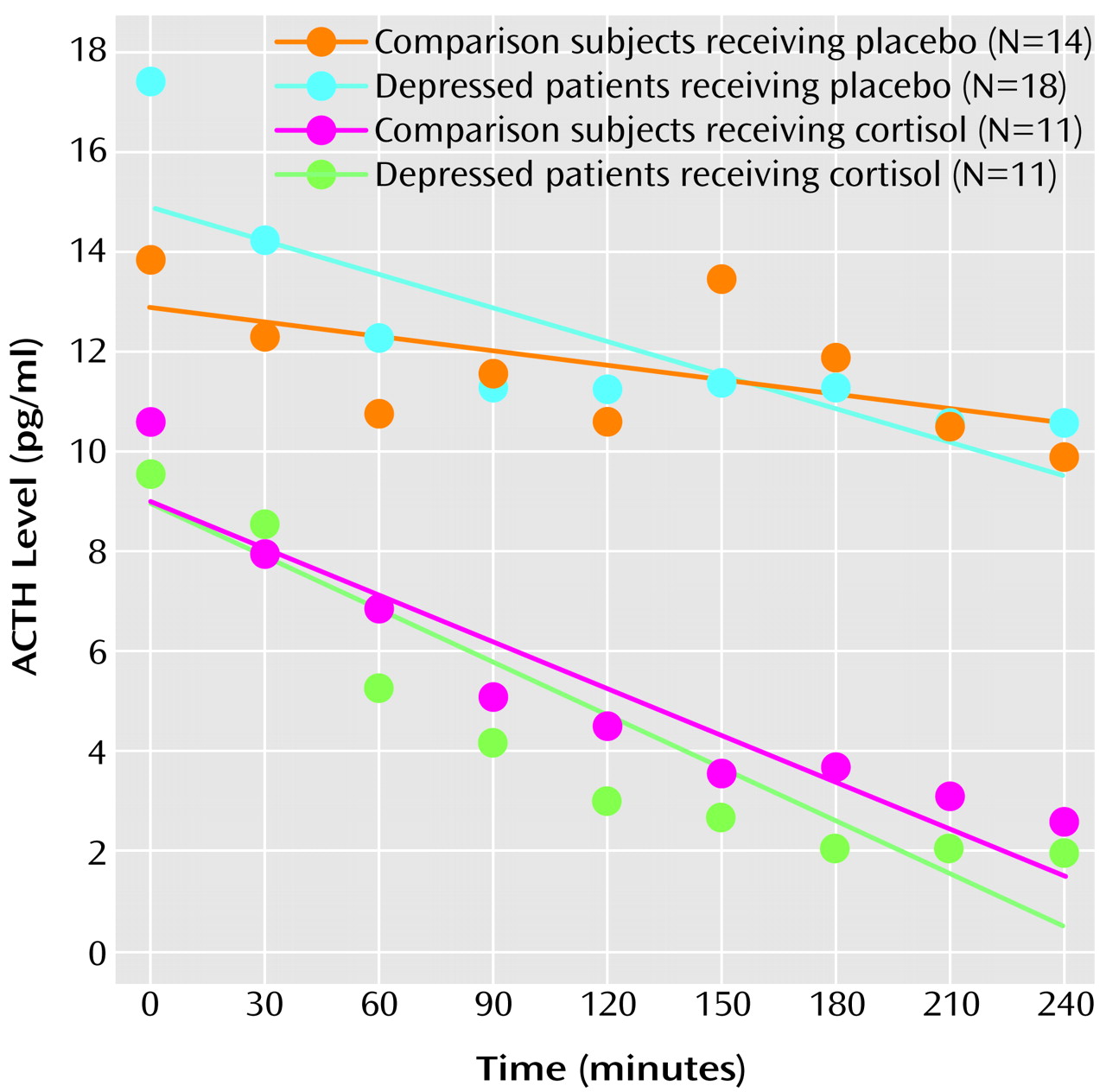
(11). Subsequently, at 7:00 p.m., blood was drawn through an intravenous line to measure cortisol and ACTH levels. The subjects were then randomly assigned to receive 15 mg of cortisol (hydrocortisone sodium succinate) or placebo, infused over 2 hours beginning immediately after the 7:00 p.m. baseline blood draw. Cortisol and placebo were administered by using a double-blind procedure. Every 30 minutes from 7:30 p.m. to 11:00 p.m., blood was drawn through the intravenous line not used for the infusion to measure cortisol and ACTH levels. Some subjects were randomly assigned to receive CRF instead of cortisol or placebo, and these results are not reported here. Cortisol levels were determined by radioimmunoassay (12) and ACTH levels by immunoradiometric assay (12) in the Endocrinology Laboratory at Brigham and Women’s Hospital.
For each subject individually, ACTH levels from 7:00 p.m. to 11:00 p.m. were modeled, by using a least squares procedure, as having a linear decline, an assumption based on the known diurnal variation of HPA axis activity (13). The ACTH response to administration of cortisol or placebo was represented by the slope of the modeled linear decrease. Values of the ACTH slope were subjected to a two-way analysis of covariance (ANCOVA), in which diagnosis (major depressive disorder versus no psychiatric disorder) and treatment (cortisol versus placebo) were factors and the 7:00 p.m. baseline ACTH level, sex, and age were covariates. The hypothesis that patients with major depressive disorder have an abnormal ACTH response to cortisol was tested by examining the effect of the interaction of diagnosis and treatment.

Results

The subjects assigned to receive placebo included 18 depressed patients (11 women and seven men) and 14 comparison subjects (four women and 10 men). Those assigned to receive cortisol included 11 patients (six women and five men) and 11 comparison subjects (six women and five men). The mean ages for the placebo subjects were 40.0 years (SD=10.2) for the depressed patients and 39.7 years (SD=12.7) for the comparison subjects. The mean ages for the subjects who received cortisol were 52.6 years (SD=15.4) for the depressed patients and 40.9 years (SD=13.4) for the comparison subjects. The mean Hamilton depression scale scores were 25.6 (SD=4.3) for the depressed patients who received placebo and 24.6 (SD=3.9) for the depressed patients who received cortisol. Subjects’ weight and the area under the curve for cortisol over time after administration of cortisol did not differ significantly between groups. Infused cortisol increased serum cortisol levels to maximum values having a mean of 24.5 μg/dl (SD=5.7) in the patients and 22.5 μg/dl (SD=10.3) in the comparison subjects.
As Figure 1 shows, cortisol produced a substantial and comparable decrease in ACTH levels in both the patients and the comparison subjects, while there was relatively less change in ACTH over time in the placebo groups. Baseline ACTH levels were higher for both groups receiving placebo than for the groups receiving cortisol, reflecting considerable interindividual variability in ACTH secretion. The fit of the linear model was represented by mean R2 values of 63.2% (SD=26.5%) for the comparison subjects who received cortisol, 64.7% (SD=21.6%) for the depressed patients who received cortisol, 32.2% (SD=29.1%) for the comparison subjects who received placebo, and 31.7% (SD=30.9%) for the depressed patients who received placebo. Mean values for ACTH slope were –0.52 pg/ml per hour (SD=1.82) for the comparison subjects who received placebo, –1.28 pg/ml per hour (SD=1.69) for the depressed patients who received placebo, –1.91 pg/ml per hour (SD=1.54) for the comparison subjects who received cortisol, and –2.14 pg/ml per hour (SD=1.27) for the depressed patients who received cortisol. Analysis of the ACTH slope revealed a highly significant effect of treatment (F=33.3, df=1, 47, p<0.001), but no significant effects of diagnosis (F=0.4, df=1, 47, p=0.50) or of the interaction of diagnosis and treatment (F=0.7, df=1, 47, p=0.40).
After administration of placebo, the ACTH decline appeared steeper in the patients than in the comparison subjects. However, this difference reflected the greater proportion of women in the group of depressed patients who received placebo, compared with the comparison subjects who received placebo, since the ACTH slope was steeper for women than for men (F=8.5, df=1, 47, p<0.01). When the ACTH slopes of patients and comparison subjects receiving placebo were compared by using ANCOVA with the same covariates as in the principal analysis, the group differences were not significant (F=0.1, df=1, 27, p=0.80).

Discussion

To our knowledge, this is the first study of cortisol feedback mechanisms in depressed patients studied during the quiescent period of HPA axis activity. No differences in ACTH response to cortisol between patients with major depressive disorder and healthy comparison subjects were found. Power was computed for the statistical model employed in this study by using Cohen’s method (14) and by assuming a relatively large effect size, indicated by f=0.40. With this approach, the test of the effect of the diagnosis-by-treatment interaction on ACTH slope had a power of 0.79, close to the usual standard of 0.80. Thus, our study provides some evidence that patients with unipolar, nonpsychotic major depressive disorder do not have a defect in cortisol feedback evaluated during evening hours.
Some limitations of this study should be noted. A single dose of cortisol may not be sensitive enough for detecting group differences in the feedback response, and a dose-response study design would have advantages. Although studying cortisol feedback during the HPA axis quiescent period may be valuable, the relationship of our findings to those of other investigators is not clear because of the absence of an additional condition in which feedback is assessed at the time of peak activity. Finally, research with a very limited number of subjects with endocrine disorders has suggested that there may be a distinct phase of feedback occurring over less than 15 minutes (15). Thus, 30-minute sampling of ACTH levels may not be frequent enough to uncover abnormalities in feedback.
Figure 1. Plasma ACTH Levels in Response to Intravenous Infusion of Placebo and Cortisol in Depressed Patients and Comparison Subjectsa
aInfusion of placebo and cortisol began at 7:00 p.m. (0 minutes) and continued over 120 minutes. Symbols show group means over time. Lines represent curves predicted by a linear decrease model based on the group mean values for slope and intercept.

Footnote

Received Oct. 18, 2000; revision received April 17, 2001; accepted May 10, 2001. From the Department of Psychiatry, Washington University School of Medicine and Metropolitan St. Louis Psychiatric Center, St. Louis, Mo.; the Department of Psychiatry and Behavioral Sciences, Stanford University School of Medicine, Stanford, Calif.; and the Division of Endocrinology/Hypertension, Brigham and Women’s Hospital and Harvard Medical School, Boston. Address reprint requests to Dr. Posener, Department of Psychiatry, Washington University School of Medicine, 660 S. Euclid Ave., Box 8134, St. Louis, MO 63110; [email protected] (e-mail).Supported by NIMH grant MH-50604 (Dr. Schatzberg), a National Alliance for Research on Schizophrenia and Depression Young Investigator Award (Dr. Posener), and NIH General Clinical Research Center grants RR-00070, RR-00036, and RR-02635.The authors thank Christine M. Blasey, Ph.D., for statistical consultation.

References

1.
Sapolsky RM: Glucocorticoids and hippocampal atrophy in neuropsychiatric disorders. Arch Gen Psychiatry 2000; 57:925-935
2.
Sheline YI, Sanghavi M, Mintun MA, Gado MH: Depression duration but not age predicts hippocampal volume loss in medically healthy women with recurrent major depression. J Neurosci 1999; 19:5034-5043
3.
Young EA, Haskett RF, Murphy-Weinberg V, Watson SJ, Akil H: Loss of glucocorticoid fast feedback in depression. Arch Gen Psychiatry 1991; 48:693-699
4.
Cooney JM, Dinan TG: Preservation of hypothalamic-pituitary-adrenal axis fast-feedback responses in depression. Acta Psychiatr Scand 1996; 94:449-453
5.
Dallman MF, Akana SF, Jacobson L, Levin N, Cascio CS, Shinsako J: Characterization of corticosterone feedback regulation of ACTH secretion. Ann NY Acad Sci 1987; 512:402-414
6.
Carroll BJ, Mendels J: Neuroendocrine regulation in affective disorders, in Hormones, Behavior and Psychopathology. Edited by Sachar EJ. New York, Raven Press, 1976, pp 193-224
7.
Spitzer RL, Williams JBW, Gibbon M, First MB: Structured Clinical Interview for DSM-III-R—Patient Version (SCID-P). New York, New York State Psychiatric Institute, Biometrics Research, 1988
8.
Hamilton M: A rating scale for depression. J Neurol Neurosurg Psychiatry 1960; 23:56-62
9.
Thase ME, Hersen M, Bellack AS, Himmelhoch JM, Kupfer DJ: Validation of a Hamilton subscale for endogenomorphic depression. J Affect Disord 1983; 5:267-278
10.
Endicott J, Andreasen N, Spitzer RL: Family History Research Diagnostic Criteria, 3rd ed. New York, New York State Psychiatric Institute, Biometrics Research, 1978
11.
Posener JA, DeBattista C, Williams GH, Kraemer HC, Kalehzan BM, Schatzberg AF: 24-hour monitoring of cortisol and corticotropin secretion in psychotic and nonpsychotic major depression. Arch Gen Psychiatry 2000; 57:755-760
12.
Taylor P, Dluhy RG, Williams GH: Beta-endorphin suppresses adrenocorticotropin and cortisol levels in normal human subjects. J Clin Endocrinol Metab 1983; 57:592-596
13.
Kaneko M, Hiroshige T, Shinsako J, Dallman MF: Diurnal changes in amplification of hormone rhythms in the adrenocortical system. Am J Physiol 1980; 239:R309-R316
14.
Cohen J: Statistical Power Analysis for the Behavioral Sciences, revised ed. New York, Academic Press, 1977
15.
Fehm HL, Voigt KH, Kummer G, Lang R, Pfeiffer EF: Differential and integral corticosteroid feedback effects on ACTH secretion in hypoadrenocorticism. J Clin Invest 1979; 63:247-253

Information & Authors

Information

Published In

Go to American Journal of Psychiatry
American Journal of Psychiatry
Pages: 2083 - 2085
PubMed: 11729034

History

Published online: 1 December 2001
Published in print: December 2001

Authors

Affiliations

Charles DeBattista, D.M.H., M.D.
Gordon H. Williams, M.D.
Alan F. Schatzberg, M.D.

Metrics & Citations

Metrics

Citations

Export Citations

If you have the appropriate software installed, you can download article citation data to the citation manager of your choice. Simply select your manager software from the list below and click Download.

For more information or tips please see 'Downloading to a citation manager' in the Help menu.

Format
Citation style
Style
Copy to clipboard

There are no citations for this item

View Options

View options

PDF/ePub

View PDF/ePub

Get Access

Login options

Already a subscriber? Access your subscription through your login credentials or your institution for full access to this article.

Personal login Institutional Login Open Athens login
Purchase Options

Purchase this article to access the full text.

PPV Articles - American Journal of Psychiatry

PPV Articles - American Journal of Psychiatry

Not a subscriber?

Subscribe Now / Learn More

PsychiatryOnline subscription options offer access to the DSM-5-TR® library, books, journals, CME, and patient resources. This all-in-one virtual library provides psychiatrists and mental health professionals with key resources for diagnosis, treatment, research, and professional development.

Need more help? PsychiatryOnline Customer Service may be reached by emailing [email protected] or by calling 800-368-5777 (in the U.S.) or 703-907-7322 (outside the U.S.).

Media

Figures

Other

Tables

Share

Share

Share article link

Share