Persistent blockade of dopamine transporters by chronic cocaine abuse may lead to depletion of brain dopamine levels
(1), possibly as a result of this blockade rendering neurons unable to reuptake and recycle dopamine, which is instead metabolized in the synapse and excreted
(2). Depletion of dopamine in the putamen may contribute to the increased frequency of extrapyramidal symptoms (including tics, choreoathetoid movements, resting hand tremor, and dystonia [
3–
6]) observed in cocaine-dependent patients
(1,
7). Treatment with neuroleptic medications that block dopamine 2 (D
2) receptors also decreases striatal dopaminergic neurotransmission, increases the frequency of extrapyramidal symptoms
(8), and is associated with striatal enlargement. This enlargement normalizes after patients are switched to atypical antipsychotic medications, which have lower affinities for the D
2 receptor
(9).
Given the greater frequency of extrapyramidal symptoms in cocaine abusers, the evidence that chronic exposure to cocaine depletes striatal dopamine, and the association of striatal enlargement with neuroleptic blockade of D
2 receptors, we hypothesized that caudate and putamen volumes would be larger in subjects with cocaine dependence. Given the role of the putamen in motor control and the higher concentration of dopamine transporters in the putamen relative to the caudate
(10), we anticipated that this effect would be greater for the putamen.
Method
Healthy and cocaine-dependent subjects were administered the SCID
(11) and underwent a physical and neurologic examination and routine laboratory testing that included a urine toxicology screening analysis. Twenty-five subjects (10 women and 15 men; 14 African American and 11 Caucasian; mean age=35.6 years, SD=5.7, range=20–45) who met criteria for chronic cocaine dependence without a comorbid axis I disorder participated. On average, these subjects had smoked 4.5 g (SD=5.9) of crack cocaine per week for 15.3 years (SD=6.5) and consumed 3.7 (SD=5.2) alcoholic drinks per day, typically in the context of cocaine use. One cocaine-dependent subject also met criteria for alcohol dependence. Subjects with an abuse or dependence history with any other class of drug were excluded. Cocaine-dependent subjects all had urine toxicology results that were positive for the cocaine metabolite benzoylecgonine upon study admission; subjects were scanned 15.3 days (SD=12.0) after their last cocaine use.
The 20 healthy comparison subjects (11 women and nine men; five African American and 15 Caucasian; mean age=35.0 years, SD=6.8, range=22–48) participating were free of any axis I disorder and consumed 0.3 (SD=0.7) alcoholic drinks per day. All subjects were free of current or previous psychotropic medication exposure. Twenty-four of the cocaine-dependent subjects and 17 of the healthy subjects were right-handed.
All subjects provided written informed consent for participation in this study, which was approved by the Yale and VA Human Investigations Committees.
All subjects were scanned with the same 1.5-T Signa (GE Medical Systems, Milwaukee) magnetic resonance scanner. Volumes of the total brain, caudate, and putamen were measured from a coronal series acquired by using three-dimensional spoiled gradient recalled echo in the steady state (acquisition matrix=256 × 192, number of excitations=1, flip angle=45°, TE=5 msec, TR=24 msec, field of view=24 cm2, slice thickness=1.9 mm [slice thickness=3 mm for 10 healthy and four cocaine-dependent subjects]).
All scans were reoriented in MEDx (Sensor Systems, Sterling, Va.) such that the axial plane was parallel to the intercommissural plane. Quantitative measurements of brain morphology were then performed in ANALYZE (Mayo Foundation, Rochester, Minn.) by a rater blind to diagnosis (L.K.J.). Volumes were calculated by multiplying area measurements by slice thickness. The caudate, including head and body, and putamen were quantified by manually outlining these structures on each coronal slice in which they were visible
(12). Total brain volume was measured by thresholding images to exclude ventricular cerebrospinal fluid and then manually outlining brain tissue, including the pons and cerebellum, on sequential coronal slices. Ten brains measured twice by the same rater revealed intrarater reliabilities (intraclass correlation coefficients [ICCs]
[13]) of 0.98 for the left and right caudate, 0.95 for the left putamen, and 0.97 for the right putamen. Measurement of five brains by the same rater indicated an intrarater ICC of 0.99 for total brain volume.
Group differences in demographic variables and total brain volume were assessed by using chi-square analyses and t tests for independent groups. Group differences in caudate and putamen morphology were assessed by dividing the volume of each structure by the corresponding total brain volume to obtain a measure of volume proportional to total brain volume and performing repeated measures analysis of covariance (ANCOVA), with diagnosis as a between-subjects variable, side as a within-subjects variable, and age and gender as covariates. The relationship between age and caudate and putamen volumes was examined by using partial correlation analysis that controlled for diagnosis and gender. Within the cocaine-dependent group, the relationships between caudate and putamen volumes and both reported cocaine use in the past 6 months (bags per week) and time since last cocaine use were also examined.
Results
Cocaine-dependent and healthy subjects did not differ in age, handedness, gender, number of tobacco cigarettes smoked per day, or total brain volume (cocaine-dependent subjects: mean=1359.5 cm3, SD=126.8; healthy subjects: mean=1385.0 cm3, SD=166.0). Healthy subjects had two more years of education (mean=14.4 years [SD=2.0] versus mean=12.4 years, [SD=2.1]; t=3.43, df=43, p=0.001) and reported lower alcohol intake than did the cocaine-dependent subjects (t=2.85, df=43, p=0.007). The ratio of African American to Caucasian subjects was greater among cocaine-dependent subjects than among healthy subjects (χ2=4.38, df=1, p=0.04). Age was significantly inversely correlated with right and left caudate volume (r=–0.42, df=41, p=0.004, and r=–0.52, df=41, p=0.0003, respectively) and right and left putamen volume (r=–0.40, df=41, p=0.008, and r=–0.40, df=41, p=0.008).
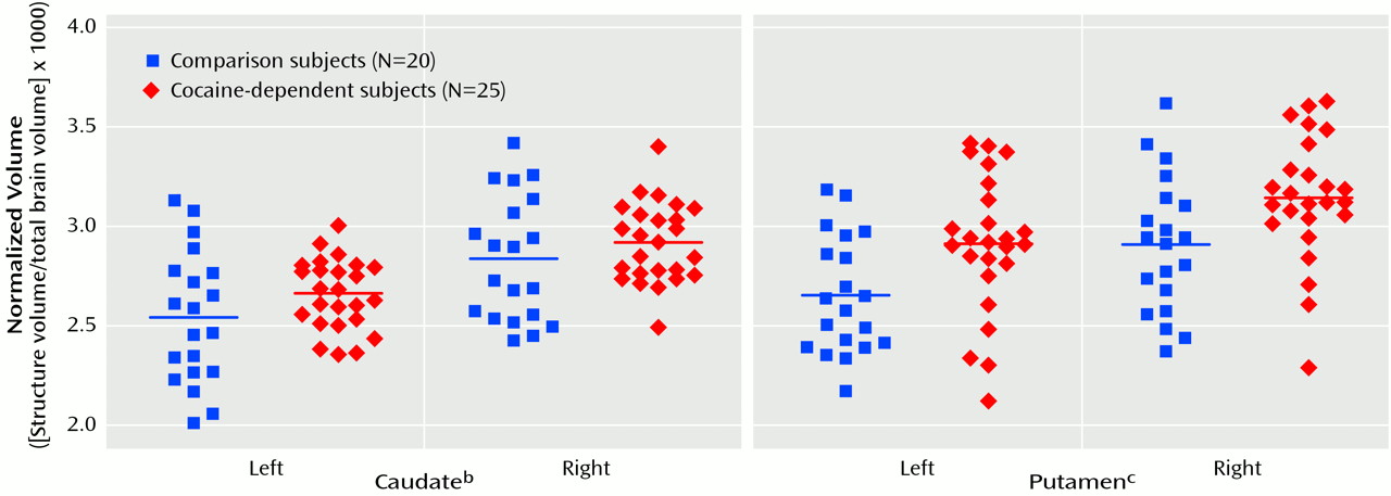
An ANCOVA of the caudate/total brain volume ratio and the putamen/total brain volume ratio that controlled for age and gender revealed a significant effect of diagnosis (
Figure 1). In the cocaine-dependent subjects, normalized caudate volumes were 4.33% larger on the left and 2.46% larger on the right relative to those of the healthy comparison subjects (left: mean=2.65 [SD=0.18] versus 2.54 [SD=0.33], respectively; right: mean=2.91 [SD=0.20] versus 2.84 [SD=0.31]). Normalized putamen volumes in the cocaine-dependent subjects were also 9.77% larger on the left and 8.59% larger on the right relative to those of the healthy comparison subjects (left: mean=2.92 [SD=0.36] versus 2.66 [SD=0.30], respectively; right: mean=3.16 [SD=0.32] versus 2.91 [SD=0.34]). The right-greater-than-left effect of side was significant for the putamen but not for the caudate (
Figure 1). There were no significant interactions. When amount of daily alcohol use was entered as a third covariate into the ANCOVA, the effect of cocaine dependence on caudate volume was no longer significant (F=3.23, df=1, 40, p=0.08), whereas the effect on putamen volume remained significant (F=6.44, df=1, 40, p=0.02). Similarly, when race was entered as a third covariate, the effect of cocaine dependence on caudate volume was no longer significant (F=3.65, df=1, 40, p=0.06), whereas the effect on putamen volume remained significant (F=5.19, df=1, 40, p=0.03). Reported cocaine use over the past 6 months and time since last cocaine use were not significantly correlated with caudate or putamen volumes.
Discussion
In this study, evidence of caudate and putamen enlargement was observed in a group of patients with chronic cocaine dependence. The degree of enlargement was greater for the putamen, consistent with its role in motor control and extrapyramidal symptoms and with the greater concentration of dopamine transporters in the putamen relative to the caudate
(1,
7,
10).
A mediating factor in the enlargement of the putamen and caudate in subjects with chronic cocaine dependence may be the depletion of striatal dopamine and consequent decreases in striatal dopaminergic neurotransmission. Evidence for depletion of striatal dopamine following chronic exposure to cocaine has been observed in both in vivo
(14) and postmortem
(1) studies of human chronic cocaine abusers and in some
(15–
17) but not all
(18) studies of rats chronically exposed to cocaine. Discrepancies in some of the preclinical data likely reflect differences between species or patterns and methods of cocaine administration. The hypothesis that the relative striatal enlargement in these patients may stem from cocaine-induced depletion of striatal dopamine is consistent with the elevated frequency of extrapyramidal symptoms observed in the cocaine-dependent patients and the enlarged striatal volumes in patients receiving neuroleptics that block D
2 receptors and reduce striatal dopaminergic neurotransmission
(9). However, other potential etiologies of this enlargement, such as drug-induced increases in perfusion of the striatum
(9), cannot be excluded by these data. The well-documented deficits in cerebral blood flow associated with chronic cocaine use argue against this latter interpretation
(19).
The right-greater-than-left asymmetry observed for the putamen and the age-related decline in the volumes of both the putamen and caudate are consistent with previous observations in healthy subjects
(20). Limitations of this study include the acquisition of slightly thicker slices in 10 healthy and four cocaine-dependent subjects, which may have contributed additional variance to the morphometric measures. In addition, since extrapyramidal symptoms were not measured in these subjects, the relationship between degree of extrapyramidal symptoms and degree of striatal enlargement could not be examined. Cocaine-dependent subjects consumed significantly greater amounts of alcohol than did healthy subjects, which is a reflection of the common co-use of alcohol with cocaine
(21). When this difference was controlled for, the effect of cocaine dependence on normalized putamen volumes remained significant, whereas this effect on normalized caudate volumes was no longer significant. Similarly, when the difference in racial composition across diagnostic groups was controlled for, the effect of cocaine dependence on normalized putamen volumes remained significant, whereas this effect on normalized caudate volumes was no longer significant. Our failure to observe a relationship between amount or recency of cocaine use and volume of the caudate or putamen may reflect the relative inaccuracy of self-report data. Last, these data cannot address the important question of whether the observed enlargement of striatal structures is reversible, as it appears to be when due to neuroleptic exposure
(9). Answering such a question will require longitudinal follow-up of patients with documented abstinence from cocaine.