Full access
Clinical Case Conference
Published Online: 1 April 2002

Psychological Versus Biological Clinical Interpretation: A Patient With Prion Disease

Publication: American Journal of Psychiatry
Conversion disorder is a diagnosis based on a psychological construct that currently has no known neurobiologic substrate (1). This diagnosis is intended to be reserved for individuals who show deficits that do not conform to known neuroanatomy or neurophysiology. Conversion disorder is usually thought to relate to “secondary gain,” a remnant of a hypothesis that conversion symptoms were the result of the primitive resolution of unconscious conflicts, also known as “primary gain.” Today, secondary gain is used more as a psychosocial assessment and attempts to take into account how illness behavior, or the “sick role,” may be used to focus family, medical, and societal resources. Conversion symptoms are also considered by most clinicians to be an expression of intense distress, resulting from trauma that the emotionally fragile individual can only express somatically. Thus, along with the diagnosis of conversion disorder is the search for the insult or trauma precipitating the expression of the somatic symptoms.
Prion diseases are a heterogeneous group of neurologic disorders that occur in familial forms affecting animals and humans. This can be through iatrogenic transmission or after ingestion of or exposure to affected tissues, or it can occur sporadically. These disorders are characterized by a confusing and invariably fatal array of clinical manifestations, including relatively rapid and progressive dementia, cerebellar ataxia, as well as the speech and visual abnormalities (2) seen in the most common form of human prion disease, Creutzfeldt-Jakob disease (3). Additionally, early in the disease course, nonspecific diffuse EEG abnormalities are found in most patients; later, diagnostic sharp-wave patterns are observed in 75%–85% of the patients with Creutzfeldt-Jakob disease. Finally, diagnostic confirmation relies on the neuropathologic examination of brain tissue obtained by biopsy or at autopsy, with demonstration of characteristic spongiform changes in the parenchyma.
This case reflects the continuing dilemma within psychiatry to reconcile confusing clinical presentations and resolve discordant psychological and neurobiological constructs of axis I diagnoses. In retrospect, it also outlines the interaction of neuropathologic lesions, symptom profiles, and personality, and highlights the level of difficulty discerning the underlying etiology of complex clinical presentations.

Case Presentation

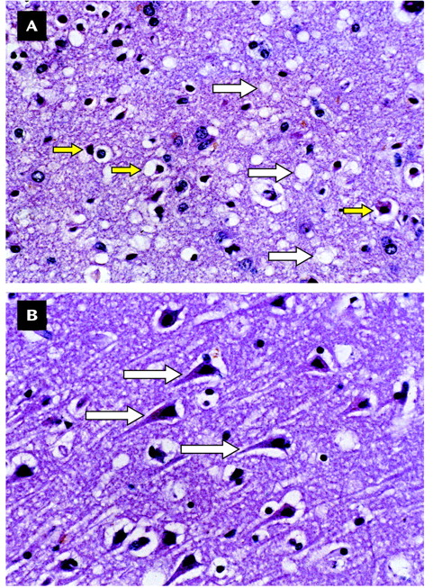
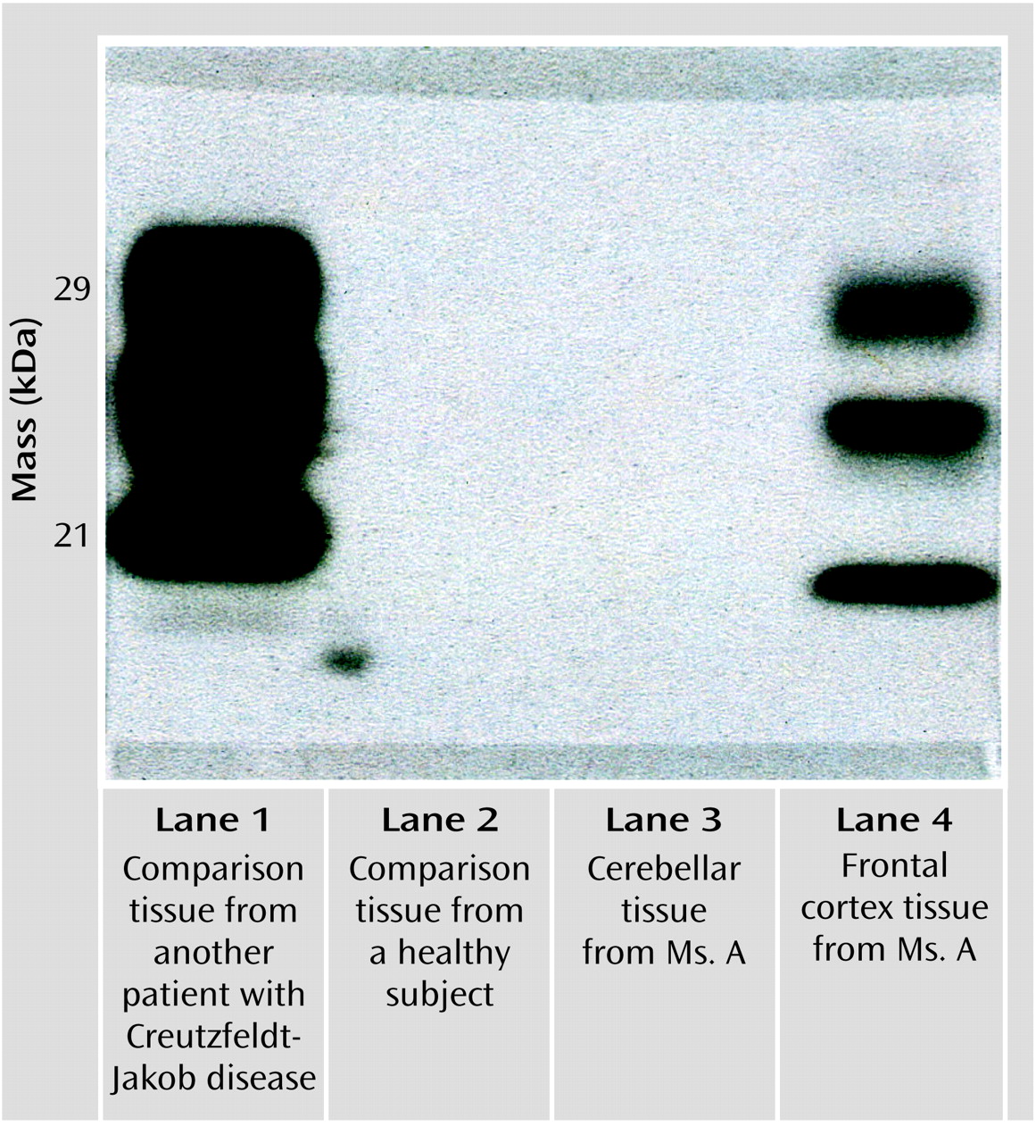
Ms. A was a 49-year-old Caucasian woman who had never married and who had been a communications product engineer. Before onset of her symptoms, she had never seen a psychiatrist, never been diagnosed with a psychiatric disorder, and never been treated with a psychotropic medication. Her medical history was notable only for a febrile illness that may have been an encephalitis of uncertain etiology after a trip to South America when she was in her 20s. There was no family history of psychiatric illness, epilepsy, or neurological or neurodegenerative disorders.
Ms. A had grown up as the youngest of three children. She described her relationship with her two brothers as an adult as “distant.” Her relationship with her parents was reported to have been “good,” although additional details were lacking, and both parents were deceased. She had no history of physical or sexual abuse, but she felt that she had been excessively teased by her brothers while growing up, which left residual resentment even late into her adult life. She did not date frequently in either her teens or 20s. She had a number of heterosexual romantic relationships but described these as superficial and brief. She noted that she had always been sexually interested in women, but until just before the onset of the symptoms leading to hospitalization, she had never had a same-sex romantic relationship. The revelation of her homosexuality to her family was poorly received. She described it as “cold,” and she felt criticized and ashamed.
Ms. A had begun her employment with a communications company as a telephone operator and had advanced over the next 20 years until she became a high-level manager in research and development. Ultimately, she was recruited into an upper-level management position with a competitor in the field. She traveled frequently to Europe as part of her new job. During this transition in her career, she met a woman with whom she hoped to have a fulfilling romantic relationship and thus estranged herself from her family.
In the context of these psychosocial stressors and after a flight from Europe, she developed an acute onset of lower-back pain while lifting her laptop computer out of an overhead bin. Soon after she saw a chiropractor for lower-back pain; upon examination, the practitioner noted some weakness of the lower extremities. She received no benefit from the chiropractic visits and began to note the onset of new symptoms. These included a tremor “all over [her] body” and weakness in “big muscle groups.” This made ambulation difficult and was the beginning of a slow deterioration in gait. She made an appointment with her primary care physician. A workup included magnetic resonance imaging (MRI) of her spine, which showed a mild lumbar right-paramedian bulge that was judged to be inadequate to explain her symptoms. Her laboratory tests included a CBC, a full metabolic panel, including liver function tests—all of which were found to be within normal ranges—a measurement of erythrocyte sedimentation rate (4), and a rheumatoid factor check (negative result). These results were obtained almost 2 months after Ms. A’s original lower-back strain.
Ms. A’s medical and emotional needs became overly burdensome to her partner, which led to the end of their romantic relationship. Ms. A continued to experience weakness in her lower extremities and ambulation difficulties. Her physician ordered an MRI of the brain, which was normal except for evidence of chronic sinusitis in the left maxillary sinus. An MRI of her cervical spine was also performed, and the results were found to be normal. Additionally, a lumbar puncture was performed as the diagnostic considerations expanded to include multiple sclerosis, encephalitis, neurosyphilis, subacute sclerosing panencephalitis, and acute idiopathic polyneuritis. Results of tests regarding CSF glucose, cell cytology, protein, gram stain, immunoglobulin M, immunoglobulin G, fluorescent treponemal antibody absorption, and immunoglobin banding were all negative.
Despite reassurance that these symptoms would likely remit, Ms. A continued to experience more weakness and ataxia. She was seen by another internist and diagnosed with atypical multiple sclerosis. She was given a trial of methylprednisolone sodium succinate and noted a brief period of symptom improvement. She was seen by a neurologist 1 month later, and an EEG was ordered. Again, the result was unrevealing.
Five months after her initial symptoms, Ms. A complained of diplopia, difficulty swallowing, and frequency of urination in addition to her symptoms of leg weakness, tremor, and gait difficulties. She saw another neurologist whose differential diagnosis included Creutzfeldt-Jacob disease. She was then seen by an associate of a national authority on prion disease, who felt that Creutzfeldt-Jakob disease was a “low-probability” diagnosis, but a measurement of blood mercury level was recommended, which was also found to be negative.
At this point Ms. A’s treating physician began to seriously consider that these symptoms might have a psychological component, which led to a psychiatric evaluation. About 6 months from the onset of her symptoms, Ms. A completed a neurocognitive and psychological battery of tests. The results were as follows: score of 30/30 on the Mini-Mental State Examination (MMSE), a score of 33/33 on the Blessed Dementia Scale (5), and a Sensory Perceptual Screening (6) examination performed without errors. She completed the Wechsler Memory Scale—Revised (7). Her score fell within the average to high-average range, and she was judged to have high-average intellectual abilities. Also of note was that her receptive and expressive language abilities were found to be intact, her conversational speech was fluid and nonphasic, her digit repetition was high average, her oral calculations were average, and her attention and concentration were normal. The only abnormal finding in this test battery was on the MMPI-2, which showed elevated depression.
Although Ms. A complained of weakness and an unstable gait, results of formal neurologic examinations continued to be completely normal. In addition, Ms. A’s complaints of waxing and waning strength and difficulty walking were difficult to explain. At this time, she was given axis I diagnoses of major depression and conversion disorder. Ms. A was given fluoxetine for depression and buspirone for anxiety. She was thereafter discharged to a residential psychiatric facility.
Nearly 8 months after the first appearance of lower-back strain, Ms. A was seen by a specialist in dissociative disorders and psychosomatic illness. He hypnotized Ms. A and noted that she was highly hypnotizable and that, under hypnosis, her symptoms improved. She continued to decompensate and was transferred from the residential facility to a skilled nursing facility secondary to functional deterioration and an inability to complete activities of daily living without assistance. This deterioration continued until Ms. A was again hospitalized, this time on the Behavioral Medicine Unit at Stanford University Hospital. At that time, she was confined to a wheelchair. She complained of waxing and waning dysarthria and difficulty swallowing, an ataxic-like gait, and tremors that would move over her entire body, which she would refer to as “convulsions.” She also had occasional complaints of diplopia, for which she would compensate by closing one eye, and frequent squinting. She continued to experience subjective weakness in her lower extremities, which varied in degree, although it was always present.
At this time, both Ms. A and her family agreed that her mood was normal. Her score on the MMSE at admission was 24/30; she appeared to be poorly motivated to complete the test. Her insight was judged to be good, and she was reported to say that she was motivated to determine the cause of her disability so she could return to work. She admitted that being ill had brought her family back to her and that her ex-lover was again in frequent contact, both of which pleased her deeply. Upon examination her tremors were noted to wax and wane depending on the content of her conversation. Her diagnoses at admission were conversion disorder and major depression, single episode, moderate, nonpsychotic, and in remission with a regimen of fluoxetine and buspirone. Psychological testing was ordered, as was a follow-up neurology consultation.
Upon completion of neurological and psychometric evaluations, Ms. A’s primary diagnosis remained conversion disorder. Supporting this diagnosis were fluctuating and at times bizarre symptoms: waxing and waning memory deficits, an inability to ambulate without assistance despite adequate strength, back pain, eye squinting, and tremor, but no evidence of spasticity or cerebellar abnormalities. There was also some concern over factitious elements, such as a new complaint of life-long auditory and visual hallucinations.
Ultimately, it was decided to proceed with a behaviorally oriented rehabilitation program. This involved aggressive physical therapy, occupational therapy, and participation in groups and activities, with the focus on treating Ms. A’s functional disability. Furthermore, with the intent of both confirming and treating the conversion disorder, it was explained to Ms. A that in the setting of such an intensive rehabilitation program, her functional ability should improve if it was simply related to an underlying medical etiology, such as the apparent severe muscle deconditioning she had experienced. It was also explained to Ms. A that if she did not improve after undergoing such an intensive rehabilitation program, it would be owing to either a lack of participation or motivation on her part or because her condition was purely the result of a psychological disturbance. That is to say, improvement would confirm a medical etiology. This behavioral approach to treating conversion disorder was based on a review of the literature and the apparent success of the use of this “double-bind” model (3).
The neurology service was consulted soon after Ms. A was hospitalized. They found her to have normal conjugate eye movements without nystagmus, although she complained of diplopia. Her palate lifted symmetrically, her gag reflex was intact, and a fluctuating dysarthria in her speech was described as “clear” at times. She was noted to have decreased muscle bulk on the anterior tibialis muscles bilaterally and a slight contracture of the left Achilles tendon, both attributed to deconditioning. Her muscle strength was intact, with bilaterally symmetric deep tendon reflexes. She had intermittent tremors of her trunk and extremities, both at rest and with movement, without an apparent pattern. She had no cerebellar abnormalities; e.g., heel-to-shin and finger-to-nose movements were intact. While standing Ms. A was unable to place her left heel to the floor, was unsteady, and was unable to stand unassisted. She appeared to be cognitively intact, although her affect ranged from tearful to “flattened.” Noting her prior workup and neurologic evaluations, the neurology service assessed the observed tremors and other symptoms as highly elaborated and “functional” in nature. There was noted a lack of cooperation with the funduscopic examination, which was complicated by Ms. A’s frequent blinking. The neurology service emphasized that at that time there was no evidence of nervous system disease, concurred with a diagnosis of conversion disorder, and recommended continued behaviorally based treatments.
With great encouragement, Ms. A was at times able to ambulate somewhat better and at other times was only able to ambulate with two-person support. Her speech would fluctuate in terms of intelligibility, her tremors would wax and wane, and her ability to perform on the MMSE would change as well. Some of the variability was clearly associated with anxiety about issues and concerns expressed in the content of her speech. She clearly responded to decreased nursing attention with vocalizations but not always with words, sometimes moaning plaintively. While eating she was observed to throw her head back, causing her to choke on her food; however, with consistent reinforcement, she would keep her chin down while chewing and have no difficulty. Because of concerns over her apparent swallowing difficulties, a videofluoroscopic study was performed and an occupational therapy swallowing examination completed. Both showed normal swallowing reflexes but noted that she would tilt her head back and choke at times. This was considered to be volitional behavior.
There were also attempts at managing her condition with psychotropic medication. Ms. A had had a good response to fluoxetine for her depression, and this treatment was continued and the dose increased. The buspirone therapy was continued, and olanzapine was added at a dose up to 10 mg, given at night, mostly for episodes of agitation and concern that Ms. A was at times psychotic. None of these medication changes appeared to change her course of illness.
Approximately 6 weeks after Ms. A’s admission, the psychiatry service noted waxing and waning primitive reflexes. In addition, she began to exhibit progressively more disinhibited behavior, such as throwing food and smearing it on her face, along with choking sounds. She developed an exaggerated startle response to innocuous stimuli.
Another MRI and EEG were performed, and again the neurology service was consulted. Ms. A had an essentially normal examination. The service noted slurred speech but felt it was functional and that no evidence of dysarthria was present. Ms. A knew the date but appeared to be disoriented to place. The neurology service did not note any disconjugate gaze but raised questions about subtle right nasolabial fold flattening, oral dyskinesias, and uncoordinated tongue movements. There also was a concern over a subtle right lower-extremity rigidity that had not been noted previously. Additionally, mild, right-greater-than-left intention tremor was noted.
Ms. A’s repeat MRI of the head resulted in a normal scan without evidence of ventricular enlargement or expanded sulci. The neurology team noted that the buspirone may have caused her tremor, referring to it as tardive dyskinesia, and thought both fluoxetine and olanzapine may have contributed to it as well. They recommended simplification of the medication regimen.
Earlier in her hospitalization, Ms. A had been administered an MMPI-2. The results of the entire neuropsychological battery were now considered to be invalid because of her excessive endorsement of symptoms. In addition, a Rorschach test given Ms. A was deemed unscorable. She tended to tell long, convoluted stories about the inkblots and could not be redirected. She used what appeared to be neologisms (e.g., “torcle bug” and “buldar”), but when she was questioned, she would laugh and say that she had made them up by combining words. Ms. A was dysarthric and difficult to understand. She tended to avoid responding to questions with direct answers and appeared to be somewhat disinhibited; e.g., she asked the female examiner out on a date.
Given the lack of information gained from administration of these tests, the examiner requested copies of the raw data from Ms. A’s previous neuropsychological testing. Her MMPI-2 results, taken 4 months earlier, were notable only for depression and lacked the expected elevation in somatic preoccupation, denial, repression, and endorsement of neurological symptoms that is often found in individuals with conversion disorder.
Subsequently, psychological testing was readministered, and this time it was considered to be valid. Ms. A was given the shorter Millon Clinical Multiaxial Inventory—III (8), the Rorschach test, and the Thematic Apperception Test. The results of the Millon Clinical Multiaxial Inventory—III were considered to be valid, but Ms. A appeared to answer it with a bias toward magnifying her symptoms, as it indicated the presence of a significant mental disorder. Her profile suggested a personality disorder with narcissistic, avoidant, and paranoid features. Her Rorschach test results had 19 scorable responses and, this time, was notable for linear responses and an absence of neologisms. Ms. A exhibited considerable effort and demonstrated complexity in her thought process, as well as an ability to synthesize information. She had some difficulty with perceptual accuracy and demonstrated a tendency to become easily disorganized.
Ms. A continued to fare poorly, her grooming was very poor, and her ability to perform simple activities of daily living was severely impaired. Despite aggressive behavioral strategies to treat a presumptive conversion disorder, no progress was seen. Repeated neurologic consultations were inconclusive. An MRI and EEG showed no abnormality. After exhaustive review of Ms. A’s care, strategies to improve her function, and attempts to define a possible medical etiology, it was reluctantly agreed that no progress had been made in the prior 6 weeks of intensive therapies. Plans were made to have her moved to a skilled nursing facility.
Ms. A’s response to this proposed move was dramatic. There was an increase in nonverbal vocalizations, choking, disinhibited outbursts, difficulty following directions, and tearfulness. She began to look more abulic, with more frequent and severe difficulties with chewing and swallowing. With the expected discharge date looming, another EEG was ordered.
It was decided to place a nasogastric tube for feeding. However, less than 24 hours after insertion, it was taken out by Ms. A. Another was placed, with a similar result. Faced with the difficulties of having Ms. A undergo even a minor procedure for the presumed conversion disorder, her unwillingness to allow the nasogastric tube to remain in place, and ambivalence about proceeding to percutaneous endoscopic gastrostomy placement in an individual with a psychosomatic illness, we decided to initiate another trial of oral feedings. That day Ms. A ate both lunch and dinner without significant difficulty.
Ms. A was found later that night in cardiopulmonary arrest. It occurred almost 90 minutes after dinner, which had been observed by nursing staff and described as uneventful. After dinner that night, Ms. A had been heard choking; although her oropharynx had been examined and was found to be clear of food and debris. She was found cyanotic approximately 30 minutes later, without respiration or pulse. Suction performed during cardiopulmonary resuscitation revealed small bits of food—apparent evidence of aspiration. Ms. A was presumed to have asphyxiated because of an obstructed airway. It was thought possible at the time that Ms. A may have moved some food into her chair and later put it into her mouth and choked, suffering asphyxiation, cardiopulmonary collapse, and near-death. She was resuscitated and sent to the intensive care unit, where about 36 hours later she was declared brain dead by EEG.
Ironically, the EEG ordered earlier in the week was read at the same time as the EEG from the ICU. The former was noted to be abnormal and interpreted as showing excessive theta activity in bilateral temporal areas that was consistent with an encephalopathy. The EEG report specifically mentioned the possibility of a rapid neurodegenerative process in the differential. Ms. A was removed from life support after a lengthy meeting with her family, and she expired shortly thereafter.
The family granted permission for an unrestricted autopsy. On general autopsy, Ms. A was found to have acute and organizing bronchopneumonia, with fragments of vegetable material in the bronchus consistent with prior aspiration. The results of gross examination of the external brain and coronal sections was found to be entirely normal, with no evidence of inflammation, neoplasm, atrophy, or white matter changes. However, microscopic examination revealed changes indicative of prion disease; namely, sections of the frontal, hippocampal, and occipital cortices showed moderate neuronal loss, reactive astrocytosis, and variably sized vacuoles within the neuropil, also know as spongiform change (Figure 1A). No kuru-type plaques were seen; no inflammation was observed. The underlying white matter showed patchy myelin loss and spongiform change. In addition, many of the sections revealed evidence of acute ischemic/hypoxic injury, with shrunken neurons displaying hyperchromatic, indistinct nuclei and eosinophilic cytoplasm (Figure 1B). Sections of the midbrain and brainstem also showed spongiform change, as well as neuronal dropout and gliosis in the substantia nigra and olivary nuclei. A section of the cerebellum showed a normal-appearing granular cell layer, loss and acute ischemic/hypoxic injury of the Purkinje neurons, and mild spongiform change in the molecular layer. The underlying cerebellar white matter had mild spongiform change, and the dentate nucleus showed gliosis and neuronal loss.
In compliance with an ongoing surveillance program sponsored by the American Association of Neuropathologists and the Centers for Disease Control and Prevention, tissue was sent for immunodiagnostic studies to collaborators at the newly formed National Prion Disease Pathology Surveillance Center in Cleveland. Western immunoblot analysis revealed the presence of the pathogenic, protease-resistant isoform of prion protein, which confirmed the diagnosis of prion disease (Figure 2). Of interest, this sensitive detection method (9) discerned the protease-resistant isoform of prion protein, as evidenced by the unique three-band pattern revealed after digestion with proteinase K in the comparison tissue from another patient with Creutzfeldt-Jakob disease (lane 1) and in the frontal cortex from Ms. A (lane 4). It found no protease-resistant isoform of prion protein in the cerebellar tissue (lane 3). This might suggest a sampling or technical error or that levels of the prion protein in the cerebellum were possibly too low for detection.

Discussion

First, we present a brief review of Creutzfeldt-Jakob disease, its common presentations, and important features in the differential diagnosis of prion disease. Then we discuss three points related to this case. One, the course of illness until the few weeks before the patient expired clearly implicated a psychosomatic illness, although she was later found to have a rapid neurodegenerative illness. Second, we discuss symptom elaboration as an illness behavior and consider how it can impede the diagnosis of neurologic or medical illnesses. Third, we discuss whether the degeneration of the frontal cortex—the associated personality changes, disinhibition, and cognitive decline—may have been responsible for the illness behavior that clouded the clinical picture. We conclude by discussing whether it is possible to consider a neurobiological basis for conversion disorder given the current diagnostic criteria.
Prion diseases affecting humans include Creutzfeldt-Jakob disease, which can occur in a sporadic or familial form or be transmitted iatrogenically, Gerstmann-Straussler-Scheinker syndrome, kuru, and fatal familial insomnia. A newly recognized variant form of Creutzfeldt-Jakob disease has been described in the United Kingdom and is associated with ingestion of contaminated beef products (10, 11). The prion (proteinaceous infectious particles) theory (12) posits that a protein molecule serves as an infectious agent in causing transmissible prion diseases.
In this patient’s case, given her negative family history and pathologic/immunodiagnostic findings, the diagnosis was most likely the sporadic form of Creutzfeldt-Jakob disease that accounts for approximately 85% of all cases. The neuropathologic hallmarks of sporadic Creutzfeldt-Jakob disease are neuronal loss, gliosis without inflammation, and spongiform change. Acute hypoxic/ischemic damage to CNS tissues can cause similar changes, necessitating confirmation of prion disease in such cases with additional immunodiagnostic techniques, such as immunohistochemistry or Western blot analysis using antibodies against the prion protein.
The etiology of sporadic Creutzfeldt-Jakob disease is not completely understood, but it is thought to involve either somatic mutations in the prion protein gene that might result in the formation of the protease-resistant isoform of prion protein gene product or spontaneous conversion of the normally expressed prion protein into the protease-resistant isoform. This abnormal form is thought to somehow elicit a chain reaction, spread to other neurons, and ultimately cause diffuse neuronal injury (13).
A new variant of Creutzfeldt-Jakob disease could be considered with this patient as it is generally seen in young adults (mean age=28 years, range=16–52) with behavioral changes such as agitation, aggression, anxiety, apathy, depression, emotional lability, insomnia, poor concentration, paranoid delusion, recklessness, or withdrawal (14) and with an absence of neurologic signs or abnormal EEG studies early in the illness course (15). While our patient did have a history of foreign travel, including time spent in the United Kingdom, direct exposure to infected material is not suspected. To date, new-variant Creutzfeldt-Jakob disease has not been found in the United States. The neuropathologic features of new-variant Creutzfeldt-Jakob disease include widespread kuru-type plaques in the cerebrum and cerebellum and prominent spongiform changes in the basal ganglia and thalamus, with less involvement in the remainder of the cerebral cortex. These changes were not present in this patient. In addition, unique banding patterns have been reported for the protease-resistant isoform of prion protein obtained from patients with sporadic, iatrogenic, and new-variant Creutzfeldt-Jakob disease as well as for other familial types of prion disease (9, 13, 16); our patient’s banding pattern was consistent with sporadic Creutzfeldt-Jakob disease.
Among patients with the sporadic form of Creutzfeldt-Jakob disease, a prodrome of so-called nonspecific symptoms is seen in about a quarter of patients and involves complaints of asthenia, insomnia, or anorexia. A rapid rate of mental deterioration and dementia is seen in about 20% of cases, while nearly half have significant cognitive decline over a period of weeks or months. Some patients with Creutzfeldt-Jakob disease show distinct neurologic deficits involving cerebellar function (33%), behavioral disturbance (29%), and visual-system disturbances (19%) (17). Extrapyramidal rigidity, myoclonus, and characteristic periodic EEG complexes tend to be present relatively late in the illness, although nonspecific EEG changes, e.g., diffuse slow-wave forms, often are present much earlier. The mean duration of illness is 8 months, and 90% of the patients are dead within 2 years (2, 17). The time of our patient’s onset of lower-back pain until her death was 12 months, but her presentation was atypical. The EEG changes seen late in the course were not the characteristic spike-and-wave complexes. Despite impairments in our patient’s day-to-day function, no clear and unambiguous neurologic signs were present until just before her death.
Neuropsychiatric presentations are not uncommon in patients with prion diseases. Behavioral abnormalities and deficits of higher cortical function are relatively common at the onset of illness. One case report (18) described a 40-year-old man with significant personality change, paranoia, hypersexuality, and apparent confabulation. Of interest is the report of a neurologic examination, which occurred at admission to a psychiatric unit; it described resting and intention tremors involving the arms, trunk, and head that were exacerbated when he was startled and improved when he was distracted. Differential diagnostic considerations in that case included conversion disorder, factitious disorder, psychotic major depression, bipolar disorder not otherwise specified, and psychosis not otherwise specified. The results of repeat neuropsychological testing showed a progressive decline in cognition across all domains. Projective testing done at one point during the course of illness was significant for the patient’s “bizarre” responses, “unusual” personalizations, and “excessive” affect and included responses to images on cards as though they were real. A single photon emission computed tomography (SPECT) scan performed during hospitalization showed left frontal, left temporal, and biparietal hypoperfusion. A variety of medication trials and bilateral ECT were all without lasting benefit. The most striking similarities of that case report to that of our patient are the confusing nature of the clinical picture and the results of an extensive workup that was initially unremarkable. Both our case and the published report highlight the need for repeat testing, even in cases that appear to be the result of idiopathic psychiatric disturbance.
Retrospectively, the correlation of the clinical picture in our case with the spongiform changes noted in the postmortem examination clarifies some of the presenting complaints. There was significant involvement in the substantia nigra. The patient had tremors that occurred both at rest and sometimes became worse with intention, which is not characteristic of injury to the substantia nigra as in Parkinson’s disease but is perhaps explained by the histologic evidence of cerebellar involvement. It is interesting to note that although the patient had an intention tremor, she maintained the ability to perform heel-to-shin and finger-to-nose exercises and showed no striking dysmetria. Alternatively, these signs may have been evidence of dystonia, which can be difficult to differentiate from suspected psychogenic tremor (19, 20). The weakness and gait abnormalities the patient exhibited are of unclear etiology but may have had to do with the involvement of the premotor area concerned with planning and executing movement or the motor cortex itself, or it may have represented dystonia (21).
Our patient also exhibited other findings described by the consulting neurology service, such as an apparent dyskinesia of the tongue. Near her death, we observed a flattened affect and at least one frontal release sign, as well as functional evidence of disinhibition and loss of appropriate social behaviors. This may have been evidence of basal ganglia dysfunction (which is affected in over half of the patients with sporadic Creutzfeldt-Jakob disease [22]) and frontal lobe, especially orbitofrontal cortical, dysfunction. The onset of depression as part of our patient’s early presentation is, in hindsight, completely concordant with significant frontal cortical and/or basal ganglia dysfunction associated with Creutzfeldt-Jakob disease.
Ms. A’s last MRI was performed about 4 weeks before her death and was reported as normal, which was concordant with the gross anatomical examination on autopsy, in which no striking atrophy was noted. It is disappointing, in retrospect, that other imaging was not attempted in this patient, although it is unclear what this would have contributed in terms of diagnosis, and many possible findings would have been nonspecific. The use of diffusion-weighted images may have allowed us to visualize localized abnormalities, and we might have expanded the differential diagnosis (23). SPECT studies have been used to demonstrate localized hypometabolism and are useful in locating affected brain structures for brain biopsy (24). We hypothesize that Ms. A would have had functional deficits in the anterior cingulate (related to attentional and motivational impairments), in orbitofrontal hypometabolism (related to impulsivity and impaired social graces), and in frontoparietal hypometabolism (related to the inability to plan and execute motor sequences). Basal ganglia abnormalities were also likely and would have contributed to these deficits.
When a patient has symptoms that are felt to be “functional” by the consulting neurologist, we begin to consider somatoform disorders and look for secondary gain as well as some inciting trauma or insult to the individual. Three components—symptoms that do not conform to our understanding of the anatomy and physiology of the CNS, inciting intrapsychic conflict, and issues of secondary gain, all in the absence of a clear neurologic diagnosis—argue for a diagnosis of conversion or other somatoform disorder (1). The case described presented all three components until late in the course of illness.
The psychosocial and psychodynamic precursors to Ms. A’s illness were felt to have been severely traumatic enough that the development of conversion symptoms as a means of coping with these stressors fit a model of psychosomatic illness (20, 25). The psychological trauma was her recent “coming out”—that is, her exploration and declaration of her homosexuality. Her family’s unsympathetic reception of her declaration was a serious disappointment. The recent change in her career had placed her in a novel environment in which she was not as secure as she had been at her prior job, a job she had held for over 20 years. These circumstances were clearly linked temporally with the onset of her symptoms and her functional decline.
The patient’s illness appeared to provide secondary gain by returning to her the only person she had loved romantically, to her family, and to relief from the stress of working. The role of secondary gain in conversion disorder is unclear (1, 20), although clinically it is often regarded as a factor in maintaining or exacerbating conversion symptoms and their associated morbidity (1).
In addition, Ms. A was readily hypnotized, and an improvement in her symptoms occurred under hypnosis. We noted exacerbation of her symptoms with duress, and fluctuation in symptoms, such as her gait disturbance, were seen throughout the day and from day to day (21). This was also true of the patient’s complaints of double vision and her tendency to squint or close one eye, her dysarthric-like speech, and difficulty swallowing. These inconsistencies focused our attention on the apparent psychosomatic nature of her deficits.
Because of the swallowing concerns, Ms. A was examined by occupational therapists, and she underwent a videofluoroscopic examination. Both examinations suggested that her swallow reflexes and musculature were intact and that it was an apparent volitional extension of the neck that precipitated the choking episodes. These findings suggested nonorganic deficits with an increasing need to recruit help from others for even the most basic activities.
Beyond the apparent nonanatomical and nonphysiologic nature of the patient’s complaints, the inciting trauma, and issues of secondary gain, there was a 6-month period of reasonably exhaustive evaluation. This included the usual laboratory studies—a measure of sedimentation rate, a heavy metal screening, a measure of rheumatoid factor, a lumbar puncture, neuroimaging, an EEG, and a trial of intravenous steroids—in the belief that the patient may have suffered from an atypical presentation of multiple sclerosis. There were repeated neurologic evaluations, which did not demonstrate reproducible or understandable deficits. Creutzfeldt-Jakob disease was considered and dismissed as unlikely earlier in the course of illness because of an atypical presentation.
Psychometric testing suggested that Ms. A had developed significant psychiatric disturbance and now exhibited primitive defenses. Her profile suggested a personality disorder with narcissistic, avoidant, and paranoid features. This change to more primitive defenses leading to significant somatization of distress was concordant with the clinical presentation.
Within the last few weeks of the patient’s life, however, the personality changes evident on the Millon Clinical Multiaxial Inventory—III and projective tests—disinhibition, apparent cognitive decline, and fluctuating frontal release signs—taken together made it imperative that the medical workup continue and clearly implicated a neurologic disorder. An MRI and EEG were repeated, but continued to be normal. When a diagnostic EEG was performed just before Ms. A’s death, its reading was unfortunately delayed. Thus, the diagnosis for this patient is clearly neurologic; however, there was evidence to suggest that elaboration of symptoms occurred during the course of her illness that may have been related to psychological processes occurring in a diseased brain, mimicking (or modeling) the conversion process.
This elaboration of symptoms may best be understood as a variation of “the sick role.” The sick role allows an individual to seek care, to be ill, and to focus his or her—and in some cases—their family’s or significant other’s attention and energy toward stabilization and recovery from illness. The behaviors around receiving care have been referred to as “illness behavior.” The nature of the symptom profile is subjective and is prone to minimization and elaboration. This situation can lead to identification of such elaboration as pathology. In this case, the patient’s need for assistance and her significant disability did elicit a caring response, and this undoubtedly was powerfully reinforcing. While this illness behavior seemed compelling in maintaining a psychosomatic interpretation of Ms. A’s illness, this pattern of behavior is seen among patients receiving care in the hospital for any medical illness (26).
Given the changes seen in Ms. A’s personality, it would be expected that she would have had a diminished capacity to use her usual coping strategies at a time of extreme duress. Because of this, we believe that she communicated distress through an elaboration of symptoms (illness behavior) during the course of her illness. We base this idea partly on the psychometric findings but also on the important psychosocial stressors that preceded the onset of symptoms. In our opinion, this illness behavior as well as her somatization of distress contributed to a confusing clinical picture, especially given the lack of evidence of a medical or neurologic illness until just before death.
Is it possible that a neurologic or psychiatric illness may provide a neurobiologic substrate for the expression of conversion symptoms or impair psychological defenses leading to use of more primitive defenses, such as in the elaboration of symptoms? Conversion disorder is more common among those with low educational achievement (25). This raises the question of whether these individuals have subtle or more marked cognitive or learning disorders indicative of significant developmental structural or cytoarchitectural abnormalities. Individuals with posttraumatic stress disorder (PTSD) have a higher incidence of conversion symptoms than other psychiatrically ill comparison subjects (27), and PTSD is associated with significant changes in limbic structures, especially the hippocampus (28). Finally, the most robust finding of comorbidity with conversion disorder is with major depression (4, 25, 29). The neurobiologic basis for affective disorders has been directed to limbic (the hippocampus [30] and amygdala [31]) and frontal cortical structures, such as the subgenual area of the frontal cortex and anterior cingulate (32, 33). How structural changes associated with a developmental abnormality, mood disorder, PTSD, or other neurologic illness might predispose toward frank conversion or pathologic illness behavior is speculative.
One interesting case report (34) suggesting a neurobiologic correlate of conversion was seen in an individual with hysterical paralysis. These authors noted that the intent to move the affected limb resulted in a change in metabolic activity that shifted from motor cortex to anterior cingulate and orbitofrontal cortex, suggesting that this change in the motor activation pathway was responsible for the paralysis. While provocative, the best such case reports may provide is a perspective that allows neurobiologic correlates to exist for psychological phenomena such as conversion disorder.
Despite such fascinating glimpses into what may be a neurobiologic understanding of conversion, given current diagnostic criteria, conversion is considered to be an entirely psychological phenomenon. It cannot be diagnosed in the context of a neurologic illness, with the exception of pseudoseizures in a patient with known epilepsy. As previously discussed, however, we can consider the boundary between symptom elaboration and frank conversion to be somewhat blurred in some cases. In the case of Ms. A, her disability always appeared to far exceed any identifiable deficits due to deconditioning, for example. The changes in personality, cognition, impulse control, and an increasing level of distress over her deficits in self-care, ambulation, and eating, and the associated loss of independence could have provided both a dynamic as well as a neurobiologic vulnerability for illness behavior that was pathologic.
Although the biological and psychological models of psychiatric illness have not clearly intersected, we are beginning to discern subtle alterations in function and in structures of the brain that appear to be involved in the expression of psychopathology. It is clear diagnostically that we cannot consider this patient to have had a conversion disorder; however, this case sheds light on how our perception of conversion as entirely psychological or neurodegeneration as entirely biological may obscure subtle interactions that can confound diagnosis.

Conclusions

A remarkable aspect of this case was the nearly unanimous opinion by the neurology and psychiatry services that this patient’s presentation was conversion disorder and was not organic until after she had coded and was in the intensive care unit. This case provides an excellent teaching example of why the physician needs to sustain a suspension of belief or disbelief and seriously consider in every patient whether an entirely psychological or entirely biological explanation will identify the current diagnosis and appropriate interventions.
The belief that this patient had a conversion disorder also appeared to dissuade the consulting neurologists from reconsidering some of their findings. Signs such as an apparent dyskinesia of the tongue, a flattened affect, at least one frontal release sign, functional evidence of disinhibition, and loss of appropriate social behaviors may have been minimized by the consulting service.
This case report is, to the best of our knowledge, the first report in the literature of a patient, who, upon death, was diagnosed with Creutzfeldt-Jakob disease and who had symptoms that were thought to represent a primary diagnosis of a psychosomatic illness. This report also contains, to our knowledge, the first serial psychometric examination of a patient with Creutzfeldt-Jakob disease. Despite the use of different psychometric instruments, there was a clear pattern of development of impaired defenses and primitive personality characteristics that were associated with clinical decline and progression of the disease.
In addition to the visible effects of the neurodegenerative process on motor control and cerebellar function, we argued that the neurodegeneration of frontal cortices and subcortical structures affected Ms. A’s personality and possibly created a vulnerability for the elaboration of symptoms concordant with pathologic illness behavior. More subtle effects that may have affected interhemispheric communication, dominance, or interference of parietal motor control may have contributed to this biologic vulnerability as well (35, 36). This case provides a small window into the neurobiology of personality and perhaps into conversion itself. It also reveals the power of our biases and the intersection of neurobiology and psychological models of psychiatric illness.
Figure 1. Sections of the Frontal Cortex Displaying Prion-Induced Spongiform Changes With Neuropil Vacuolization (thick arrows) and Neuronal Atrophy (thin arrows) (A) and Acute Neuronal Hypoxic/Ischemic Changes (arrows) (B) in a Patient With Creutzfeldt-Jakob Diseasea
aHematoxylin and eosin staining was used; original magnification: 400×.
Figure 2. Western Immunoblot Analysis Confirming the Presence in the Brain of Protease-Resistant Prion Protein in a Pattern Consistent With Creutzfeldt-Jakob Diseasea
aAll tissues were pretreated with proteinase K (for detailed methods, see reference 9).

Footnote

Received April 18, 2000; revision received Oct. 9, 2001; accepted Oct. 17, 2001. From the Depression Research Clinic, Department of Psychiatry and Behavioral Sciences, Stanford University School of Medicine. Address reprint requests to Dr. Solvason, Depression Research Clinic, Department of Psychiatry and Behaviorial Sciences, Stanford University School of Medicine, 401 Quarry Rd., Rm. 2137, Stanford, CA 94305-5723 ; [email protected] (e-mail). The authors thank Dr. P. Parchi and colleagues at the National Prion Disease Pathology Surveillance Center for performing and interpreting the Western immunoblots displayed in Figure 2.

References

1.
Sadock BJ, Sadock VA (eds): Kaplan and Sadock’s Comprehensive Textbook of Psychiatry, 7th ed. Baltimore, Lippincott Williams & Wilkins, 2000
2.
Brown P, Cathala F, Castaigne P, Gajdusek DC: Creutzfeldt-Jakob disease: clinical analysis of a consecutive series of 230 neuropathologically verified cases. Ann Neurol 1986; 20:597-602
3.
Teasell R, Shapiro A: Strategic-behavioral intervention in the treatment of chronic nonorganic motor disorders. Am J Phys Med Rehabil 1994; 73:44-50
4.
Bowman ES, Markand ON: Psychodynamics and psychiatric diagnoses of pseudoseizure subjects. Am J Psychiatry 1996; 153:57-63
5.
Blessed G, Tomlinson BE, Roth M: The association between quantitative measure of dementia and of senile change in the cerebral gray matter of elderly subjects. Br J Psychiatry 1968; 114: 797-811
6.
Reitan R: Aphasia and sensory-perceptual deficits in adults. Tucson, Ariz, Neuropsychology Press, 1984
7.
Wechsler D: Wechsler Memory Scale—Revised. San Antonio, Tex, Psychological Corporation, 1987
8.
Millon T, Millon C, Davis RD: Millon Clinical Multiaxial Inventory—III. Minneapolis, National Computer System, 1994
9.
Parchi P, Castellani R, Capellari S, Ghetti B, Young K, Chen SG, Farlow M, Dickson DW, Sima AA, Trojanowski JQ, Petersen RB, Gambetti P: Molecular basis of phenotypic variability in sporadic Creutzfeldt-Jakob disease. Ann Neurol 1996; 39:767-778
10.
Belay ED: Transmissible spongiform encephalopathies in humans. Annu Rev Microbiol 1999; 53:283-314
11.
Will RG, Ironside JW, Zeidler M, Cousens SN, Estibeiro K, Alperovitch A, Poser S, Pocchiari M, Hofman A, Smith PG: A new variant of Creutzfeldt-Jakob disease in the UK. Lancet 1996; 347:921-925
12.
Prusiner SB: Novel proteinaceous infectious particles cause scrapie. Science 1982; 216:136-144
13.
Dearmond SJ, Prusiner SB: Prion diseases, in Greenfield’s Neuropathology. Edited by Graham DI, Lantos PL. London, Arnold Press, 1997, pp 235-280
14.
Zeidler M, Johnstone EC, Bamber RW, Dickens CM, Fisher CJ, Francis AF, Goldbeck R, Higgo R, Johnson-Sabine EC, Lodge GJ, McGarry P, Mitchell S, Tarlo L, Turner M, Ryley P, Will RG: New variant Creutzfeldt-Jakob disease: psychiatric features. Lancet 1997; 350:908-910
15.
Zeidler M, Stewart GE, Barraclough CR, Bateman DE, Bates D, Burn DJ, Colchester AC, Durward W, Fletcher NA, Hawkins SA, Mackenzie JM, Will RG: New variant Creutzfeldt-Jakob disease: neurological features and diagnostic tests. Lancet 1997; 350:903-907
16.
Collinge J, Sidle KC, Meads J, Ironside J, Hill AF: Molecular analysis of prion strain variation and the aetiology of “new variant” CJD. Nature 1996; 383:685-690
17.
Brown P, Gibbs C: Human spongiform encephalopathy: the National Institutes of Health series of 300 cases of experimentally transmitted disease. Ann Neurol 1994; 35:513-529
18.
Moellentine CK, Rummans TA: The varied neuropsychiatric presentations of Creutzfeldt-Jakob disease. Psychosomatics 1999; 40:260-263
19.
Kulisevsky J, Berthier ML, Avila A, Gironell A, Escartin AE: Unrecognized Tourette syndrome in adult patients referred for psychogenic tremor. Arch Neurol 1998; 55:409-414
20.
Factor SA, Podskalny GD, Molho ES: Psychogenic movement disorders: frequency, clinical profile, and characteristics. J Neurol Neurosurg Psychiatry 1995; 59:406-412
21.
Hayes MW, Graham S, Heldorf P, de Moore G, Morris JG: A video review of the diagnosis of psychogenic gait: appendix and commentary. Mov Disord 1999; 14:914-921
22.
Bradley WG, Daroff RB, Fenichel GM, Marsden CD (eds): Neurology in Clinical Practice, 2nd ed, vol II: The Neurological Disorders. Boston, Butterworth-Heinemann, 1996, pp 1293-1306
23.
Na DL, Suh CK, Choi SH, Moon HS, Seo DW, Kim SE, Na DG, Adair JC: Diffusion-weighted magnetic resonance imaging in probable Creutzfeldt-Jakob disease: a clinical-anatomic correlation. Arch Neurol 1999; 56:951-957
24.
Case records of the Massachusetts General Hospital: weekly clinicopathological exercises: case 28-1999: a 68-year-old woman with rapidly progressive dementia and a gait disorder (clin conf). N Engl J Med 1999; 341:901-908
25.
Binzer M, Andersen PM, Kullgren G: Clinical characteristics of patients with motor disability due to conversion disorder: a prospective control group study. J Neurol Neurosurg Psychiatry 1997; 63:83-88
26.
Binzer M, Eisemann M, Kullgren G: Illness behavior in the acute phase of motor disability in neurological disease and in conversion disorder: a comparative study. J Psychosom Res 1998; 44:657-666
27.
Andreski P, Chilcoat H, Breslau N: Post-traumatic stress disorder and somatization symptoms: a prospective study. Psychiatry Res 1998; 79:131-138
28.
Yehuda R: Linking the neuroendocrinology of post-traumatic stress disorder with recent neuroanatomic findings. Semin Clin Neuropsychiatry 1999; 4:256-265
29.
Binzer M, Eisemann M: Childhood experiences and personality traits in patients with motor conversion symptoms. Acta Psychiatr Scand 1998; 98:288-295
30.
Bremner JD, Narayan M, Anderson ER, Staib LH, Miller HL, Charney DS: Hippocampal volume reduction in major depression. Am J Psychiatry 2000; 157:115-117
31.
Sheline YI, Gado MH, Price JL: Amygdala core nuclei volumes are decreased in recurrent major depression. Neuroreport 1998; 9:2023-2028; correction, 9:2436
32.
Ongur D, Drevets WC, Price JL: Glial reduction in the subgenual prefrontal cortex in mood disorders. Proc Natl Acad Sci USA 1998; 95:13290-13295
33.
Drevets WC, Ongur D, Price JL: Reduced glucose metabolism in the subgenual prefrontal cortex in unipolar depression. Mol Psychiatry 1998; 3:190-191
34.
Marshall JC, Halligan PW, Fink GR, Wade DT, Frackowiak RS: The functional anatomy of a hysterical paralysis. Cognition 1997; 64:B1-B8
35.
Drake ME Jr: Conversion hysteria and dominant hemisphere lesions. Psychosomatics 1993; 34:524-530
36.
Yazici KM, Kostakoglu L: Cerebral blood flow changes in patients with conversion disorder. Psychiatry Res 1998; 83:163-168

Information & Authors

Information

Published In

Go to American Journal of Psychiatry
American Journal of Psychiatry
Pages: 528 - 537
PubMed: 11925288

History

Published online: 1 April 2002
Published in print: April 2002

Authors

Affiliations

H. Brent Solvason, Ph.D., M.D.
Penelope Zeifert, Ph.D.
Benjamin H. Flores, M.D.

Metrics & Citations

Metrics

Citations

Export Citations

If you have the appropriate software installed, you can download article citation data to the citation manager of your choice. Simply select your manager software from the list below and click Download.

For more information or tips please see 'Downloading to a citation manager' in the Help menu.

Format
Citation style
Style
Copy to clipboard

There are no citations for this item

View Options

View options

PDF/ePub

View PDF/ePub

Get Access

Login options

Already a subscriber? Access your subscription through your login credentials or your institution for full access to this article.

Personal login Institutional Login Open Athens login
Purchase Options

Purchase this article to access the full text.

PPV Articles - American Journal of Psychiatry

PPV Articles - American Journal of Psychiatry

Not a subscriber?

Subscribe Now / Learn More

PsychiatryOnline subscription options offer access to the DSM-5-TR® library, books, journals, CME, and patient resources. This all-in-one virtual library provides psychiatrists and mental health professionals with key resources for diagnosis, treatment, research, and professional development.

Need more help? PsychiatryOnline Customer Service may be reached by emailing [email protected] or by calling 800-368-5777 (in the U.S.) or 703-907-7322 (outside the U.S.).

Media

Figures

Other

Tables

Share

Share

Share article link

Share