Several studies have used in vivo and postmortem methods to examine whether the lateralization of the planum temporale is disturbed in schizophrenia
(1–
6). Methodological differences between studies, such as the application of different definitions of planum temporale and planum temporale borders
(1–
5), may have contributed to the conflicting results. However, this has not been clarified. Barta et al.
(6) found that different methods of surface rendering may yield different results with regard to planum temporale asymmetry and that the examination of planum temporale surface and planum temporale volume may produce different results in the same subjects. All volumetric planum temporale studies that detected asymmetry disturbances in patients with schizophrenia examined the total planum temporale
(1,
4,
5), and studies that examined the
anterior planum temporale failed to detect significant differences between patients and comparison subjects
(2,
3).
To test the hypothesis that different definitions of planum temporale borders may influence the results with regard to a possible disturbance of planum temporale asymmetry in schizophrenia, we attempted to examine the anterior, posterior, and total planum temporale volumes simultaneously. Three frequently used definitions of the posterior border of the planum temporale proper were applied
(4).
Results
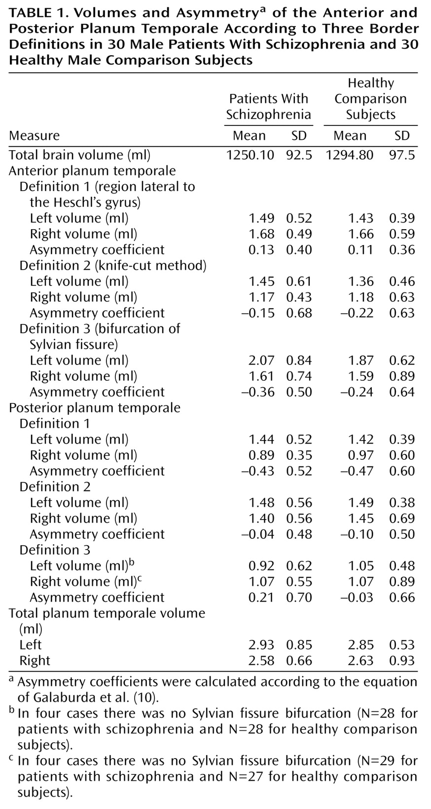
An overview of the results is given in
Table 1. The groups did not differ significantly with respect to age (t=–0.97, df=58, p=0.92).
Regarding the total planum temporale, the two-factor ANOVA revealed no significant differences between the groups (F=0.69, df=1, 58, p=0.41). The total left planum temporale was significantly larger than the total right planum temporale (F=5.7, df=1, 58, p=0.02), without significant interaction between groups and hemispheres (F=0.26, df=1, 58, p=0.61).
Regarding the anterior planum temporale, the two-factor ANOVA found no significant volume differences between patients and healthy subjects for any of the three definitions (definition 1: F=1.0, df=1, 58, p=0.32; definition 2: F=1.1, df=1, 58, p=0.30; definition 3: F=1.3, df=1, 58, p=0.29).
Regarding the posterior planum temporale, the two-factor ANOVA found no significant volume differences between patients and healthy subjects for any of the three definitions (definition 1: F=0.02, df=1, 58, p=0.89; definition 2: F=0.05, df=1, 58, p=0.82; definition 3: F=0.19, df=1, 54, p=0.66).
No significant differences between men with and without schizophrenia emerged regarding asymmetry coefficients for any of the three definitions.
Discussion
To our knowledge, this investigation is the largest study to date on planum temporale volumes of male patients with schizophrenia. Our data suggest that the definition of planum temporale borders does not influence the results concerning possible disturbances of planum temporale asymmetry in schizophrenia. Therefore, results may reflect real morphological brain differences between our group of patients and those examined by others. There are four studies that examined the volume of the planum temporale in schizophrenia in vivo by MRI
(2–
5) and one that performed an in vitro examination of postmortem tissue
(1).
Falkai et al.
(1), who detected planum temporale asymmetry disturbances, examined the brains of chronically ill hospitalized patients. In contrast, patients in the Maudsley family study
(2), which did not find significant differences in the asymmetry of the planum temporale between patients and control subjects, had a shorter illness duration than those examined by Kwon et al.
(4) and Falkai et al.
(1). These findings are consistent with the assumption that patients with a more chronic course of disease may show more brain abnormalities.
In conclusion, our results may support the neurodevelopmental hypothesis of more severe brain alterations in a subgroup of more severely ill patients with schizophrenia. Possible correlations between factors of chronicity, severity of the schizophrenic disturbances, and planum temporale measurements need to be evaluated in further studies.


