Full access
Article
Published Online: 1 December 2003

MRI Assessment of Gray and White Matter Distribution in Brodmann’s Areas of the Cortex in Patients With Schizophrenia With Good and Poor Outcomes

Abstract

OBJECTIVE: High-resolution magnetic resonance imaging (MRI) was used to compare cortical gray and white matter and CSF volumes in schizophrenia patients with poor outcomes, schizophrenia patients with good outcomes, and healthy comparison subjects. METHOD: T1-weighted, 1.2-mm-thick MR images were acquired for 37 patients with schizophrenia and 37 healthy, age- and sex-matched comparison subjects. The patients were assigned to subgroups with poor outcomes (N=13) and good outcomes (N=24) on the basis of clinical characteristics. Poor-outcome patients were those who were continuously hospitalized or completely dependent on others for basic needs, were unemployed, and had severe negative symptoms and severe formal thought disorder. The MR images were reoriented to standard position parallel to the anterior commissure-posterior commissure line and segmented into CSF, gray matter, and white matter tissue types. The tissue types were assigned to Brodmann’s areas by using the Perry postmortem histological atlas, and tissue-type volumes in the three subject groups were compared. RESULTS: Compared to the healthy subjects, the overall patient group had a significantly smaller mean cortical gray matter volume and significantly larger mean CSF volume, especially in the frontal lobe and left temporal lobe. The smaller frontal lobe volume in schizophrenia was confirmed for unadjusted volumes and for volumes with adjustment for whole brain volumes. Compared to patients with good outcomes, patients with poor outcomes (Kraepelinian schizophrenia) had significantly smaller gray matter volumes in the temporal and occipital lobes, but no difference between groups was found for total frontal lobe volume. Only 21% of the healthy subjects had volumes 0.5 standard deviations below the mean for healthy subjects in any area of the frontal or temporal lobes, compared with 62% of poor-outcome patients. CONCLUSIONS: Poor outcome in patients with schizophrenia may be associated with a more posterior distribution (posteriorization) of gray matter deficits across widely distributed cortical regions.
Cortical gray matter deficits are a frequent finding in schizophrenia. Global gray matter volumes approximately 2%–4% smaller than normal have been reported in meta-analyses of volumetric magnetic resonance imaging (MRI) studies in schizophrenia (1, 2). Significantly smaller than normal regional volumes in schizophrenia have been reported for the frontal cortex (312), insula (13, 14), and superior temporal gyrus (6, 8, 15–19). One recent voxel-based morphometry study found focal gray matter density deficits in the right supramarginal gyrus and in the orbitofrontal, superior temporal, occipitotemporal, precuneate, posterior cingulate, and insular cortices bilaterally in patients with schizophrenia or schizophreniform disorder (20). These widespread deficits have been hypothesized to indicate an abnormality at a supraregional level of brain organization (2126). These findings have been difficult to interpret because only a few of the various brain regions have been examined in each study. Without specific statistical contrasts of frontal and temporal areas with other cortical areas, it is difficult to conclude that the effect in one lobe or one Brodmann’s area is greater than that in another. Further, the considerable variation in structural findings in the cortex in schizophrenia might also reflect the etiologic heterogeneity of subjects with schizophrenia in the studies or a sequential developmental change with illness progression. Distinguishing patients with schizophrenia according to outcome may be one strategy to address this etiologic heterogeneity. Three classifications to subtype schizophrenia according to outcome or severity have been proposed (27). One such classification reliably divides patients into Kraepelinian (poor-outcome) and non-Kraepelinian (good-outcome) schizophrenia subtypes on the basis of longitudinal course (28). The course of poor-outcome schizophrenia is characterized by severe dysfunctions in self-care with continuous and complete dependence on others for obtaining and maintaining the basic necessities of life, including food, clothing, and shelter. Compared to patients with good-outcome schizophrenia, patients with poor-outcome schizophrenia have been shown to exhibit more severe negative symptoms, more severe formal thought disorder, and poor response to treatment, while no significant differences between the two groups in the severity of delusions, hallucinations, and bizarre behavior have been reported (29). Our group observed that poor-outcome patients exhibited relatively larger ventricular size, implying greater cortical deficits, compared with good-outcome patients (30). Several studies have associated severity of negative symptoms with smaller prefrontal volumes (31, 32). In contrast, severity of formal thought disorder has been associated with abnormalities in left temporal lobe volumes (16; see reference 33 for review). Smaller temporal lobe volumes were seen in more chronic state hospital patients than in less chronic patients (8). This pattern of prefrontal deficits with negative symptoms and temporal abnormalities with thought disorder is similar to the functional deficits in the frontal lobe/occipital lobe ratio associated with negative symptoms in schizophrenia (see reviews, references 34, 35) and temporal lobe metabolic increases with hallucinations (e.g., references 36, 37). Thus, differences in the anteroposterior gradient of regional size may be a pattern in schizophrenia that is perhaps most marked in the poor-outcome group. It is also possible that poor-outcome patients have volume deficits in both the frontal and temporal cortices—the “two-hit” hypothesis—while good-outcome patients have changes in only one of the two areas.
In the present study we made a comprehensive analysis of Brodmann’s areas in the frontal, temporal, and occipital lobes in a group of patients with schizophrenia and healthy comparison subjects. Our goal was to examine the patterns of volume deficits in individual Brodmann’s areas and their association with outcome. While the data could have been divided into several papers, one on each lobe, we felt that presenting all the data together was more efficient and less biased. Rather than to rely entirely on 78 t tests, one for each Brodmann’s area in each hemisphere, we developed groupings of areas to examine frontal-posterior gradients, specific prefrontal deficits, and temporal deficits in multivariate analyses of variance (MANOVAs). These groupings of Brodmann’s areas were based on already reported deficits in both functional activity and structural volume. First, based on studies by ourselves and others (38) showing less activity in the frontal lobe, compared with the occipital areas, we hypothesized that a frontal-to-posterior contrast of Brodmann’s areas between patients with schizophrenia and healthy comparison subjects, as well as within the schizophrenia group between the poor- and good-outcome subgroups, would reveal more marked gradients of gray matter size and/or sulcal enlargement as manifest by larger than normal CSF volume in the patients, especially those with poor outcome. We further specifically tested whether volume deficits are uniform throughout the frontal lobe and whether these deficits differentially affect verbal and spatial processing areas, as we did in earlier functional studies using the same method for assessment of Brodmann’s areas (39). Second, the extensive study of temporal lobe activity and volume differences, perhaps specifically in Brodmann’s area 22 or in the medial temporal regions, suggested the need to assess specificity through detailed analysis of the temporal lobe that contrasted these areas with others in the same lobe with visual functions and with areas in the occipital lobe with similar visual functions. Third, we examined whether deficits in volume in both frontal and temporal areas are associated with poorer outcomes. These three types of analyses are presented below as economical ways of organizing our approach to a complete if complex data set.

Method

Subjects

Thirty-seven patients with schizophrenia (27 men, 10 women; age: mean=43.0 years, SD=11.9, range=20–66; education: mean=12.6 years, SD=1.9) were recruited from the Mount Sinai Medical Center, New York City; the Bronx Veteran Affairs Medical Center in the Bronx, New York; and Pilgrim State Psychiatric Center in Brentwood, New York. Thirty-six patients were right-handed and one was ambidextrous on the basis of evaluation with the Edinburgh Handedness Inventory (40). The patients were evaluated with the Comprehensive Assessment of Symptoms and History (41) and were given a diagnosis of schizophrenia (N=36) or schizoaffective disorder (N=1) according to the DSM-IV criteria. The mean age at onset of psychotic symptoms for these 37 patients was 21.0 years (SD=6.9), and the mean age for beginning antipsychotic medications was 25.6 years (SD=10.9). The patients’ mean number of months of hospitalization was 40.4 (SD=65.0). The age at onset of illness, age at first use of antipsychotic medications, and hospitalization data were not available for one patient. A battery of rating scales, including the Positive and Negative Syndrome Scale (42), was administered. The patients’ mean scores on the Positive and Negative Syndrome Scale subscales were as follows: mean=21.0, SD=9.8, for the positive subscale; mean=18.0, SD=7.2, for the negative subscale; and mean=33.3, SD=12, for general psychopathology. Positive and Negative Syndrome Scale scores were not obtainable for one patient.
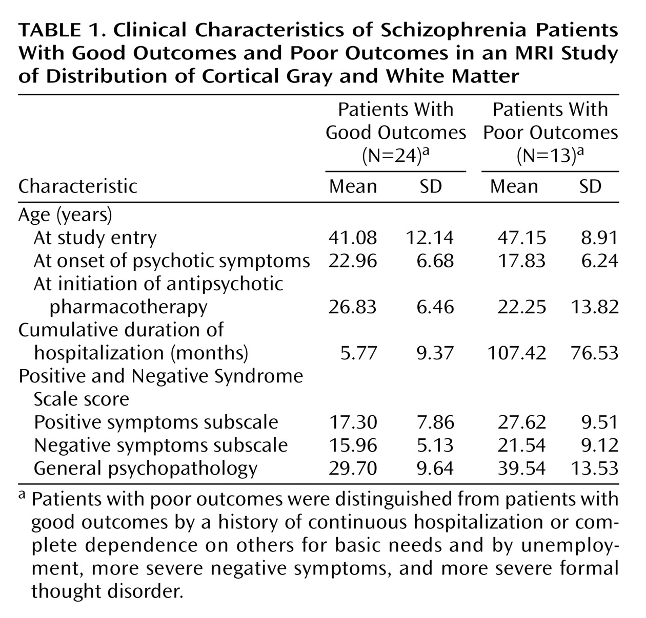
The patients with schizophrenia were assigned to poor-outcome (Kraepelinian) (N=13) and good-outcome (non-Kraepelinian) (N=24) subgroups on the basis of the clinical criteria of Keefe et al. (29) (Table 1). The poor-outcome subgroup was characterized by continuous hospitalization or complete dependence on others to meet basic needs, unemployment, and no evidence of remission of symptoms (30). Good-outcome patients did not differ from poor-outcome patients in age at first use of antipsychotic pharmacotherapy (t=1.37, df=34, p=0.18). However, poor-outcome patients had an earlier age at onset of the illness (t=2.22, df=34, p=0.03) and significantly more months of hospitalization (t=6.50, df=34, p<0.0001). The Positive and Negative Syndrome Scale subscale scores of the healthy comparison subjects and the good- and poor-outcome patients were compared by using MANOVA, with the subscale scores as repeated measures. The significant main effect of diagnosis (F=29.38, df=1, 34, p<0.00001) indicated that poor-outcome patients had significantly higher scores on all three Positive and Negative Syndrome Scale subscales. There was no significant interaction between diagnosis and Positive and Negative Syndrome Scale subscale scores (F=0.62, df=2, 68, p=0.54).
Thirty-seven healthy volunteers approximately matched in age and sex to the patients (23 men, 14 women; age: mean=44.1 years, SD=13.7, range=22–85; education: mean=16.0 years, SD=3.2) were recruited by word of mouth or advertisement. The proportion of female subjects in the three study groups was similar (38% in the comparison group, 25% in the good-outcome patient group, and 31% in the poor-outcome patient group) (χ2=1.12, df=2, p=0.57). Thirty-four comparison subjects were right-handed and two were left-handed on the basis of evaluation with the Edinburgh Handedness Inventory (40). All comparison subjects were assessed with the Comprehensive Assessment of Symptoms and History interview to confirm the absence of psychiatric illness in themselves and their first-degree relatives.

Image Acquisition

T1-weighted MRI scans were acquired with the 1.5-T Signa 5x system (GE Medical Systems, Milwaukee) with repetition time=24 msec, echo time=5 msec, flip angle=40°, slice thickness=1.2 mm, pixel matrix=256×256, field of view=23 cm, and total slices=128.

Brodmann’s Area Measurements and Quantification of Tissue Type

Perry et al. provided a unpublished coronal atlas (R.H. Perry, personal communication, 1993) composed of 33 axial maps of the Brodmann’s areas based on microscopic examination of the entire hemisphere of a postmortem brain. Our earlier studies in which the Perry atlas was used (4345) described the method in greater detail. Coronal slices perpendicular to the anterior commissure-posterior commissure line were reconstructed in a 256×256 pixel matrix. First, we determined the front (first slice containing the cortical ribbon) and back of the brain (last slice containing the cortical ribbon) and identified 33 evenly spaced slices such that the first slice began 1/34th of the distance from front to back. For each temporal lobe, we identified the temporal pole and the most posterior extent of the sylvian fissure and divided the space into 13 equally spaced slices. The brain edge was obtained on the approximately circular 33 nontemporal slices and 26 (13 in each hemisphere) temporal slices by depositing points visually on the tips of the gyri and then fitting a spline curve to the points. Each slice was then divided into 20 radial sectors on each hemisphere surface and 10 midline sectors (44). Brodmann’s areas were then assessed for the gray matter, white matter, and CSF pixels within each sector (see later discussion of segmentation method); means were weighted according to the number of sectors in each region of interest and proportionately combined to obtain a single measure. Some of the smaller Brodmann’s areas were combined (e.g., 3-1-2-5) for simplicity.
For gray matter, white matter, and CSF quantification, the coronal images for both the temporal and frontal lobes were segmented into gray matter, white matter, and CSF by using cutoff values individually determined in each subject by examining the within-brain-edge histogram of axial MRI values. Nearly every subject had a clear point of rarity between gray matter and white matter, and 75% of subjects had a point of rarity for gray matter and CSF. For those few subjects missing this second trough, we multiplied the numerical gray scale position (on a 0–255 scale, typically 120–150) by 0.32, the average proportion of the gray scale position that the CSF peak occupied (e.g., 150 × 0.32=47).
Four complementary approaches to validating our gray matter/white matter segmentation were carried out. First, we validated the MRI intensity value histogram peak and trough selection with two raters. The intraclass correlation coefficients (ICCs) for the gray matter, white matter, and CSF components for the two tracers were 0.98, 0.99, and 0.92, respectively, for the 28 subjects included in the pilot data analysis. Second, we used the stereology approach to visually identify gray matter and white matter (46). We counted points on the frontal coronal sections in 20 individuals and obtained a 0.87 correlation between gray matter volume by visual inspection point-counting and gray matter volume obtained by using automated segmentation. Third, we compared the left and right hemisphere gray matter, white matter, and CSF segmentation values as a rough validator of how our segmentation method identified a reliable whole-brain subject trait from an independent brain area; these ICCs were 0.85, 0.74, and 0.86, respectively. This analysis demonstrates that the final result from segmentation yielded statistically significant separation of the tissue types. Fourth, to assess test-retest reliability over time, we examined MR image pairs obtained 4–6 weeks apart for 16 patients with schizophrenia. The second image in each pair was coregistered to the first (47). Images were resectioned and the brain edges traced on the first image in each pair. These edges were then overlaid on the second image, and the segmentation program was run by using the gray matter, white matter, and CSF thresholds from the first image. This approach provides the worst-case reliability covering all methods, since it examines segmentation on the second MR image with the edges traced independently on the first and uses threshold values from the first image. We obtained frontal gray matter, CSF, and white matter reliability measures for the middle frontal gyral region (Brodmann’s areas 44 plus 45 plus 46); these ICC values were 0.88, 0.77, and 0.75, respectively. Note that measurement of the second scan was entirely automated with coregistration by algorithm and by using the edges and thresholds obtained from the first scan. Interrater reliability for the areas of the coronal slices was 0.98.

Statistical Methods

Three strategies were used in assessing the data. First, we tested specific interregional hypotheses by using groupings based on Brodmann’s areas with repeated measures analysis of variance (ANOVA) and MANOVA. From our review of imaging literature, we noted the scarcity of interregional contrasts and the lack of examination of the specificity of group contrasts to a particular area. Four general interregional or intralobe contrasts seemed appropriate as described in the introduction. Data from 39 Brodmann’s areas identified by Perry (areas 1-2-3-5, 4, 6, 7a and 7b, 8, 9, 10, 11, 12, 17, 18, 19, 20, 21, 22, 23, 24, 25, 27, 28, 29, 30, 31, 32, 34, 35, 36, 37, 38, 39, 40, 41, 42, 43, 44, 45, 46, and 47) were obtained. Subgroups of these variables developed on a theoretical basis were entered into multiway repeated measures MANOVAs. The patients’ diagnostic categories were used as an independent group factor for the whole population (e.g., schizophrenia patients versus healthy volunteers or good-outcome patients versus poor-outcome patients). The repeated measures were region (e.g., frontal, temporal, and occipital lobe), selected sets of Brodmann’s areas (e.g., dorsolateral prefrontal [44-45-46] and occipital [17-18-19]), hemisphere (right, left), and tissue type (CSF, gray matter, white matter). Group-by-region and higher-order interactions were examined to establish regional differences. Follow-up simple interactions were performed to identify the strongest sources of group interactions. This approach, which provided tests of hypothesized group differences, can help minimize type I statistical error involved with t tests for each area, group contrast, and hemisphere. This approach is similar to strategies used for comparing batteries of neuropsychological test results in patients with schizophrenia and healthy subjects where the tests are grouped on the basis of type of function or where frontal and temporal tasks are contrasted (48). We computed absolute volumes in mm and relative volumes as the ratio of the area of the region of interest to the volume of brain. While ratio measures may not adequately correct for normal variation in intracranial volume, they are more commonly reported. We explored the use of brain volume as a covariate, as recommended elsewhere (49), but obtained very similar results to the ratio data (see the first part of the Results section) and therefore retained the ratio results for comparability. We report both overall F and Rao’s R values from Statistica (50). Second, we report exploratory t test analyses for every Brodmann’s area (averaged across hemispheres as an analogue of significance probability mapping, which is often used in group contrasts). Third, to test the “two-hit hypothesis” of poor outcome, we assessed whether patients with volumetric deficits in two areas had been assigned to the poor-outcome group. For comparison of proportions of patient groups, the exact probability from binomial proportion tables was used.

Results

Dorsolateral Frontal, Temporal, and Occipital Lobes

Comparison of patients with schizophrenia and healthy subjects

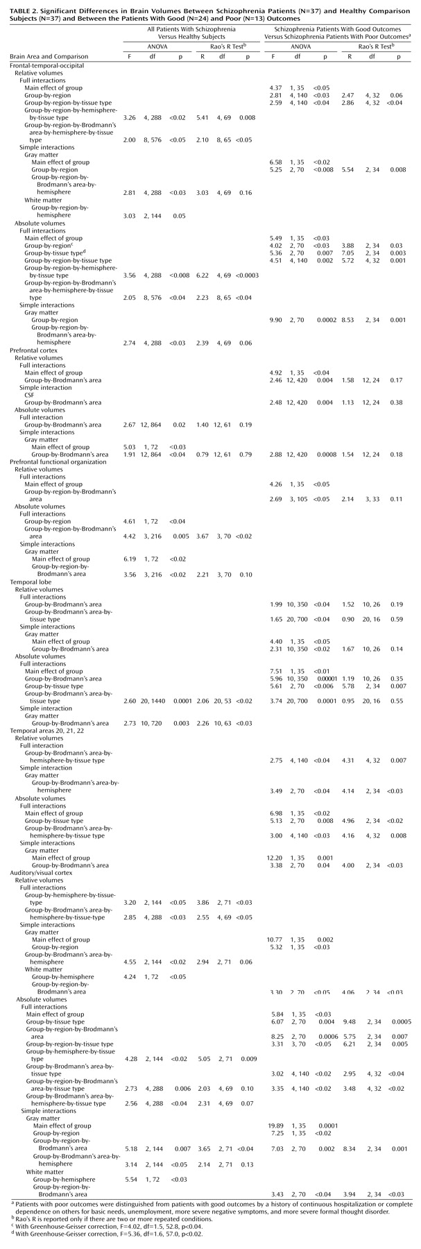
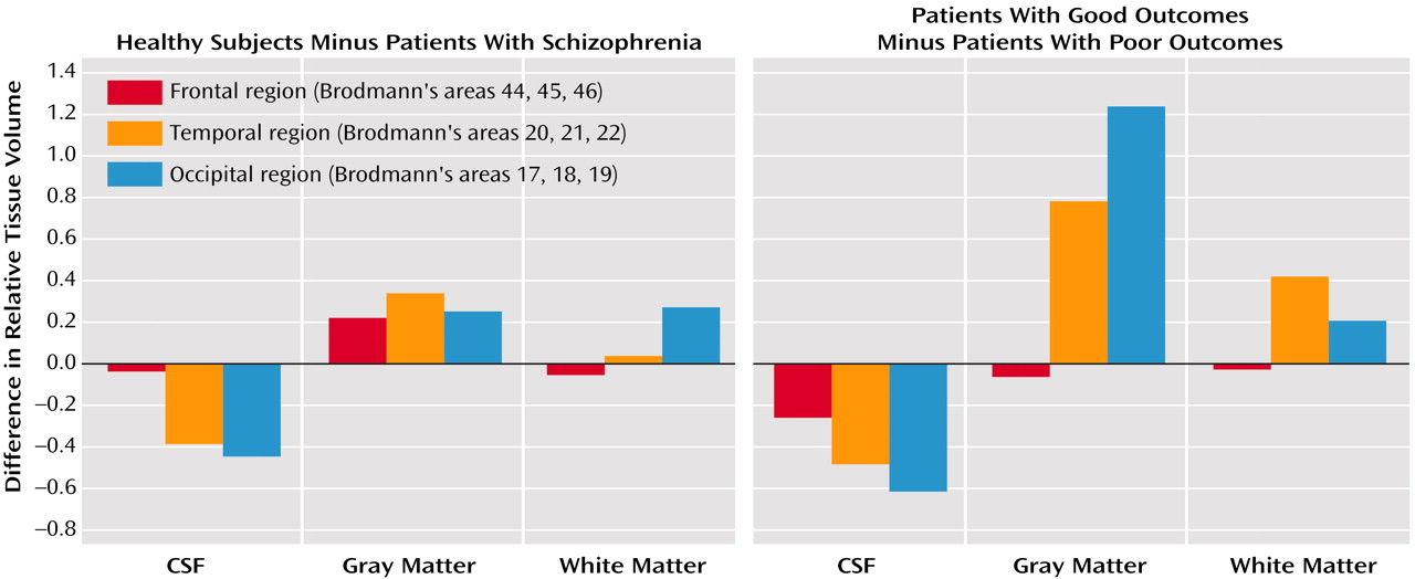
A summary analysis of relative tissue volume in the healthy comparison subjects compared with that in all patients with schizophrenia and of gray matter, white matter, and CSF volumes for the dorsolateral frontal (Brodmann’s areas 44, 45, 46), temporal (Brodmann’s areas 20, 21, 22), and occipital (Brodmann’s areas 17, 18, 19) lobes for the right and left hemispheres in those regions showed that the patients had less tissue in the left hemisphere (Figure 1, left panel), especially in the temporal region (all ANOVA effects and follow-up simple interactions are reported in Table 2). This difference was confirmed for both gray and white matter considered separately but not for CSF.
A similar analysis using data on absolute volumes found that patients had larger CSF volumes and smaller gray and white matter volumes than the healthy comparison subjects and that this difference was more marked for gray matter in the temporal and occipital regions and for the left hemisphere. The largest effects were for gray matter in Brodmann’s areas 17 and 18 in the occipital lobe and Brodmann’s area 20 in the temporal lobe.

Comparison of good-outcome and poor-outcome patients

A summary analysis comparing relative tissue volumes and gray matter, white matter, and CSF volumes for the dorsolateral frontal (Brodmann’s areas 44, 45, 46), temporal (Brodmann’s areas 20, 21, 22), and occipital (Brodmann’s areas 17, 18, 19) lobes in good-outcome and poor-outcome patients revealed smaller volumes across gray and white matter in the poor-outcome patients. This difference was more pronounced in the temporal and occipital regions than in the frontal region. CSF space was greater in poor-outcome patients across all three regions, while the smaller gray and white matter volumes were evident in the temporal and occipital cortices (Figure 1, right panel). Almost identical results were obtained in a summary analysis with age and brain volume as covariates, with significant group-by-region (F=4.03, df=1, 35, p<0.03), group-by-tissue-type (F=5.36, df=2, 70, p=0.007), and group-by-region-by-tissue-type (F=4.51, df=4, 140, p=0.002) interactions.
The same analysis using data for absolute volumes revealed smaller volumes in the poor-outcome patients across gray matter, white matter, and CSF. This effect was more prominent in temporal and occipital regions than in the frontal regions, more prominent in gray than white matter, and least prominent in CSF. The largest group differences in volume were for temporal and occipital lobe gray matter; white matter differences were half as great and CSF differences less than one-fourth as great.

Prefrontal Cortex

Comparison of patients with schizophrenia and healthy subjects

A summary analysis of relative tissue volume and of gray matter, white matter, and CSF volumes for the right and left hemispheres of the prefrontal cortex (Brodmann’s areas 8, 9, 10, 11, 12, 32, 24, 25, 6, 44, 45, 46, 47) in the healthy comparison subjects and in all patients with schizophrenia revealed no significant differences between the two groups. A similar analysis using data on absolute volumes found smaller total volumes in the patients with schizophrenia, and this difference was most marked in Brodmann’s areas 8, 9, and 25. The simple interaction tests revealed smaller gray matter volume in patients with schizophrenia, compared with healthy subjects, especially for Brodmann’s area 9 and, to a lesser degree, for Brodmann’s areas 8, 32, 24, 6, and 45, while no significant differences between groups were found for CSF or white matter volumes. An analysis of covariance with whole brain volume as a covariate confirmed the group-by-Brodmann’s-area interaction (F=2.67, df=12, 864, p<0.002; Rao’s R=1.40, df=12, 61, p=0.19).

Comparison of good-outcome and poor-outcome patients

A summary analysis comparing relative tissue volumes and gray matter, white matter, and CSF volumes for the right and left hemispheres of the prefrontal cortex (Brodmann’s areas 8, 9, 10, 11, 12, 32, 24, 25, 6, 44, 45, 46, 47) in good-outcome and poor-outcome patients with schizophrenia found smaller volumes in poor-outcome patients, especially in Brodmann’s areas 8, 9, and 6. The simple interaction tests found smaller CSF volumes in poor-outcome patients, especially in Brodmann’s area 6 and, to a lesser degree, in Brodmann’s areas 9, 32, and 8. The simple interaction tests for white and gray matter volumes produced no significant interactions.
The same analysis using data on absolute volumes revealed no differences between the groups. The simple interaction tests found smaller gray matter volume in the good-outcome group, especially in Brodmann’s area 6, and no differences in white matter or CSF volumes.

Functional Organization of the Frontal Lobe

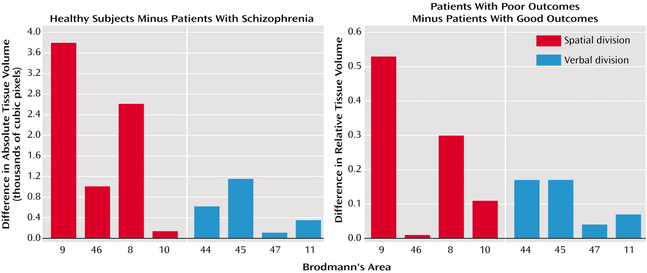
Following a model suggested by P. Goldman-Rakic (personal communication, 2000) based on organizational features of the prefrontal cortex, we contrasted data for spatial/object processing areas (Brodmann’s areas 9, 46, 8, and 10) with verbal areas (Brodmann’s areas 44, 45, 47, and 11) in a four-way ANOVA.

Comparison of patients with schizophrenia and healthy subjects

A summary analysis comparing relative tissue volume and gray matter, white matter, and CSF volumes for the right and left hemispheres for the frontal lobe spatial (Brodmann’s areas 9, 46, 8, and 10) and verbal (Brodmann’s areas 44, 45, 47, and 11) functional divisions in patients with schizophrenia and healthy subjects showed no significant differences between the groups. The simple interaction tests for CSF, gray matter, and white matter volumes also did not reach significance.
A similar analysis using data for absolute volumes (Figure 2, left panel) revealed smaller volumes in the patients with schizophrenia compared with the healthy subjects. The simple interaction tests showed smaller gray matter volumes in the patients with schizophrenia, with no significant differences in white matter or CSF volumes.

Comparison of good-outcome and poor-outcome patients

A summary analysis comparing relative tissue volume (Figure 2, right panel) and gray matter, white matter, and CSF volumes for the right and left hemispheres for the frontal lobe spatial (Brodmann’s areas 9, 46, 8, and 10) and verbal (Brodmann’s areas 44, 45, 47, and 11) functional divisions in good-outcome and poor-outcome patients with schizophrenia showed smaller tissue volume in the good-outcome patients than in poor-outcome patients. No significant differences were found in simple interaction tests for gray matter, white matter, and CSF volumes.
The same analyses using data on absolute volumes produced no significant interactions.

Temporal Lobe

Comparison of patients with schizophrenia and healthy subjects

Both summary and simple analyses comparing relative tissue volume and gray matter, white matter, and CSF volumes for the temporal lobe (Brodmann’s areas 42, 41, 22, 21, 20, 36, 28, 35, 27, 29, 30) for the right and left hemispheres in patients with schizophrenia and healthy subjects found no significant differences between the two groups.
A similar analysis using data for absolute volumes showed that the patients had smaller gray matter volumes in Brodmann’s areas 22, 21, and 20 and smaller white matter and larger CSF volumes in Brodmann’s area 36. Simple interaction tests confirmed smaller gray matter volumes in patients with schizophrenia in Brodmann’s areas 22, 21, and 20, and, to a lesser degree, in Brodmann’s areas 36 and 28. No significant differences between groups were found for CSF or white matter volumes.

Comparison of good-outcome and poor-outcome patients

A summary analysis comparing relative tissue volume and gray matter, white matter, and CSF volumes for the temporal lobe (Brodmann’s areas 42, 41, 22, 21, 20, 36, 28, 35, 27, 29, 30) for the right and left hemispheres in good-outcome and poor-outcome patients with schizophrenia revealed smaller gray matter volumes, especially in Brodmann’s area 22, in the poor-outcome group. Simple interaction tests confirmed smaller gray matter volumes in the poor-outcome group, especially in Brodmann’s area 22, and, to a lesser degree, in Brodmann’s areas 21, 20, 36, and 28, while tests for differences in white matter and CSF volumes did not reach significance.
A similar analysis using data on absolute volumes found smaller gray matter volumes in poor-outcome patients, especially in Brodmann’s areas 22, 21, and 20, and smaller white matter volumes in Brodmann’s area 21.

Temporal Lobe Brodmann’s Areas 20, 21, and 22

Comparison of patients with schizophrenia and healthy subjects

Both summary and simple analyses comparing relative and absolute tissue volumes and gray matter, white matter, and CSF volumes for the temporal lobe Brodmann’s areas 20, 21, and 22 for the right and left hemispheres in the patients with schizophrenia and the healthy subjects found no significant differences between the two groups.

Comparison of good-outcome and poor-outcome patients

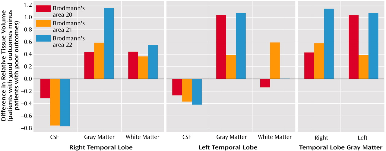
Both summary and simple analyses comparing relative tissue volume and gray matter, white matter, and CSF volumes for the temporal lobe Brodmann’s areas 20, 21, and 22 for the right and left hemispheres in good-outcome and poor-outcome patients with schizophrenia revealed smaller gray and white matter volumes and larger CSF volumes in the poor-outcome group, with the most marked effect in Brodmann’s area 22 bilaterally for gray matter (Figure 3, left and center panels). Simple interaction tests for gray matter volumes confirmed the smaller volumes in the poor-outcome group, which were most marked in Brodmann’s area 22 (Figure 3, right panel), while results for white matter and CSF volumes did not reach significance.
The same analysis using data for absolute volumes showed smaller gray and white matter volumes in the poor-outcome group and no between-group differences in CSF volumes. Simple interaction tests confirmed the smaller gray matter volumes in the poor-outcome group, which were most prominent in Brodmann’s area 22. A simple interaction test for white matter volume did not reach significance (main effect of diagnosis: F=3.85, df=1, 35, p=0.06; group-by-Brodmann’s area-by-hemisphere interaction: F=2.99, df=2, 70, p=0.06; Rao’s R=2.91, df=2, 34, p=0.07). No significant between-group differences were found in CSF volume.

Auditory and Visual Cortex Regions

Comparison of patients with schizophrenia and healthy subjects

A summary analysis comparing relative tissue volume and white matter, gray matter, and CSF volumes for primary, secondary, and association exemplary areas for visual (Brodmann’s areas 17, 18, 19) and auditory (Brodmann’s areas 41, 42, 22) cortex regions for the right and left hemispheres in patients with schizophrenia and healthy subjects showed larger CSF volumes in the patients with schizophrenia. Simple interaction tests for gray matter alone revealed smaller volumes in the patients with schizophrenia. Simple interaction tests for white matter found smaller volumes in the right hemisphere and larger volumes in the left hemisphere in patients with schizophrenia. Simple interaction tests for CSF volume produced no significant results.
A similar analysis using data on absolute volumes showed smaller gray and white matter volumes as well as larger CSF volumes in the visual cortex in the left hemisphere in the patients with schizophrenia. Simple interaction tests confirmed smaller gray matter volume across the visual cortex region and in Brodmann’s area 22 in the auditory cortex in the patients with schizophrenia. Simple interaction tests for white matter alone confirmed smaller volumes in the left hemisphere in the patients with schizophrenia, while no significant interactions for CSF volumes were uncovered.

Comparison of good-outcome and poor-outcome patients

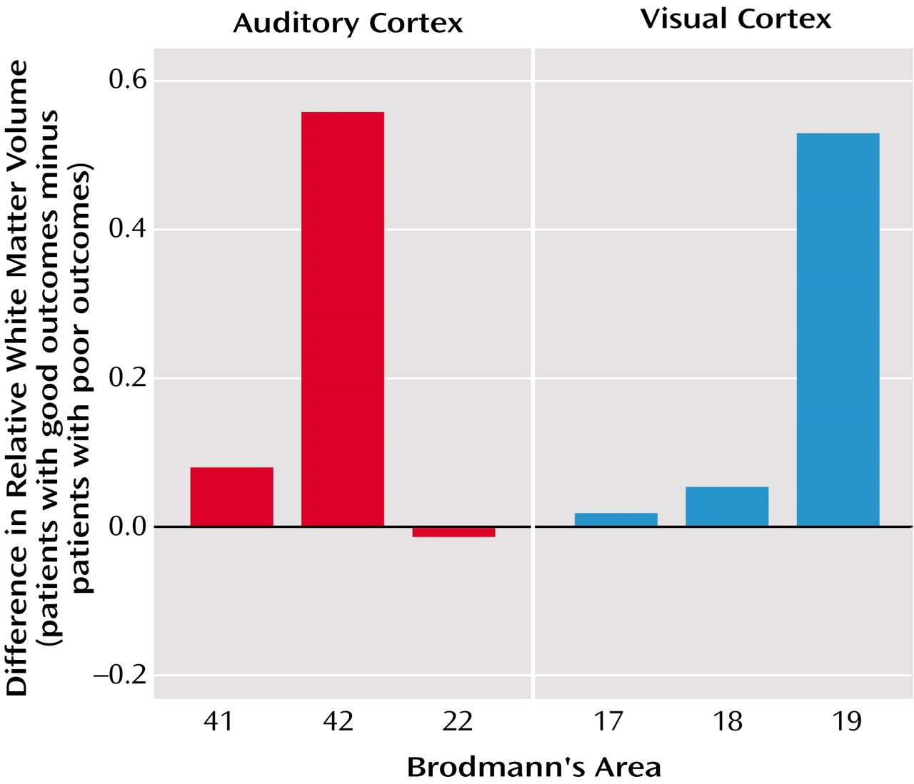
A summary analysis comparing relative tissue volume and white matter, gray matter, and CSF volumes for the primary, secondary, and association exemplary areas for visual (Brodmann’s areas 17, 18, 19) and auditory (Brodmann’s areas 41, 42, 22) cortex regions for the right and left hemispheres in patients with good-outcome and poor-outcome schizophrenia revealed tendencies toward smaller white and especially gray matter volumes and larger CSF volumes in the poor-outcome group that did not reach significance (group-by-tissue-type interaction: F=2.88, df=2, 70, p=0.06 and Rao’s R=3.49, df=2, 34, p<0.05; group-by-region-by-Brodmann’s-area interaction: F=2.80, df=2, 70, p=0.07 and Rao’s R=2.35, df=2, 34, p=0.11). Simple interaction tests confirmed smaller gray matter volumes (more prominent in the visual cortex) and smaller white matter volumes (Figure 4) (in Brodmann’s area 19 in the visual cortex and Brodmann’s area 42 in the auditory cortex) in the poor-outcome group. Simple interaction tests for CSF volumes found no intergroup differences.
The same analysis using data on absolute volumes showed smaller white and especially gray matter volumes and higher CSF volumes in the poor-outcome group, and this effect was more prominent in the visual cortex. Simple interaction tests confirmed smaller gray matter volumes (especially in the visual cortex) and smaller white matter volumes (most marked across the visual cortex and in Brodmann’s area 42 in the auditory cortex) in the poor-outcome group.

Tests of the Frontal-Temporal Two-Hit Hypothesis

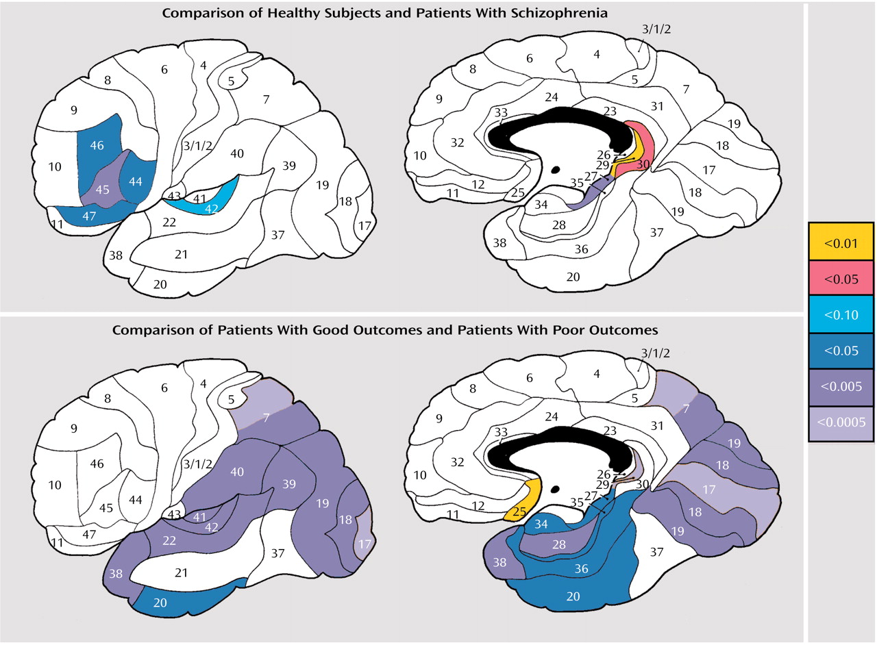
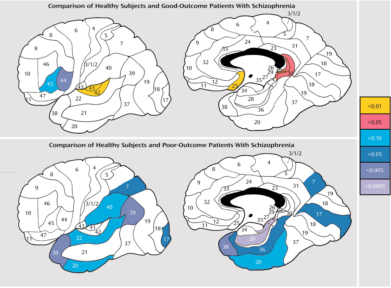
Exploratory maps comparing findings for the healthy subjects with those for the patients with schizophrenia and comparing findings for the patients with good outcome with those for the patients with poor outcome are shown in Figure 5 and Figure 6. Relative volumes in the dorsolateral prefrontal region (Brodmann’s areas 44, 45, 46, 47) and in the temporal lobe (Brodmann’s area 22, i.e., superior temporal gyrus, and Brodmann’s area 20) were expressed as “0” if they were more than 0.5 standard deviations below the mean for the 37 healthy comparison subjects. For the first comparison, we based the variable selection on our earlier analysis in a separate sample (39) and compared individuals scoring 0 for frontal area 46 and 0 for either temporal area 22 or temporal area 20. We found that while only four of the 37 healthy comparison subjects had two hits (11%), five of the 13 poor-outcome patients did (39%) (p=0.02, test of proportions). It is interesting to note that the patients with two hits had an older age at onset (25.0 years versus 19.3 years) (t=2.53, df=34, p<0.02), first received neuroleptics at a later age (30.4 years versus 22.8 years) (t=2.41, df=34, p<0.03), and had more years of education (13.4 years versus 11.9 years) (t=2.29, df=34, p<0.03) than the patients with one hit. When relative volumes for all dorsolateral areas (a 0 for any of areas 44, 45, 46, or 47) and all temporal areas (a 0 for any of areas 20, 21, 22, or 38) were combined, only eight of 37 healthy subjects (21%) had two hits, compared with eight of 13 poor-outcome patients (62%) (p<0.005, exact probability test) and 11 of 24 good-outcome patients (45%) (p<0.03, exact probability test). The difference in proportions for the poor-outcome and good-outcome patients was not significant (p=0.16, exact probability test).

Correlation of Gray and White Matter Volumes With Symptom Severity

We examined on an exploratory basis the correlation between relative gray and white matter volumes in the Brodmann’s areas and symptom severity assessed with the Positive and Negative Syndrome Scale subscales. Of the 312 possible correlations (involving 39 Brodmann’s areas, two hemispheres, two scales, two tissues), a total of 18 correlations, or about 5% of the correlations, were significant (p<0.05, exact probability test). However, of the 11 significant correlations of volume with negative symptom scores, eight were for posterior areas (occipital and parietal) and three were for anterior areas, while all seven significant correlations of volume with positive symptom scores were for anterior areas (orbitofrontal and dorsolateral prefrontal) and none were for posterior areas (p<0.001, exact probability test).

Discussion

Our findings are consistent with the majority of structural MRI studies of schizophrenia (33, 51) in that patients with schizophrenia were found to have abnormalities in the prefrontal gray matter, especially the dorsolateral regions 44, 45, 46, and 47, and in the temporal lobes, especially the superior temporal gyrus, region 22. Our data support the specificity of both dorsolateral and superior temporal gyrus findings and do not suggest generalized, nonspecific volumetric deficits.
The superior temporal gyrus, corresponding to Brodmann’s area 22, has been found to have smaller volumes than other temporal lobe regions in schizophrenia in some but not all studies. In the current data, significantly smaller area 22 volumes, compared to medial or inferior temporal lobe volumes, were confirmed for poor-outcome patients but not the entire group of patients with schizophrenia. The poor-outcome patients in the current study are probably more similar clinically to the patients in whom relatively smaller volumes were found in area 22 in a previous study (16), but this original study did not include a stringent requirement for an ANOVA diagnostic-group-by-temporal-region interaction for relative data. Our statistical contrasts are consistent with the concept of posteriorization—smaller volumes in more posterior brain regions in patients with more severe symptoms.
Although some studies have reported that smaller gray matter volume in the frontal lobe may be associated with chronicity and severity of the illness (11, 52), our data show more posterior (temporo-parieto-occipital) abnormalities in these patients. Gur et al. (9) found smaller frontal volumes associated with symptom severity only in women, while temporal lobe volume correlations did not meet the Bonferroni criterion and were not reported (53). No correlation between severity of thought disorder and temporal lobe volumes was found in a study of patients with schizotypal personality disorder (54). Our earlier results in a separate group of patients showed smaller temporal lobe gray matter volumes in patients with schizophrenia and in patients with schizotypal personality disorder but no relationship between smaller temporal lobe volumes and symptom severity (55).

Methodological Issues Associated With Thresholding Segmentation

If our method for determining the segmentation threshold has random error, group differences would be less likely to be significant (note that the total number of pixels is CSF plus gray matter plus white matter). If the threshold is systematically too high or low across all subjects, then significance is transferred to another tissue type (e.g., lost from white matter and displaced to gray matter), although if adjacent types are complementary (e.g., larger white matter volumes and smaller gray matter volumes), some group differences might be attenuated. If threshold interacts with diagnosis (e.g., poor-outcome patients have too many CSF pixels falsely identified as gray matter), then the effects of the interactions involving CSF and gray matter volumes would be diminished, larger CSF and smaller gray matter volumes would not be found, and CSF volumes would appear to be smaller and gray matter volumes larger. Thus, the current pattern of results does not appear to be attributable to random thresholding error or a shift in the threshold of CSF and gray matter in schizophrenia.

Illness Severity and Posterior Cerebral Areas

In this study, severity of illness was associated with profoundly smaller posterior gray matter volumes, whereas frontal lobe deficits were similar in all patients with schizophrenia, regardless of outcome. Furthermore, these structural differences between poor-outcome (Kraepelinian) and good-outcome (non-Kraepelinian) groups correspond to the differences in metabolic rates between poor-outcome and good-outcome patients found in our recent study employing [18F]fluorodeoxyglucose positron emission tomography in a separate group of patients (56). Parallel to our structural findings of smaller volumes in poor-outcome patients compared to good-outcome patients, the poor-outcome group showed less metabolic activity in the superior portion of the anterior cingulate gyrus, superior and middle temporal regions, and visual association areas (inferior and posterior temporal cortex, Brodmann’s areas 18 and 21). At the same time, Kraepelinian patients had lower relative metabolic rates in the temporal lobes, while non-Kraepelinian patients’ metabolic rates were similar to those in healthy comparison subjects. However, data for structural temporal lobe deficits in our current study showed that non-Kraepelinian patients had mean values between those for the poor-outcome group and those for the healthy subjects and were less likely to have smaller volumes in both the frontal and temporal regions in the same individuals. These findings suggest that unlike the good-outcome group, patients with poor-outcome schizophrenia cannot functionally compensate for smaller temporal gray matter volumes with either greater temporal or greater frontal function. Thus, gray matter deficits in poor-outcome schizophrenia fall below the threshold of functional compensatory reserve capacities. Poor outcome may arise from the structural deficits beyond the capacities of functional compensation either in adjacent temporal regions or in prefrontal regions, and this pattern appears to be related to having two or more affected regions in both the frontal and temporal lobes.

Two-Hit Model of Poor Outcome

Two aspects of the two-hit defective compensation model need special consideration. Very few healthy subjects showed both a frontal and a temporal Brodmann’s area volume deficit, fewer than expected if the areas were entirely independent. However, while a large proportion of poor-outcome patients showed deficits in both areas, more than expected if the areas were independent, 38% of the patients did not have volumes 0.5 standard deviations below the mean for healthy subjects in both areas. While beyond the scope of the current report, further exploration of the distribution of areas of volume deficits and their conjoint probabilities in both patients and healthy subjects may reveal mechanisms of compensation, protection, and vulnerability. The proportions of good-outcome and poor-outcome patients with both a frontal and a temporal volume deficit suggest that the effect is not entirely due to greater symptom severity, since the 0.39 proportion of two-hit patients among the poor-outcome patients is much greater than the expected 0.09 proportion of patients with equally severe symptoms and both frontal and temporal lobe deficits with statistical independence of deficits in the lobes (0.30 of the population is 0.5 standard deviation low; 0.3×0.3=0.09). Second, the group of patients with two hits, which was not exactly the same as the group of patients with poor outcomes, had illness onset 7 years later, received neuroleptics 8 years later, and had 2 more years of education than the patients without two hits. It is possible that there was a slow development of the illness in the group with two hits and that the illness might not have been recognized until the second region was affected. Only longitudinal studies can fully address this surprising finding.

Laterality Findings

The finding that patients with schizophrenia had smaller temporal lobes, especially on the left side, is consistent with a meta-analysis of MRI findings (57) and our own earlier findings for a separate group of patients (55). This pattern may be associated with larger left temporal ventricular horn volumes, which have been reported to be asymmetric in some (30, 55, 58, 59) but not all (57) studies. This lateralization is most apparent in the left temporal verbal processing areas and is consistent with the many linguistic disturbances in schizophrenia.
In our study, patients with schizophrenia were divided into good- and poor-outcome groups primarily on the basis of their ability to care for themselves (29), whereas some other studies of outcome in schizophrenia have used duration of hospitalization, chronicity, or predominance of positive versus negative symptoms, making direct comparisons difficult. Contrary to our expectations of the importance of frontal lobe executive functions in the ability to care for oneself, structural frontal lobe deficits did not differentiate between those with impaired and those with preserved abilities for self-care. Poor-outcome patients also showed volumetric deficits in the parietal cortex (Brodmann’s areas 39 and 40), a region that is associated with goal-directed motor behavior (60). Thus, it is possible that basic survival skills—e.g., recognizing people, monetary value of things, climate-appropriate clothes, and home as distinct from other locations, as well as shelter-seeking behavior, and ability to perform actions involving objects, ability to translate decisions to concrete, goal-directed actions—are more important for differentiation by ability for self-care than positive symptoms or disturbance of executive functions. Our data appear to support the theory of a frontal-to-posterior dimension underlying good-to-poor outcome in schizophrenia, particularly as it relates to self-care abilities, with a posteriorization of gray matter deficits with poor outcome.
TABLE 1
TABLE 2
Figure 1. Differences in Relative Tissue Volumes in the Frontal, Temporal, and Occipital Lobes of the Left Hemisphere Between Healthy Comparison Subjects (N=37) and Schizophrenia Patients and in the Frontal, Temporal, and Occipital Regions Between the Patients With Good (N=24) and Poor (N=13) Outcomes
Figure 2. Differences in Absolute Tissue Volumes in Spatial and Verbal Frontal Processing Regions Between Healthy Comparison Subjects (N=37) and Schizophrenia Patients and Differences in Relative Tissue Volumes Between the Patients With Good (N=24) and Poor (N=13) Outcomes
Figure 3. Differences in Relative Tissue Volumes in the Right and Left Temporal Lobe and in Temporal Lobe Gray Matter Between Schizophrenia Patients With Good (N=24) and Poor (N=13) Outcomes
Figure 4. Differences in Relative White Matter Volumes in the Primary, Secondary, and Association Exemplary Areas of the Auditory and Visual Cortical Regions Between Schizophrenia Patients With Good (N=24) and Poor (N=13) Outcomes
Figure 5. Lateral and Medial Views of the Brain Showing Brodmann’s Areas With Differences in Relative Tissue Volumes Between Healthy Comparison Subjects (N=37) and Schizophrenia Patients and Between the Patients With Good (N=24) and Poor (N=13) Outcomesa
aThe color bar shows p values for t tests for each Brodmann’s area (averaged across right and left hemispheres). In the upper panel, Brodmann’s areas in which the healthy comparison subjects had larger volumes than the patients with schizophrenia are shown in purple and blue, and areas in which they had smaller volumes than the patients are shown in orange and yellow. In the bottom panel, areas in which the poor-outcome patients had smaller volumes than the patients with good outcomes are shown in purple and blue, and areas in which they had larger volumes than the good-outcome patients are shown in yellow.
Figure 6. ateral and Medial Views of the Brain Showing Brodmann’s Areas With Differences in Relative Tissue Volumes Between Healthy Comparison Subjects (N=37) and Schizophrenia Patients With Good Outcomes (N=24) and Between Healthy Subjects and Patients With Poor Outcomes (N=13)a
aThe color bar shows p values for t tests for each Brodmann’s area (averaged across right and left hemispheres). Brodmann’s areas in which the healthy comparison subjects had larger volumes than the patients with schizophrenia are shown in purple and blue, and areas in which they had smaller volumes than the patients are shown in orange and yellow.

Footnote

Presented in part at the 155th annual meeting of the American Psychiatric Association, Philadelphia, May 18–23, 2002. Received Oct. 30, 2002; revision received April 17, 2003; accepted April 25, 2003. From the Department of Psychiatry, Mount Sinai School of Medicine, New York; and the Bronx VA Medical Center, Bronx, N.Y. Address reprint requests to Dr. Mitelman, Department of Psychiatry, Box 1505, Mount Sinai Medical Center, One Gustave L. Levy Pl., New York, NY 10029; [email protected] (e-mail). Supported in part by NIMH grants MH-56489, MH-60023, MH-60384, MH-56140, and MH-42827; VA Merit Awards 2571-005 and 7609-28; NIH grant RR-00071; and a Young Investigator Award from the National Alliance for Research on Schizophrenia and Depression.

References

1.
Lawrie SM, Abukmeil SS: Brain abnormality in schizophrenia: a systematic and quantitative review of volumetric magnetic resonance imaging studies. Br J Psychiatry 1998; 172:110–120
2.
Wright IC, Rabe-Hesketh S, Woodruff PWR, David AS, Murray RM, Bullmore ET: Meta-analysis of regional brain volumes in schizophrenia. Am J Psychiatry 2000; 157:16–25
3.
Breier A, Buchanan R, Elkashef A, Munson R, Kirkpatrick B, Gellad F: Brain morphology and schizophrenia. Arch Gen Psychiatry 1992; 49:921–926
4.
Kawasaki Y, Maeda Y, Urata K, Higashima M, Yamaguchi N, Suzuki M, Takashima T, Ide Y: A quantitative magnetic resonance imaging study of patients with schizophrenia. Eur Arch Psychiatry Clin Neurosci 1993; 242:268–272
5.
Andreasen NC, Arndt S, Swayze V II, Cizadlo T, Flaum M, O’Leary D, Ehrhardt JC, Yuh WTC: Thalamic abnormalities in schizophrenia visualized through magnetic resonance image averaging. Science 1994; 266:294–298
6.
Schlaepfer TE, Harris GJ, Tien AY, Peng LW, Lee S, Federman EB, Chase GA, Barta PE, Pearlson GD: Decreased regional cortical gray matter volume in schizophrenia. Am J Psychiatry 1994; 151:842–848
7.
Buchanan RW, Vladar K, Barta PE, Pearlson GD: Structural evaluation of the prefrontal cortex in schizophrenia. Am J Psychiatry 1998; 155:1049–1055
8.
Sullivan EV, Lim KO, Mathalon D, Marsh L, Beal DM, Harris D, Hoff AL, Faustman WO, Pfefferbaum A: A profile of cortical gray matter volume deficits characteristic of schizophrenia. Cereb Cortex 1998; 8:117–124
9.
Gur RE, Cowell PE, Latshaw A, Turetsky BI, Grossman RI, Arnold SE, Bilker WB, Gur RC: Reduced dorsal and orbital prefrontal gray matter volumes in schizophrenia. Arch Gen Psychiatry 2000; 57:761–768
10.
Hirayasu Y, Tanaka S, Shenton ME, Salisbury DF, DeSantis MA, Levitt JJ, Wible C, Yurgelun-Todd D, Kikinis R, Jolesz FA, McCarley RW: Prefrontal gray matter volume reduction in first episode schizophrenia. Cereb Cortex 2001; 11:374–381
11.
Hulshoff Pol HE, Schnack HG, Bertens MGBC, van Haren NEM, van der Tweel I, Staal WG, Baaré WFC, Kahn RS: Volume changes in gray matter in patients with schizophrenia. Am J Psychiatry 2002; 159:244–250
12.
Crespo-Facorro B, Kim J, Andreasen NC, O’Leary DS, Magnotta V: Regional frontal abnormalities in schizophrenia: a quantitative gray matter volume and cortical surface size study. Biol Psychiatry 2000; 48:110–119
13.
Goldstein JM, Goodman JM, Seidman LJ, Kennedy DN, Makris N, Lee H, Tourville J, Caviness VS Jr, Faraone SV, Tsuang MT: Cortical abnormalities in schizophrenia identified by structural magnetic resonance imaging. Arch Gen Psychiatry 1999; 56:537–547
14.
Crespo-Facorro B, Kim J, Andreasen NC, O’Leary DS, Bockholt HJ, Magnotta V: Insular cortex abnormalities in schizophrenia: a structural magnetic resonance imaging study of first-episode patients. Schizophr Res 2000; 46:35–43; correction, 2001; 51:183–184
15.
Barta PE, Pearlson GD, Powers RE, Richards SS, Tune LE: Auditory hallucinations and smaller superior temporal gyral volume in schizophrenia. Am J Psychiatry 1990; 147:1457–1462
16.
Shenton ME, Kikinis R, Jolesz FA, Pollak SD, LeMay M, Wible CG, Hokama H, Martin J, Metcalf D, Coleman M, et al: Abnormalities of the left temporal lobe and thought disorder in schizophrenia: a quantitative magnetic resonance imaging study. N Engl J Med 1992; 327:604–612
17.
Flaum M, Swayze VW II, O’Leary DS, Yuh WTC, Ehrhardt JC, Arndt SV, Andreasen NC: Effects of diagnosis, laterality, and gender on brain morphology in schizophrenia. Am J Psychiatry 1995; 152:704–714
18.
Keshavan MS, Haas GL, Kahn CE, Aguilar E, Dick EL, Schooler NR, Sweeney JA, Pettegrew JW: Superior temporal gyrus and the course of early schizophrenia: progressive, static, or reversible? J Psychiatr Res 1998; 32:161–167
19.
Sullivan EV, Mathalon DH, Lim KO, Marsh L, Pfefferbaum A: Patterns of regional cortical dysmorphology distinguishing schizophrenia and chronic alcoholism. Biol Psychiatry 1998; 43:118–131
20.
Hulshoff Pol HE, Schnack HG, Mandl RC, van Haren NE, Koning H, Collins DL, Evans AC, Kahn RS: Focal gray matter density changes in schizophrenia. Arch Gen Psychiatry 2001; 58:1118–1125
21.
Wright IC, Sharma T, Ellison ZR, McGuire PK, Friston KJ, Brammer MJ, Murray RM, Bullmore ET: Supra-regional brain systems and the neuropathology of schizophrenia. Cereb Cortex 1999; 9:366–378
22.
Friston KJ, Frith CD: Schizophrenia: a disconnection syndrome? Clin Neurosci 1995; 3:89–97
23.
McGuire PK, Frith CD: Disordered functional connectivity in schizophrenia. Psychol Med 1996; 26:663–667
24.
Sigmundsson T, Suckling J, Maier M, Williams SCR, Bullmore ET, Greenwood KE, Fukuda R, Ron MA, Toone BK: Structural abnormalities in frontal, temporal, and limbic regions and interconnecting white matter tracts in schizophrenic patients with prominent negative symptoms. Am J Psychiatry 2001; 158:234–243
25.
Zipursky RB, Lim KO, Sullivan EV, Brown BW, Pfefferbaum A: Widespread cerebral gray matter volume deficits in schizophrenia. Arch Gen Psychiatry 1992; 49:195–205
26.
Harvey I, Ron MA, Du Boulay G, Wicks D, Lewis SW, Murray RM: Reduction of cortical volume in schizophrenia on magnetic resonance imaging. Psychol Med 1993; 23:591–604
27.
Roy MA, Merette C, Maziade M: Subtyping schizophrenia according to outcome or severity: a search for homogeneous subgroups. Schizophr Bull 2001; 27:115–138
28.
Keefe RSE, Frescka E, Apter SH, Davidson M, Macaluso JM, Hirschowitz J, Davis KL: Clinical characteristics of Kraepelinian schizophrenia: replication and extension of previous findings. Am J Psychiatry 1996; 153:806–811
29.
Keefe RS, Mohs RC, Losonczy MF, Davidson M, Silverman JM, Kendler KS, Horvath TB, Nora R, Davis KL: Characteristics of very poor outcome schizophrenia. Am J Psychiatry 1987; 144:889–895
30.
Davis KL, Buchsbaum MS, Shihabuddin L, Spiegel-Cohen J, Metzger M, Frecska E, Keefe RS, Powchik P: Ventricular enlargement in poor-outcome schizophrenia. Biol Psychiatry 1998; 43:783–793
31.
Baare WF, Hulshoff Pol HE, Hijman R, Mali WP, Viergever MA, Kahn RS: Volumetric analysis of frontal lobe regions in schizophrenia: relation to cognitive function and symptomatology. Biol Psychiatry 1999; 45:1597–1605
32.
Wible CG, Anderson J, Shenton ME, Kricun A, Hirayasu Y, Tanaka S, Levitt JJ, O’Donnell BF, Kikinis R, Jolesz FA, McCarley RW: Prefrontal cortex, negative symptoms, and schizophrenia: an MRI study. Psychiatry Res 2001; 108:65–78
33.
Shenton ME, Dickey CC, Frumin M, McCarley RW: A review of MRI findings in schizophrenia. Schizophr Res 2001; 49:1–52
34.
Buchsbaum MS, Hazlett EA, Haznedar MM, Spiegel-Cohen J, Wei TC: Visualizing fronto-striatal circuitry and neuroleptic effects in schizophrenia. Acta Psychiatr Scand Suppl 1999; 395:129–137
35.
Potkin SG, Alva G, Fleming K, Anand R, Keator D, Carreon D, Doo M, Jin Y, Wu JC, Fallon JH: A PET study of the pathophysiology of negative symptoms in schizophrenia. Am J Psychiatry 2002; 159:227–237
36.
Silbersweig DA, Stern E, Frith C, Cahill C, Holmes A, Grootoonk S, Seaward J, McKenna P, Chua SE, Schnorr L, et al: A functional neuroanatomy of hallucinations in schizophrenia. Nature 1995; 378:176–179
37.
DeLisi LE, Buchsbaum MS, Holcomb HH, Langston KC, King AC, Kessler R, Pickar D, Carpenter WT Jr, Morihisa JM, Margolin R, et al: Increased temporal lobe glucose use in chronic schizophrenic patients. Biol Psychiatry 1989; 25:835–851
38.
Buchsbaum M, Hazlett E: Positron emission tomography studies of abnormal glucose metabolism in schizophrenia. Schizophr Bull 1998; 24:343–364
39.
Buchsbaum MS, Nenadic I, Hazlett EA, Spiegel-Cohen J, Fleischman MB, Akhavan A, Silverman JM, Siever LJ: Differential metabolic rates in prefrontal and temporal Brodmann areas in schizophrenia and schizotypal personality disorder. Schizophr Res 2002; 54:141–150
40.
Oldfield RC: The assessment and analysis of handedness: the Edinburgh Inventory. Neuropsychologia 1971; 9:97–113
41.
Andreasen NC, Flaum M, Arndt S: The Comprehensive Assessment of Symptoms and History (CASH): an instrument for assessing diagnosis and psychopathology. Arch Gen Psychiatry 1992; 49:615–623
42.
Kay SR, Fiszbein A, Opler LA: The Positive and Negative Syndrome Scale (PANSS) for schizophrenia. Schizophr Bull 1987; 13:261–276
43.
Simeon D, Guralnik O, Hazlett EA, Spiegel-Cohen J, Hollander E, Buchsbaum MS: Feeling unreal: a PET study of depersonalization disorder. Am J Psychiatry 2000; 157:1782–1788
44.
Hazlett EA, Buchsbaum MS, Haznedar MM, Singer MB, Schnur DB, Jimenez EA, Buchsbaum BR, Troyer BT: Prefrontal cortex glucose metabolism and startle eyeblink modification abnormalities in unmedicated schizophrenia patients. Psychophysiology 1998; 35:186–198
45.
Stein DJ, Buchsbaum MS, Hof PR, Siegel BV Jr, Shihabuddin L: Greater metabolic rate decreases in hippocampal formation and proisocortex than in neocortex in Alzheimer’s disease. Neuropsychobiology 1998; 37:10–19
46.
Keshavan MS, Anderson S, Beckwith C, Nash K, Pettegrew JW, Krishnan KR: A comparison of stereology and segmentation techniques for volumetric measurements of lateral ventricles in magnetic resonance imaging. Psychiatry Res 1995; 61:53–60
47.
Woods RP, Mazziotta JC, Cherry SR: MRI-PET registration with automated algorithm. J Comput Assist Tomogr 1993; 17:536–546
48.
Hill SK, Ragland JD, Gur RC, Gur RE: Neuropsychological differences among empirically derived clinical subtypes of schizophrenia. Neuropsychology 2001; 15:492–501
49.
Arndt S, Cohen G, Alliger RJ, Swayze VW II, Andreasen NC: Problems with ratio and proportion measures of imaged cerebral structures. Psychiatry Res Neuroimaging 1991; 40:79–89
50.
Statistica, 5.5 ed. Tulsa, Okla, StatSoft, 1999
51.
Ross CA, Pearlson GD: Schizophrenia, the heteromodal association neocortex and development: potential for a neurogenetic approach. Trends Neurosci 1996; 19:171–176
52.
Staal WG, Hulshoff Pol HE, Schnack HG, van Haren NEM, Seifert N, Kahn RS: Structural brain abnormalities in chronic schizophrenia at the extremes of the outcome spectrum. Am J Psychiatry 2001; 158:1140–1142
53.
Gur RE, Turetsky BI, Cowell PE, Finkelman C, Maany V, Grossman RI, Arnold SE, Bilker WB, Gur RC: Temporolimbic volume reductions in schizophrenia. Arch Gen Psychiatry 2000; 57:769–775
54.
Dickey CC, McCarley RW, Voglmaier MM, Niznikiewicz MA, Seidman LJ, Hirayasu Y, Fischer I, Teh EK, Van Rhoads R, Jakab M, Kikinis R, Jolesz FA, Shenton ME: Schizotypal personality disorder and MRI abnormalities of temporal lobe gray matter. Biol Psychiatry 1999; 45:1393–1402
55.
Downhill JE, Buchsbaum MS, Hazlett EA, Barth S, Lees Roitman S, Nunn M, Lekarev O, Wei T, Shihabuddin L, Mitropoulou V, Silverman J, Siever LJ: Temporal lobe volume determined by magnetic resonance imaging in schizotypal personality disorder and schizophrenia. Schizophr Res 2001; 48:187–199
56.
Buchsbaum MS, Shihabuddin L, Hazlett EA, Schroder J, Haznedar MM, Powchik P, Spiegel-Cohen J, Wei T, Singer MB, Davis KL: Kraepelinian and non-Kraepelinian schizophrenia subgroup differences in cerebral metabolic rate. Schizophr Res 2002; 55:25–40
57.
Sommer I, Ramsey N, Kahn R, Aleman A, Bouma A: Handedness, language lateralisation and anatomical asymmetry in schizophrenia: meta-analysis. Br J Psychiatry 2001; 178:344–351
58.
Dauphinais ID, DeLisi LE, Crow TJ, Alexandropoulos K, Colter N, Tuma I, Gershon ES: Reduction in temporal lobe size in siblings with schizophrenia: a magnetic resonance imaging study. Psychiatry Res 1990; 35:137–147
59.
Crow TJ, Colter N, Frith CD, Johnstone EC, Owens DG: Developmental arrest of cerebral asymmetries in early onset schizophrenia. Psychiatry Res 1989; 29:247–258
60.
Freund HJ: The parietal lobe as a sensorimotor interface: a perspective from clinical and neuroimaging data. Neuroimage 2001; 14:S142-S146

Information & Authors

Information

Published In

Go to American Journal of Psychiatry
American Journal of Psychiatry
Pages: 2154 - 2168
PubMed: 14638586

History

Published online: 1 December 2003
Published in print: December 2003

Authors

Details

Serge A. Mitelman, M.D.
Lina Shihabuddin, M.D.
Adam M. Brickman, M.A.
Erin A. Hazlett, Ph.D.
Monte S. Buchsbaum, M.D.

Metrics & Citations

Metrics

Citations

Export Citations

If you have the appropriate software installed, you can download article citation data to the citation manager of your choice. Simply select your manager software from the list below and click Download.

For more information or tips please see 'Downloading to a citation manager' in the Help menu.

Format
Citation style
Style
Copy to clipboard

View Options

View options

PDF/EPUB

View PDF/EPUB

Get Access

Login options

Already a subscriber? Access your subscription through your login credentials or your institution for full access to this article.

Personal login Institutional Login Open Athens login
Purchase Options

Purchase this article to access the full text.

PPV Articles - American Journal of Psychiatry

PPV Articles - American Journal of Psychiatry

Not a subscriber?

Subscribe Now / Learn More

PsychiatryOnline subscription options offer access to the DSM-5-TR® library, books, journals, CME, and patient resources. This all-in-one virtual library provides psychiatrists and mental health professionals with key resources for diagnosis, treatment, research, and professional development.

Need more help? PsychiatryOnline Customer Service may be reached by emailing [email protected] or by calling 800-368-5777 (in the U.S.) or 703-907-7322 (outside the U.S.).

Media

Figures

Other

Tables

Share

Share

Share article link

Share