Full access
Brief Report
Published Online: 1 June 2004

Dorsolateral Prefrontal Cortical Pathology in Generalized Anxiety Disorder: A Proton Magnetic Resonance Spectroscopic Imaging Study

Publication: American Journal of Psychiatry

Abstract

OBJECTIVE: Few neuroimaging studies of generalized anxiety disorder have been conducted. The present study used proton magnetic resonance spectroscopy to assess concentrations of N-acetylaspartate, often considered a marker of neuronal viability, in generalized anxiety disorder patients. METHOD: N-Acetylaspartate/creatine resonance ratios were measured in the left and right dorsolateral prefrontal cortex and hippocampus of 15 medication-free generalized anxiety disorder patients and 15 age- and sex-matched healthy volunteers. RESULTS: Generalized anxiety disorder patients had a 16.5% higher N-acetylaspartate/creatine ratio in the right dorsolateral prefrontal cortex compared with healthy participants; 13 of 15 matched patient-comparison subject pairs displayed a difference in this direction. In addition, generalized anxiety disorder patients reporting childhood abuse had lower N-acetylaspartate/creatine ratios in the right dorsolateral prefrontal cortex than did nonabused patients. Metabolite differences were not detected in other regions. CONCLUSIONS: Generalized anxiety disorder is associated with asymmetric increases in the N-acetylaspartate/creatine ratio, a suggested marker of neuronal viability, in the prefrontal cortex. The findings also support prior research linking childhood abuse to reduced neuronal viability.
The neurobiological bases of generalized anxiety disorder are poorly characterized. Hypermetabolism in prefrontal cortical regions (1) and neuronal hypertrophy in limbic structures (2) have been observed. Proton magnetic resonance spectroscopy (1H-MRS) studies in related disorders have found topographic reductions (or no change) in prefrontal cortical or hippocampal measures of N-acetylaspartate, an amino acid considered a marker of neuronal viability (for review, see reference 3). Similarly, we recently found decreases in N-acetylaspartate/creatine ratios in prefrontal cortical regions of adult nonhuman primates who had undergone adverse rearing conditions as infants approximately 10 years earlier, suggesting possible trauma-related neurotoxicity (4).
However, to our knowledge no 1H-MRS study of generalized anxiety disorder has been reported. The current study examined neuronal viability in the dorsolateral prefrontal cortex and hippocampus of patients with generalized anxiety disorder, hypothesizing reductions in the N-acetylaspartate/creatine ratio. We also determined whether a history of childhood trauma identified a subgroup of adult generalized anxiety disorder patients with more pronounced deficits in the N-acetylaspartate/creatine ratio.

Method

Fifteen adult patients with DSM-IV generalized anxiety disorder (seven men and eight women; mean age=39.3 years, SD=13.3) and 15 healthy volunteers (mean age=39.1, SD=13.5) recruited through advertisements and clinic/physician referrals were prospectively pair-matched with respect to age and sex. As determined with the Structured Clinical Interview for DSM-IV Axis I Disorders (5), comorbid disorders in the generalized anxiety disorder group were dysthymia (N=5), social anxiety disorder (N=2), and concurrent social anxiety disorder and dysthymia (N=1). Inclusion criteria required that generalized anxiety disorder precede onset of comorbid axis I disorders. Exclusion criteria were a major depressive episode or substance abuse within 6 months of study entry; a lifetime history of psychotic, bipolar, obsessive-compulsive, posttraumatic stress, or eating disorder; substance dependence (other than nicotine); mental retardation or learning disability; autism; or significant medical or neurological conditions. Of the generalized anxiety disorder patients, six were medication-naive, and no subject had psychotropic exposure at least 4 weeks before scanning. Healthy participants had no history of axis I disorder, either personally or in first-degree relatives. All participants had unremarkable urine toxicology, urinalysis, CBC, blood chemistry, and ECG results. Timing of scans in female participants was matched relative to their menstrual phase. Groups did not differ in ethnic composition, educational level, IQ, height, weight, or use of hormonal contraception. Patients had screening and day-of-scan scores of at least 18 on the Hamilton Anxiety Rating Scale (mean=22.6, SD=3.5) and at least 45 on the Penn State Worry Questionnaire (mean=63.3, SD=8.1). Scores of healthy participants were substantially lower, and did not overlap those of patients (Hamilton anxiety scale mean=0.2 [SD=0.4]; Penn State Worry Questionnaire mean=29.1 [SD=8.6]). Participants were classified as having experienced early trauma (six generalized anxiety disorder patients, no comparison subjects) if they reported recurrent and severe prepubertal physical or sexual abuse, as determined with a modified Early Trauma Inventory (6, 7). The current study was approved by the Columbia University Institutional Review Board; all subjects provided written informed consent before participation.

1H-MRS Procedure

Scans were carried out on a 1.5-T GE Signa scanner, using the multislice 1H-MRS imaging sequence of Duyn et al. (8). Following sagittal scout images, a four-section T1-weighted axial/oblique localizer image, angulated parallel to the Sylvian fissure, was acquired with a slice thickness of 15 mm and an interslice gap of 3.5 mm matching the subsequent 1H-MRS scan. Spectroscopy was conducted with TE/TR 280/2300 msec, field of view 24 mm, 32×32 phase-encoding steps with circular k-space sampling, and 256 points along the signal acquisition domain, using nominal voxel sizes of 1.5×0.75×0.75 cm. Metabolite resonance area ratios (N-acetylaspartate/creatine, choline-containing compounds/creatine) were computed for the bilateral hippocampus and dorsolateral prefrontal cortex, with two investigators, masked to diagnosis, conducting the curve-fitting.

Results

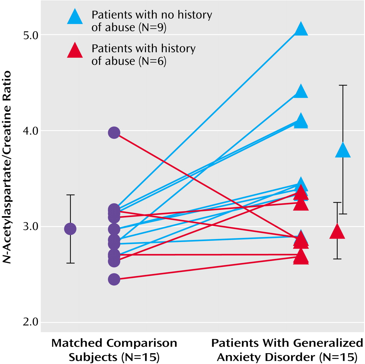
Generalized anxiety disorder patients had a 16.5% higher N-acetylaspartate/creatine ratio in the right dorsolateral prefrontal cortex than did their matched comparison subjects (mean=3.46 [SD=0.69] versus mean=2.97 [SD=0.36], respectively; t=2.58, df=14, p<0.03). Furthermore, the N-acetylaspartate/creatine ratio in the right dorsolateral prefrontal cortex was higher in 13 of 15 generalized anxiety disorder patients than in their matched comparison subjects (Figure 1). However, the six generalized anxiety disorder patients reporting childhood abuse had a lower mean N-acetylaspartate/creatine ratio (mean=2.95, SD=0.67) in the right dorsolateral prefrontal cortex than did the nine nonabused generalized anxiety disorder patients (mean=3.80, SD=0.67) (t=2.89, df=13, p<0.006); the ratio of the abused generalized anxiety disorder patients did not differ significantly from the mean N-acetylaspartate/creatine ratio of comparison subjects. The N-acetylaspartate/creatine ratio in the right dorsolateral prefrontal cortex was not significantly associated with clinical ratings. Patients and healthy subjects did not differ in right dorsolateral prefrontal cortex choline/creatine ratio (t=0.67, df=14, p=0.52). Among generalized anxiety disorder patients, N-acetylaspartate/choline in the right dorsolateral prefrontal cortex correlated with N-acetylaspartate/creatine (r=0.65 in the right dorsolateral prefrontal cortex, p=0.009), although elevations in N-acetylaspartate/choline did not reach significance (t=1.53, df=14, p=0.15). Between-group metabolite ratio differences were not found in the left dorsolateral prefrontal cortex or in either the right or left hippocampus. Within the healthy subjects, N-acetylaspartate/creatine ratios did not differ between the right and left dorsolateral prefrontal cortex.

Discussion

Contrary to our initial hypotheses, the N-acetylaspartate/creatine ratio, a measure of neuronal viability, was increased in the right dorsolateral prefrontal cortex in generalized anxiety disorder patients versus healthy comparison subjects. However, generalized anxiety disorder patients with self-reported childhood abuse had significantly lower N-acetylaspartate/creatine in this region than nonabused patients. These data suggest a potential biological subtype in generalized anxiety disorder, which is consonant with reports in major depressive disorder identifying biological subgroups based on history of early trauma (7). The direction of N-acetylaspartate/creatine abnormality in generalized anxiety disorder contrasts with that reported in posttraumatic stress disorder and mood disorders but is potentially consistent with findings suggesting prefrontal cortical hypermetabolism in generalized anxiety disorder (1) and increased cerebral blood flow in the right dorsolateral prefrontal cortex in persons with social phobia anticipating public speaking (9).
Methodological limitations of the study constrain interpretation. Ratio analyses of the N-acetylaspartate/creatine ratio may reflect changes in creatine, which is typically higher in gray matter than in white matter. Confounding effects of partial volume averaging cannot be ruled out, since tissue segmentation was not performed. Research using tissue segmentation techniques and absolute metabolite quantitation in larger samples will clarify these findings.
Figure 1. N-Acetylaspartate/Creatine Ratios in the Right Dorsolateral Prefrontal Cortex of Healthy Subjects and Age- and Sex-Matched Patients With Generalized Anxiety Disorder, by Childhood Abuse Historya
aData points off to either side represent means and standard deviations for the respective groups. N-Acetylaspartate/creatine ratios in the right dorsolateral prefrontal cortex were higher in patients with generalized anxiety disorder than in healthy comparison subjects matched for age and sex (t=2.58, df=14, p<0.03). The ratio also was lower in patients who experienced childhood abuse-related trauma than in patients reporting no trauma (t=2.89, df=13, p<0.02).

Footnote

Presented in part at the 58th annual meeting of the Society of Biological Psychiatry, San Francisco, May 15–17, 2003. Received July 22, 2003; revision received Dec. 2, 2003; accepted Dec. 4, 2003. From the Department of Biological Psychiatry, New York State Psychiatric Institute; the Departments of Psychiatry and Radiology, Columbia University College of Physicians and Surgeons, New York; the Department of Psychiatry, Mount Sinai School of Medicine, New York; and the Department of Psychiatry, Downstate Medical Center, Brooklyn, N.Y. Address reprint requests to Dr. Mathew, Department of Biological Psychiatry, New York State Psychiatric Institute/Columbia University, 1051 Riverside Dr., Unit 126, New York, NY 10032; [email protected] (e-mail). Supported in part by a grant from GlaxoSmithKline and NIMH grant R24 MH-61274. The authors thank Manuel de la Nuez, B.A.; Justine Kent, M.D.; Jose Martinez, M.S.; Mary Beth Reynoso, M.A.; Curtis Whittman, B.A.; and Ezekiel Young, M.A., for their assistance. Drs. Coplan and Gorman have received compensation and honoraria from GlaxoSmithKline for speaking and consulting.

References

1.
Wu JC, Buchsbaum MS, Hershey TG, Hazlett E, Sicotte N, Johnson JC: PET in generalized anxiety disorder. Biol Psychiatry 1991; 29:1181–1199
2.
De Bellis MD, Casey BJ, Dahl RE, Birmaher B, Williamson DE, Thomas KM, Axelson DA, Frustaci K, Boring AM, Hall J, Ryan ND: A pilot study of amygdala volumes in pediatric generalized anxiety disorder. Biol Psychiatry 2000; 48:51–57
3.
Lyoo IK, Renshaw PF: Magnetic resonance spectroscopy: current and future applications in psychiatric research. Biol Psychiatry 2002; 51:195–207
4.
Mathew SJ, Shungu DC, Mao X, Smith ELP, Perera GM, Kegeles LS, Perera T, Lisanby SH, Rosenblum LA, Gorman JM, Coplan JD: A magnetic resonance spectroscopic imaging study of adult nonhuman primates exposed to early-life stressors. Biol Psychiatry 2003; 54:727–735
5.
First MB, Spitzer RL, Gibbon M, Williams JBW: Structured Clinical Interview for DSM-IV Axis I Disorders, Patient Edition (SCID-P), version 2. New York, New York State Psychiatric Institute, Biometrics Research, 1995
6.
Bremner JD, Vermetten E, Mazure CM: Development and preliminary psychometric properties of an instrument for the measurement of childhood trauma: the Early Trauma Inventory. Depress Anxiety 2000; 12:1–12
7.
Vythilingam M, Heim C, Newport J, Miller AH, Anderson E, Bronen R, Brummer M, Staib L, Vermetten E, Charney DS, Nemeroff CB, Bremner JD: Childhood trauma associated with smaller hippocampal volume in women with major depression. Am J Psychiatry 2002; 159:2072–2080
8.
Duyn JH, Gillen J, Sobering G, van Zijl PCM, Moonen CTW: Multisection proton MR spectroscopic imaging of the brain. Radiology 1993; 188:277–282
9.
Tillfors M, Furmark T, Marteinsdottir I, Fredrikson M: Cerebral blood flow during anticipation of public speaking in social phobia; a PET study. Biol Psychiatry 2002; 52:1113–1119

Information & Authors

Information

Published In

Go to American Journal of Psychiatry
American Journal of Psychiatry
Pages: 1119 - 1121
PubMed: 15169704

History

Published online: 1 June 2004
Published in print: June 2004

Authors

Details

Sanjay J. Mathew, M.D.
Jeremy D. Coplan, M.D.
Eric L.P. Smith, Ph.D.
Harold A. Sackeim, Ph.D.
Dikoma C. Shungu, Ph.D.

Metrics & Citations

Metrics

Citations

Export Citations

If you have the appropriate software installed, you can download article citation data to the citation manager of your choice. Simply select your manager software from the list below and click Download.

For more information or tips please see 'Downloading to a citation manager' in the Help menu.

Format
Citation style
Style
Copy to clipboard

View Options

View options

PDF/EPUB

View PDF/EPUB

Get Access

Login options

Already a subscriber? Access your subscription through your login credentials or your institution for full access to this article.

Personal login Institutional Login Open Athens login
Purchase Options

Purchase this article to access the full text.

PPV Articles - American Journal of Psychiatry

PPV Articles - American Journal of Psychiatry

Not a subscriber?

Subscribe Now / Learn More

PsychiatryOnline subscription options offer access to the DSM-5-TR® library, books, journals, CME, and patient resources. This all-in-one virtual library provides psychiatrists and mental health professionals with key resources for diagnosis, treatment, research, and professional development.

Need more help? PsychiatryOnline Customer Service may be reached by emailing [email protected] or by calling 800-368-5777 (in the U.S.) or 703-907-7322 (outside the U.S.).

Media

Figures

Other

Tables

Share

Share

Share article link

Share