Full access
New Research
Published Online: 1 April 2011

Correlation of Individual Differences in Schizotypal Personality Traits With Amphetamine-Induced Dopamine Release in Striatal and Extrastriatal Brain Regions

Abstract

Objective:

Schizotypal personality traits are associated with schizophrenia spectrum disorders, and individuals with schizophrenia spectrum disorders demonstrate increased dopamine transmission in the striatum. The authors sought to determine whether individual differences in normal variation in schizotypal traits are correlated with dopamine transmission in the striatum and in extrastriatal brain regions.

Method:

Sixty-three healthy volunteers with no history of psychiatric illness completed the Schizotypal Personality Questionnaire and underwent positron emission tomography imaging with [18F]fallypride at baseline and after administration of oral d-amphetamine (0.43 mg/kg). Dopamine release, quantified by subtracting each participant's d-amphetamine scan from his or her baseline scan, was correlated with Schizotypal Personality Questionnaire total and factor scores using region-of-interest and voxel-wise analyses.

Results:

Dopamine release in the striatum was positively correlated with overall schizotypal traits. The association was especially robust in the associative subdivision of the striatum. Voxel-wise analyses identified additional correlations between dopamine release and schizotypal traits in the left middle frontal gyrus and left supramarginal gyrus. Exploratory analyses of Schizotypal Personality Questionnaire factor scores revealed correlations between dopamine release and disorganized schizotypal traits in the striatum, thalamus, medial prefrontal cortex, temporal lobe, insula, and inferior frontal cortex.

Conclusions:

The association between dopamine signaling and psychosis phenotypes extends to individual differences in normal variation in schizotypal traits and involves dopamine transmission in both striatal and extrastriatal brain regions. Amphetamine-induced dopamine release may be a useful endophenotype for investigating the genetic basis of schizophrenia spectrum disorders.
Personality traits are often related to psychopathology (1). Investigating the neural substrates of personality traits may inform etiological and pathophysiological models of psychiatric disorders. In the case of psychosis, attention has focused on the relationship between schizotypal personality traits and schizophrenia spectrum disorders. Schizotypal personality traits encompass a broad range of personality characteristics and experiences, including unusual perceptions and beliefs, social anxiety or withdrawal, and disorganized thoughts or behaviors (2). These traits cluster into positive, negative, and disorganized factors that are conceptually similar to the symptom dimensions of schizophrenia (24). The expression of schizotypal traits ranges from benign odd perceptual experiences or beliefs to severe symptoms associated with significant psychosocial impairment and schizotypal personality disorder (2). Premorbid personality in schizophrenia is marked by an excess of schizotypal traits, and schizotypal personality disorder is a risk factor for schizophrenia (57). Indicators of cerebral dysfunction observed in schizophrenia spectrum disorders, including cognitive impairment and sensory gating deficits, are correlated with schizotypal traits in psychiatrically healthy individuals, which further underscores the link between schizotypal traits and schizophrenia spectrum disorders (814).
The neural basis of individual differences in schizotypal personality traits is poorly understood. Dopamine signaling may be associated with normal variation in these traits, given that dopamine dysregulation is prominent in schizophrenia spectrum disorders. Positron emission tomography (PET) imaging with displaceable dopamine receptor ligands sensitive to endogenous dopamine levels has shown that patients with schizotypal personality disorder demonstrate exaggerated dopamine release in the striatum following d-amphetamine challenge (15). Schizophrenia patients also demonstrate increased d-amphetamine-induced dopamine release in the striatum (16, 17). Dopamine release is especially robust in schizophrenia patients who are experiencing an acute illness exacerbation and, in contrast to patients with schizotypal personality disorder, is correlated with a transient increase in positive psychotic symptoms (16).
Determining the relationship between dopamine transmission and individual differences in schizotypal traits may further our understanding of dopamine dysregulation in schizophrenia spectrum disorders. While the findings in schizophrenia provide a compelling case for a state component to hyperdopaminergia, the evidence for a trait basis is less conclusive, given that it is based largely on findings from one study of schizotypal personality disorder that included relatively few patients (15). Moreover, the extent to which dopamine signaling varies continuously with dimensional measures of schizotypy is unknown. Evidence that dopamine signaling is correlated with individual differences in schizotypy would lend considerable support to the hypothesis that there is a trait component to hyperdopaminergia and may further suggest that hyperdopaminergia is an endophenotype of schizophrenia spectrum disorders.
It is also unknown whether dopamine dysregulation in schizophrenia spectrum disorders includes extrastriatal brain regions. There are reasons to suspect that it may, given reports of elevated l-dopa uptake in the amygdala and medial prefrontal cortex in schizophrenia (18, 19) and findings from a recent meta-analysis showing that dopamine receptor occupancy by antipsychotics in the temporal cortex is strongly related to clinical efficacy (20). Examining the relationship between schizotypal personality traits and extrastriatal dopamine transmission may provide testable hypotheses on the role of extrastriatal dopamine transmission in schizophrenia spectrum disorders.
We examined the relationship between d-amphetamine-induced dopamine release determined from PET imaging with [18F]fallypride (a ligand that can quantify both striatal and extrastriatal dopamine D2/D3 receptors) and schizotypal personality traits in a large sample of healthy individuals. We hypothesized that schizotypal personality traits would be positively correlated with d-amphetamine-induced dopamine release in the striatum, and we sought to determine whether similar associations exist in extrastriatal brain regions.

Method

Participants

Sixty-three participants drawn from two studies of individual differences in d-amphetamine-induced dopamine release were included in this investigation. Study procedures were identical for the two studies, except that one group was given identical-appearing capsules containing placebo or amphetamine on PET scanning days (placebo-controlled cohort; N=48), whereas the other group was not blind to amphetamine administration (open-label cohort; N=15). Fourteen participants in the open-label cohort were included in a previous report (21). Table 1 summarizes the characteristics of the total sample and of each cohort. All participants received a physical and neurological examination that included ECG, blood chemistries, urine analysis, urine drug screen, and T1, T2, and T2 flair MRI scans. Exclusion criteria included a history of neurological or psychiatric disorder; severe past or concomitant medical illness; borderline elevated blood pressure; abnormal results on ECG, comprehensive medical panel, CBC, or urine analysis; positive finding on 10-panel urine drug screen; brain abnormalities revealed on MRI scanning; use of psychotropic medication during the preceding 6 months; history of substance abuse or dependence; lifetime use of cocaine or amphetamines; any illicit drug use in the previous 2 months; and pregnancy or lactation. Axis I psychopathology was ruled out using the Structured Clinical Interview for DSM-IV Axis I Disorders (22). Axis II psychopathology was not formally assessed. The study was approved by the Vanderbilt University Institutional Review Board, and written informed consent was obtained from each participant.
TABLE 1. Characteristics of Participants in a Study of Open-Label or Placebo-Controlled d-Amphetamine-Induced Dopamine Release
   d-Amphetamine Administration
VariableTotal Sample (N=63)Open-Label (N=15)Placebo-Controlled (N=48)
 N%N%N%
Male3250.8746.72552.1
Ethnicity      
    White5384.112804185.4
    African American69.5213.348.3
    Other46.316.736.3
 MeanSDMeanSDMeanSD
Agea (years)23.13.726.13.322.13.3
Schizotypal Personality Questionnaire      
    Total score8.18.18.78.77.97.9
    Cognitive-perceptual factor1.01.61.31.81.01.5
    Paranoid factor3.03.03.23.12.92.9
    Negative factor3.33.84.34.73.03.5
    Disorganized factor2.83.42.74.42.93.1
a Significant difference between groups, p<0.001.

Procedures and Assessments

Study and image acquisition procedures have been described previously (21). Prior to scanning, participants completed the Schizotypal Personality Questionnaire (2). The focus of this investigation is on the total score; however, factor-analytic studies indicate that the Schizotypal Personality Questionnaire can be separated into cognitive-perceptual, paranoid, negative, and disorganized factors (3). Exploratory analyses of the factor scores were undertaken to determine whether dopamine release is related to a specific dimension of schizotypy.
Participants underwent two PET scans with [18F]fallypride. The first was a baseline scan; the second occurred on a different day, 3.5 hours after administration of oral d-amphetamine (0.43 mg/kg) or placebo. As noted, 15 participants were not blind to d-amphetamine administration, and 48 participants received identical-appearing capsules on scan days that contained either placebo or d-amphetamine. Physiological measures (blood pressure, heart rate, temperature, and respirations) were monitored on scan days, and participants completed a brief screen of possible side effects. Participants also completed a brief neurological screen at baseline and at the end of the scan protocol. Blood samples for CBC and a comprehensive medical panel were also obtained at baseline and at completion of the scan protocol.

PET Image Acquisition and Data Preprocessing

PET imaging was performed at Vanderbilt University Medical Center on either a GE Discovery LS scanner (N=30) or, after the center's scanner was upgraded, a GE Discovery STE system (N=33). All participants received their baseline and d-amphetamine scans on the same scanner. To assess the validity of combining data across scanners, we compared dopamine release in each of the anatomical regions of interest (described below) between scanners. No differences were observed in any region of interest. Moreover, voxel-wise analysis comparing dopamine release between the two scanners did not identify any clusters after whole brain correction at t=2.5 (lowest cluster-level p value >0.90).
Three-dimensional emission acquisitions and transmission attenuation correction scans were performed following a 5.0 mCi slow bolus injection of [18F]fallypride (specific activity >3,000 Ci/mmol). Serial scans started simultaneously with the bolus injection of [18F]fallypride and were obtained for approximately 3.5 hours. The extended scanning time allowed for stable kinetic model fits in both striatal and extrastriatal brain regions. The initial scan sequence coincided with the start of the [18F]fallypride injection and included the following frames: eight for 15 seconds, six for 30 seconds, five for 1 minute, two for 2.5 minutes, three for 5 minutes, and three for 10 minutes. After the initial scan sequence, a 10-minute transmission scan was obtained, and then the participant was allowed a break. Approximately 85–90 minutes after the injection, a second scan sequence of two frames of 25 minutes each followed by a second transmission scan was obtained. The participant was then allowed a second break, and at approximately 165–170 minutes, a 40-minute emission scan followed by a third transmission scan was obtained. Serial PET scans were coregistered using a mutual-information rigid-body algorithm to minimize potential modeling errors due to head motion within and between scans (23). Consistent with our prior studies with [18F]fallypride (for example, reference 24), parametric binding potential (BPND) images of dopamine D2/D3 receptor density were calculated using the full (four-parameter) reference region model (25) with the cerebellum serving as the reference region. Previous studies in our lab (26) have shown that this method produces BPND estimates that closely agree with those derived from Logan plots (27) using a metabolite-corrected plasma input function.
A high-resolution T1-weighted MRI scan was also obtained for each participant, and PET and MRI scans were coregistered to one another (23). After coregistration, each participant's BPND image was warped to a canonical brain that had been normalized to the MNI152 template brain and resampled to 2 mm3. Parametric images of dopamine release, in percent, were created by subtracting each participant's d-amphetamine scan from his or her baseline scan and dividing the difference by the baseline scan using the ImCalc function in SPM2 (http://www.fil.ion.ucl.ac.uk/spm). In addition, dopamine release values for several anatomically defined regions of interest were extracted from the parametric images of dopamine release by calculating the mean of the voxels within each region of interest. The regions of interest included the left and right striatum, thalamus, amygdala, and hippocampus. Dopamine release values were averaged across hemispheres to produce one value for each region of interest. The striatum regions of interest were taken from the Laboratory of Neuro Imaging, UCLA (LONI) Probabilistic Brain Atlas (28) and partitioned into limbic, associative, and sensorimotor functional subdivisions using previously described criteria (29, 30). Briefly, the striatum atlas was divided into five regions of interest: ventral striatum, dorsal caudate rostral to the anterior commissure (AC), dorsal putamen rostral to the AC, postcommissural caudate, and postcommissural putamen. The limbic subdivision comprised the ventral striatum, the associative striatum was the weighted average of the pre- and postcommissural dorsal caudate and precommissural putamen, and the sensorimotor subdivision consisted of the postcommissural putamen. Dopamine release in the entire striatum, weighted by the size of each subdivision, was also calculated. The thalamus region of interest was derived from the International Consortium for Brain Mapping (ICBM) Deep Nuclei Probabilistic Atlas (http://www.loni.ucla.edu/Atlases), thresholded at 80% to avoid partial volume effects. The hippocampus and amygdala regions of interest were created using the WFU (Wake Forest University) PickAtlas and manually edited using criteria previously described by our group to avoid partial volume effects from adjacent structures (24).

Statistical Analysis

The relationship between Schizotypal Personality Questionnaire score and dopamine release was examined with region-of-interest and voxel-wise analyses. First, Schizotypal Personality Questionnaire score was correlated with dopamine release in the regions of interest. The correlations for the striatum and striatum subdivisions were thresholded at a p value of 0.05, given our a priori hypothesis that dopamine release in the striatum is correlated with overall schizotypal traits. The significance threshold was set to p=0.016 for extrastriatal regions to correct for the number of structures examined. For the voxel-wise analysis, multiple regression analysis was used, with Schizotypal Personality Questionnaire score entered as a predictor of dopamine release at each voxel. Given our hypothesis, we first examined the extent to which dopamine release in the striatum was related to schizotypal traits by restricting the voxel-wise analysis to the LONI Probabilistic Brain Atlas striatum map using the small-volume-correction tool in SPM2. Only clusters within the LONI Probabilistic Brain Atlas striatum mask that exceeded the cluster-wise corrected threshold at a voxel-wise p value of 0.05 are reported. Next, we examined positive correlations throughout the brain. Only clusters exceeding the whole brain cluster-wise corrected alpha of 0.05 for voxel-wise t=2.5 are reported (31). The voxel-wise analysis was masked to exclude voxels with mean BPND values below 0.40 on the amphetamine scan. Significant clusters were converted to Talairach coordinates using ICBM_SPM2Tal (32). The estimated smoothness of the statistical parametric map generated for the voxel-wise regression analysis in the x, y, and z planes, respectively, was 6.6 mm, 7.5 mm, and 6.4 mm. Age, sex, and cohort (open-label or placebo-controlled) were included as nuisance covariates in both the region-of-interest and voxel-wise analyses. Scanner was not included as a covariate for two reasons. First, all participants in the open-label cohort were scanned on the Discovery LS scanner, so inclusion of scanner as a covariate would have been redundant given that cohort and scanner were not independent. Second, as noted above, neither voxel-wise nor region-of-interest analyses revealed any significant differences in dopamine release between scanners.

Results

Overall Schizotypal Traits and Dopamine Release: Region-of-Interest Analysis

Correlations between dopamine release and Schizotypal Personality Questionnaire scores in the total sample and in the placebo-controlled subgroup are presented in Table 2. For the striatum, overall schizotypal traits were correlated with dopamine release in the whole striatum and associative subdivision. No correlations reached the corrected alpha level (p=0.016) in the extrastriatal regions, although dopamine release in the amygdala was correlated with the Schizotypal Personality Questionnaire score at the uncorrected alpha level. The results were virtually identical when the analysis was restricted to the placebo-controlled cohort. The mean peak plasma d-amphetamine level was 72 ng/ml (SD=19). Consistent with our previous report on the open-label cohort (21), peak plasma d-amphetamine level was unrelated to striatal dopamine release. Baseline BPND values and dopamine release for each region of interest are presented in Table S1 in the data supplement that accompanies the online edition of this article.
TABLE 2. Correlation Between d-Amphetamine-Induced Dopamine Release and Schizotypal Personality Traits: Region-of-Interest Analysisa
 Schizotypal Personality Questionnaire
   Factor
 Total ScoreCognitive-Perceptual SubscoreParanoid SubscoreNegative SubscoreDisorganized Subscore
Region of Interestrprprprprp
Total sample (N=63)          
Whole striatum0.260.0490.060.6310.160.2270.140.2770.350.006
Limbic0.220.0940.020.9030.140.2850.120.3530.280.031
Associative0.270.0380.110.4130.180.1700.180.1610.340.008
Sensorimotor0.200.1280.020.8660.100.4400.070.6200.330.010
Amygdala0.290.0250.170.1980.220.0940.220.0860.260.042
Hippocampus0.080.569–0.020.9100.130.3430.090.4920.030.804
Thalamus0.210.1000.080.5440.150.2670.170.2080.290.025
Placebo-controlled cohort (N=48)          
Whole striatum0.320.0290.250.0920.260.0860.200.1880.360.013
Limbic0.340.0230.260.0780.270.0680.210.1630.340.023
Associative0.290.0480.230.1230.240.1160.200.1740.340.023
Sensorimotor0.270.0740.210.1720.210.1720.130.3930.330.026
Amygdala0.350.0180.260.0790.270.0660.240.1070.370.011
Hippocampus0.210.1600.130.3930.190.2040.120.4420.250.096
Thalamus0.290.0490.240.1150.210.1720.190.2180.410.004
a Partial correlation after covarying for age, gender, and, for total the sample, cohort (i.e., open-label or placebo-controlled).

Overall Schizotypal Traits and Dopamine Release: Voxel-Wise Analysis

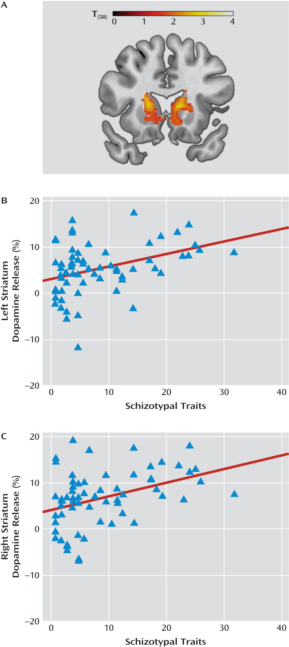
The results of the voxel-wise analysis are presented in Table 3 and Figures 1 and 2. Small-volume correction within the striatum revealed positive correlations bilaterally in the striatum. The clusters were centered in the head of the caudate but extended into the ventral striatum (see Figure 1). The corresponding correlations between mean dopamine release extracted from each cluster and Schizotypal Personality Questionnaire score, after controlling for age, gender, and cohort, were r=0.41 (p=0.001) for the left striatum and r=0.40 (p=0.002) for the right striatum. The findings were unchanged when the analysis was restricted to the placebo-controlled cohort (left striatum: r=0.45, p=0.002; right striatum: r=0.36, p=0.014).
TABLE 3. Correlations Between d-Amphetamine-Induced Dopamine Release and Schizotypal Personality Traits: Voxel-Wise Analysis
 Talairach Coordinates  
Brain RegionxyzVolume (Voxels)aPeak t value
Schizotypal Personality Questionnaire, total score     
Right caudateb102239613.20
Left caudateb–180264683.05
Left middle frontal gyrus (Brodmann's area 9/10)–3340201643.50
Left supramarginal gyrus/inferior parietal lobule (Brodmann's area 40)–57–53291533.43
Schizotypal Personality Questionnaire, disorganized factor subscore     
Right medial frontal gyrus (Brodmann's area 9/10)344172555.21
Right temporal lobe (Brodmann's areas 20, 21, 22)45–12–37595.15
Left inferior/middle temporal gyrus (Brodmann's areas 20, 21, 37)–50–45104354.38
Left insula/inferior frontal gyrus (Brodmann's areas 13, 37)–3015–138144.30
Right insula/inferior frontal gyrus (Brodmann's areas 13, 47)38579314.30
Left inferior/middle temporal gyrus (Brodmann's areas 20, 21)–56–35–162134.27
Right thalamus6–462024.22
Right superior frontal gyrus (Brodmann's area 10)2847271854.19
Right caudate/putamen1013114423.40
Left caudate/putamen–181393223.51
a Voxel size=2×2×2 mm.
b Small-volume correction.
FIGURE 1. Correlation Between Schizotypal Traits and d-Amphetamine-Induced Dopamine Release in the Striatuma
a In panel A, the voxel-wise analysis restricted to the striatum revealed positive correlations between Schizotypal Personality Questionnaire score and dopamine release in the striatum bilaterally. Image thresholded at p=0.05 (small-volume correction). Scatterplots depict the correlation between Schizotypal Personality Questionnaire score and dopamine release in the left (panel B; R2=0.143) and right (panel C; R2=0.152) striatum clusters.
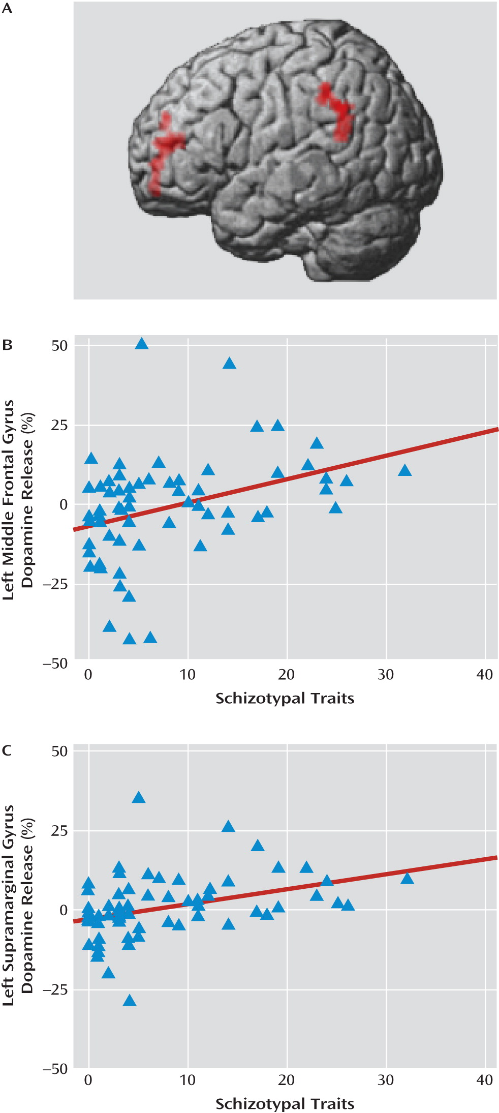
FIGURE 2. Correlation Between Schizotypal Traits and d-Amphetamine-Induced Dopamine Release in the Cortexa
a Panel A shows that Schizotypal Personality Questionnaire score was correlated with dopamine release in the left middle frontal gyrus and the inferior parietal lobule. Image thresholded at p=0.05 (whole brain cluster-level corrected). Scatterplots depict the correlation between dopamine release and schizotypal traits in the left middle frontal gyrus (panel B; R2=0.129) and the left supramarginal gyrus (panel C; R2=0.142).
Whole brain analysis identified two additional clusters (see Table 3 and Figure 2). They included a region within the left middle frontal gyrus corresponding to Brodmann's area 9/10 and the left supramarginal gyrus within the inferior parietal lobule. The corresponding correlations between mean dopamine release extracted from each cluster and Schizotypal Personality Questionnaire score, after controlling for age, gender, and cohort, were r=0.44 (p=0.0004) and r=0.46 (p=0.0002) for the left middle frontal and supramarginal gyrus clusters, respectively. The findings were unchanged when the analysis was restricted to the placebo-controlled cohort (left middle frontal gyrus: r=0.48, p=0.001; left supramarginal gyrus: r=0.55, p=0.0001).
No inverse correlations between dopamine release and Schizotypal Personality Questionnaire score were identified in the striatum or whole brain.

Dopamine Release and Specific Dimensions of Schizotypy

We examined the relationship between Schizotypal Personality Questionnaire factor scores and dopamine release in the regions of interest to determine whether a particular facet of schizotypy was related to dopamine release (see Table 2). No statistical correction was applied given the exploratory nature of this analysis. Robust correlations were observed between disorganized schizotypal traits and dopamine release in the whole striatum and associated subdivisions, in the amygdala, and in the thalamus. Similar results were observed in the placebo-controlled cohort. The other factor scores were not correlated with dopamine release in either the entire sample or the placebo-controlled cohort.
Given the widespread correlations observed between dopamine release and disorganized traits in the region-of-interest analysis, we performed an exploratory voxel-wise multiple regression analysis regressing disorganized factor scores on dopamine release with age, sex, and cohort as nuisance covariates. The results were thresholded at a whole brain cluster-wise corrected alpha of 0.05 for voxel-wise t=2.5. Dopamine release was correlated with disorganized traits in several subcortical and cortical regions (see Table 3; also see Figure S1 in the online data supplement), including the left and right striatum; the right thalamus and pregenual cingulate/medial prefrontal cortex; the left and right temporal cortex; the superior frontal gyrus; and the left and right insula. All of the clusters remained significant when the analysis was restricted to the placebo-controlled cohort (all cluster p values <0.003).

Discussion

Dopamine release in striatal and extrastriatal brain regions is correlated with individual differences in schizotypal traits. Our findings suggest that the link between d-amphetamine-induced dopamine release and schizophrenia spectrum disorders extends to normal variation in schizotypal personality traits. These results parallel associations previously reported between schizotypal traits and relative impairments in cognition and sensory gating in samples with similar Schizotypal Personality Questionnaire scores (814). Moreover, the correlations between dopamine release and schizotypal traits reported here are similar in magnitude to those reported from previous investigations of the association between psychometrically measured schizotypy and behavioral measures of cognition or sensory gating.
These findings may further our understanding of dopamine dysregulation in schizophrenia spectrum disorders. Extrapolating dopamine release based on the correlation we observed for the striatum region of interest, we obtain predicted dopamine release values of 9%–13% for Schizotypal Personality Questionnaire scores between 30 and 40 (the range reported in patients with schizophrenia and schizotypal personality disorder [13, 3335 ]). This is similar to the 10%–12% increase observed in schizotypal personality disorder and remitted schizophrenia patients, but substantially less than the 20%–22% increase reported in acutely ill schizophrenia patients (15). Although caution is warranted when making comparisons between studies that used different radioligands, slightly different d-amphetamine doses, and different delivery routes (intravenous versus oral), the similarity in mean striatal dopamine release between the control sample (N=57) reported by Abi-Dargham et al. (15) (7%–7.5%) and the present study (∼6%) supports the validity of this comparison. Thus, our findings support the hypothesis that the modest elevation in dopamine release observed in schizotypal personality disorder and remitted schizophrenia is a stable trait indicator related to schizotypy, while the robust increases reported in acutely ill schizophrenia patients is probably a state component superimposed on a trait-wise elevation in dopamine transmission (15).
Identifying the neural basis of individual differences in personality traits associated with psychiatric illnesses is similar to imaging genetics approaches examining relationships between brain structure/function and putative psychiatric disorder risk genes in healthy individuals. By providing further support for a trait basis for dopamine dysfunction, our findings suggest that d-amphetamine-induced dopamine release may represent an endophenotype of schizophrenia spectrum disorders. Evidence that unaffected relatives of patients with schizophrenia demonstrate elevated presynaptic dopamine synthesis capacity in the striatum also implicates hyperdopaminergia as an endophenotype for schizophrenia (36). Identification of gene variants associated with psychostimulant-induced dopamine release may provide clues to the genetic basis of schizophrenia spectrum disorders.
Imaging studies of dopamine release in clinical studies have been limited to the striatum; however, there are reasons to suspect that hyperdopaminergia in schizophrenia spectrum disorders extends beyond the striatum (20). Our findings showing a relationship between schizotypal traits and dopamine release in prefrontal regions may at first glance appear to contradict the cortical hypodopaminergia hypothesis of schizophrenia (37). However, the evidence supporting cortical hypodopaminergia in schizophrenia is indirect and inconsistent. PET studies of cortical dopamine D1 receptors in schizophrenia have reported increased, decreased, and unaltered levels (3840). Moreover, inferences about dopamine function based on differences in receptor levels observed between patients and comparison subjects under normal physiological conditions may not generalize to stimulant challenge. We find little evidence that baseline BPND is associated with dopamine release in our data. Thus, it is possible that hypodopaminergia inferred from differences in baseline BPND may be unrelated to amphetamine-induced dopamine release, or may actually co-occur with stimulant-induced hyperdopaminergia. Studies of extrastriatal amphetamine-induced dopamine release in schizophrenia spectrum disorders are clearly warranted.
Multimodal imaging may inform the relationship between schizotypy, especially disorganized traits, brain function, and dopamine signaling. The face validity of disorganized factor questions suggests that they may be related to subtle limitations in executive cognitive functions. Individual differences in disorganized schizotypal traits are correlated with executive function, and abnormal prefrontal cortical functioning during task performance is correlated with disorganized symptoms in schizophrenia patients (9, 33, 4143). The prefrontal cortex influences dopamine function directly by altering midbrain dopamine cell firing and indirectly through presynaptic innervation of striatal dopamine terminals (44, 45). Consequently, alterations in prefrontal cortical function may alter the response of both cortical and subcortical dopamine systems to amphetamine challenge.
This study has several limitations. Our findings require replication in a sample with a broader range of schizotypal traits in order to better characterize the relationship between specific facets of schizotypy and dopamine release. Also, because we did not rule out axis II psychopathology during screening, it is possible that some participants met criteria for schizotypal personality disorder; this is unlikely, however, given the range of Schizotypal Personality Questionnaire scores in this sample. It is also unlikely that our results are related to other dimensions of psychopathology, such as anxiety and depression, given that neither is associated with dopamine release (46, 47). Interview-based measures may be more sensitive to schizotypal traits than self-report questionnaires, which raises the possibility that different results might have been obtained had we used interview-based methods (48). Finally, combining study subjects across amphetamine administration protocols and PET scanners is not ideal. However, our findings were largely unchanged when analyses were restricted to the placebo-controlled cohort, and we did not find any differences in dopamine release between scanners.
Footnote

Received Feb. 3, 2010; revisions received June 26 and Oct. 5, 2010; accepted Oct. 25, 2010.

Supplementary Material

File (ajp_168_04_418_01.pdf)

References

1.
Widiger TA, Mullins-Sweatt SN: Five-factor model of personality disorder: a proposal for DSM-V. Annu Rev Clin Psychol 2009; 5:197–220
2.
Raine A: The SPQ: a scale for the assessment of schizotypal personality based on DSM-III-R criteria. Schizophr Bull 1991; 17:555–564
3.
Compton MT, Goulding SM, Bakeman R, Clure-Tone EB: Confirmation of a four-factor structure of the Schizotypal Personality Questionnaire among undergraduate students. Schizophr Res 2009; 111:46–52
4.
Gruzelier JH: The factorial structure of schizotypy, part I: affinities with syndromes of schizophrenia. Schizophr Bull 1996; 22:611–620
5.
Woods SW, Addington J, Cadenhead KS, Cannon TD, Cornblatt BA, Heinssen R, Perkins DO, Seidman LJ, Tsuang MT, Walker EF, McGlashan TH: Validity of the prodromal risk syndrome for first psychosis: findings from the North American Prodrome Longitudinal Study. Schizophr Bull 2009; 35:894–908
6.
Kendler KS, Gruenberg AM, Strauss JS: An independent analysis of the Copenhagen sample of the Danish Adoption Study of Schizophrenia, V: the relationship between childhood social withdrawal and adult schizophrenia. Arch Gen Psychiatry 1982; 39:1257–1261
7.
Ott SL, Roberts S, Rock D, Allen J, Erlenmeyer-Kimling L: Positive and negative thought disorder and psychopathology in childhood among subjects with adulthood schizophrenia. Schizophr Res 2002; 58:231–239
8.
Chen WJ, Hsiao CK, Lin CC: Schizotypy in community samples: the three-factor structure and correlation with sustained attention. J Abnorm Psychol 1997; 106:649–654
9.
Noguchi H, Hori H, Kunugi H: Schizotypal traits and cognitive function in healthy adults. Psychiatry Res 2008; 161:162–169
10.
Bedwell JS, Kamath V, Compton MT: The relationship between interview-based schizotypal personality dimension scores and the Continuous Performance Test. Schizophr Res 2009; 108:158–162
11.
Wang J, Miyazato H, Hokama H, Hiramatsu K, Kondo T: Correlation between P50 suppression and psychometric schizotypy among non-clinical Japanese subjects. Int J Psychophysiol 2004; 52:147–157
12.
Matsui M, Yuuki H, Kato K, Takeuchi A, Nishiyama S, Bilker WB, Kurachi M: Schizotypal disorder and schizophrenia: a profile analysis of neuropsychological functioning in Japanese patients. J Int Neuropsychol Soc 2007; 13:672–682
13.
Cadenhead KS, Light GA, Geyer MA, Braff DL: Sensory gating deficits assessed by the P50 event-related potential in subjects with schizotypal personality disorder. Am J Psychiatry 2000; 157:55–59
14.
Croft RJ, Lee A, Bertolot J, Gruzelier JH: Associations of P50 suppression and desensitization with perceptual and cognitive features of "unreality" in schizotypy. Biol Psychiatry 2001; 50:441–446
15.
Abi-Dargham A, Kegeles LS, Zea-Ponce Y, Mawlawi O, Martinez D, Mitropoulou V, O'Flynn K, Koenigsberg HW, Van Heertum R, Cooper T, Laruelle M, Siever LJ: Striatal amphetamine-induced dopamine release in patients with schizotypal personality disorder studied with single photon emission computed tomography and [123I]iodobenzamide. Biol Psychiatry 2004; 55:1001–1006
16.
Laruelle M, Abi-Dargham A, Gil R, Kegeles L, Innis R: Increased dopamine transmission in schizophrenia: relationship to illness phases. Biol Psychiatry 1999; 46:56–72
17.
Breier A, Su TP, Saunders R, Carson RE, Kolachana BS, de Bartolomeis A, Weinberger DR, Weisenfeld N, Malhotra AK, Eckelman WC, Pickar D: Schizophrenia is associated with elevated amphetamine-induced synaptic dopamine concentrations: evidence from a novel positron emission tomography method. Proc Natl Acad Sci USA 1997; 94:2569–2574
18.
Kumakura Y, Cumming P, Vernaleken I, Buchholz HG, Siessmeier T, Heinz A, Kienast T, Bartenstein P, Grunder G: Elevated [18F]fluorodopamine turnover in brain of patients with schizophrenia: an [18F]fluorodopa/positron emission tomography study. J Neurosci 2007; 27:8080–8087
19.
Lindstrom LH, Gefvert O, Hagberg G, Lundberg T, Bergstrom M, Hartvig P, Langstrom B: Increased dopamine synthesis rate in medial prefrontal cortex and striatum in schizophrenia indicated by L-(beta-11C) DOPA and PET. Biol Psychiatry 1999; 46:681–688
20.
Stone JM, Davis JM, Leucht S, Pilowsky LS: Cortical dopamine D2/D3 receptors are a common site of action for antipsychotic drugs: an original patient data meta-analysis of the SPECT and PET in vivo receptor imaging literature. Schizophr Bull 2009; 35:789–797
21.
Riccardi P, Li R, Ansari MS, Zald D, Park S, Dawant B, Anderson S, Doop M, Woodward N, Schoenberg E, Schmidt D, Baldwin R, Kessler R: Amphetamine-induced displacement of [18F]fallypride in striatum and extrastriatal regions in humans. Neuropsychopharmacology 2006; 31:1016–1026
22.
First MB, Spitzer RL, Gibbon M, Williams JBW: Structured Clinical Interview for DSM-IV Axis I Disorders (SCID), Clinician Version. Washington, DC, American Psychiatric Press, 1996
23.
Maes F, Collignon A, Vandermeulen D, Marchal G, Suetens P: Multimodality image registration by maximization of mutual information. IEEE Trans Med Imaging 1997; 16:187–198
24.
Zald DH, Woodward ND, Cowan RL, Riccardi P, Ansari MS, Baldwin RM, Cowan RL, Smith CE, Hakyemez H, Li R, Kessler RM: The interrelationship of dopamine D2-like receptor availability in striatal and extrastriatal brain regions in healthy humans: a principal component analysis of [18F]fallypride binding. Neuroimage 2010; 51:53–62
25.
Lammertsma AA, Bench CJ, Hume SP, Osman S, Gunn K, Brooks DJ, Frackowiak RS: Comparison of methods for analysis of clinical [11C]raclopride studies. J Cereb Blood Flow Metab 1996; 16:42–52
26.
Kessler RM, Mason NS, Jones C, Ansari MS, Manning RF, Price RR: [18F]N-ally-5-fluorproplepidepride (fallypride): radiation dosimetry, quantification of striatal and extrastriatal dopamine receptors in man. Neuroimage 2000; 11:S32
27.
Logan J, Fowler JS, Volkow ND, Wolf AP, Dewey SL, Schlyer DJ, MacGregor RR, Hitzemann R, Bendriem B, Gatley SJ: Graphical analysis of reversible radioligand binding from time-activity measurements applied to [N-11C-methyl]-(–)-cocaine PET studies in human subjects. J Cereb Blood Flow Metab 1990; 10:740–747
28.
Shattuck DW, Mirza M, Adisetiyo V, Hojatkashani C, Salamon G, Narr KL, Poldrack RA, Bilder RM, Toga AW: Construction of a 3D probabilistic atlas of human cortical structures. Neuroimage 2008; 39:1064–1080
29.
Martinez D, Slifstein M, Broft A, Mawlawi O, Hwang DR, Huang Y, Cooper T, Kegeles L, Zarahn E, Abi-Dargham A, Haber SN, Laruelle M: Imaging human mesolimbic dopamine transmission with positron emission tomography, part II: amphetamine-induced dopamine release in the functional subdivisions of the striatum. J Cereb Blood Flow Metab 2003; 23:285–300
30.
Mawlawi O, Martinez D, Slifstein M, Broft A, Chatterjee R, Hwang DR, Huang Y, Simpson N, Ngo K, Van HR, Laruelle M: Imaging human mesolimbic dopamine transmission with positron emission tomography, I: accuracy and precision of D(2) receptor parameter measurements in ventral striatum. J Cereb Blood Flow Metab 2001; 21:1034–1057
31.
Poline JB, Worsley KJ, Evans AC, Friston KJ: Combining spatial extent and peak intensity to test for activations in functional imaging. Neuroimage 1997; 5:83–96
32.
Lancaster JL, Tordesillas-Gutierrez D, Martinez M, Salinas F, Evans A, Zilles K, Mazziotta JC, Fox PT: Bias between MNI and Talairach coordinates analyzed using the ICBM-152 brain template. Hum Brain Mapp 2007; 28:1194–1205
33.
Daneluzzo E, Bustini M, Stratta P, Casacchia M, Rossi A: Schizotypal Personality Questionnaire and Wisconsin Card Sorting Test in a population of DSM-III-R schizophrenic patients and control subjects. Compr Psychiatry 1998; 39:143–148
34.
Vollema MG, Sitskoorn MM, Appels MC, Kahn RS: Does the Schizotypal Personality Questionnaire reflect the biological-genetic vulnerability to schizophrenia? Schizophr Res 2002; 54:39–45
35.
Koenigsberg HW, Reynolds D, Goodman M, New AS, Mitropoulou V, Trestman RL, Silverman J, Siever LJ: Risperidone in the treatment of schizotypal personality disorder. J Clin Psychiatry 2003; 64:628–634
36.
Huttunen J, Heinimaa M, Svirskis T, Nyman M, Kajander J, Forsback S, Solin O, Ilonen T, Korkeila J, Ristkari T, McGlashan T, Salokangas RK, Hietala J: Striatal dopamine synthesis in first-degree relatives of patients with schizophrenia. Biol Psychiatry 2008; 63:114–117
37.
Davis KL, Kahn RS, Ko G, Davidson M: Dopamine in schizophrenia: a review and reconceptualization. Am J Psychiatry 1991; 148:1474–1486
38.
Abi-Dargham A, Mawlawi O, Lombardo I, Gil R, Martinez D, Huang Y, Hwang DR, Keilp J, Kochan L, Van Heertum R, Gorman JM, Laruelle M: Prefrontal dopamine D1 receptors and working memory in schizophrenia. J Neurosci 2002; 22:3708–3719
39.
Okubo Y, Suhara T, Suzuki K, Kobayashi K, Inoue O, Terasaki O, Someya Y, Sassa T, Sudo Y, Matsushima E, Iyo M, Tateno Y, Toru M: Decreased prefrontal dopamine D1 receptors in schizophrenia revealed by PET. Nature 1997; 385:634–636
40.
Karlsson P, Farde L, Halldin C, Sedvall G: PET study of D1 dopamine receptor binding in neuroleptic-naive patients with schizophrenia. Am J Psychiatry 2002; 159:761–767
41.
Yoon JH, Minzenberg MJ, Ursu S, Walters R, Wendelken C, Ragland JD, Carter CS: Association of dorsolateral prefrontal cortex dysfunction with disrupted coordinated brain activity in schizophrenia: relationship with impaired cognition, behavioral disorganization, and global function. Am J Psychiatry 2008; 165:1006–1014
42.
Moritz S, Andresen B, Naber D, Krausz M, Probsthein E: Neuropsychological correlates of schizotypal disorganization. Cogn Neuropsychiatry 1999; 4:343–349
43.
Matheson S, Langdon R: Schizotypal traits impact upon executive working memory and aspects of IQ. Psychiatry Res 2008; 159:207–214
44.
Deutch AY: The regulation of subcortical dopamine systems by the prefrontal cortex: interactions of central dopamine systems and the pathogenesis of schizophrenia. J Neural Transm Suppl 1992; 36:61–89
45.
Sesack SR, Carr DB: Selective prefrontal cortex inputs to dopamine cells: implications for schizophrenia. Physiol Behav 2002; 77:513–517
46.
Schneier FR, Abi-Dargham A, Martinez D, Slifstein M, Hwang DR, Liebowitz MR, Laruelle M: Dopamine transporters, D2 receptors, and dopamine release in generalized social anxiety disorder. Depress Anxiety 2009; 26:411–418
47.
Parsey RV, Oquendo MA, Zea-Ponce Y, Rodenhiser J, Kegeles LS, Pratap M, Cooper TB, Van HR, Mann JJ, Laruelle M: Dopamine D(2) receptor availability and amphetamine-induced dopamine release in unipolar depression. Biol Psychiatry 2001; 50:313–322
48.
Kendler KS, Thacker L, Walsh D: Self-report measures of schizotypy as indices of familial vulnerability to schizophrenia. Schizophr Bull 1996; 22:511–520

Information & Authors

Information

Published In

Go to American Journal of Psychiatry
American Journal of Psychiatry
Pages: 418 - 426
PubMed: 21159728

History

Received: 3 February 2010
Revision received: 26 June 2010
Revision received: 5 October 2010
Accepted: 25 October 2010
Published online: 1 April 2011
Published in print: April 2011

Authors

Details

Neil D. Woodward, Ph.D.
From the Psychiatric Neuroimaging Program, Department of Psychiatry; the Department of Psychology; the Institute of Imaging Sciences; and the Department of Radiology, Vanderbilt University School of Medicine.
Ronald L. Cowan, M.D., Ph.D.
From the Psychiatric Neuroimaging Program, Department of Psychiatry; the Department of Psychology; the Institute of Imaging Sciences; and the Department of Radiology, Vanderbilt University School of Medicine.
Sohee Park, Ph.D.
From the Psychiatric Neuroimaging Program, Department of Psychiatry; the Department of Psychology; the Institute of Imaging Sciences; and the Department of Radiology, Vanderbilt University School of Medicine.
M. Sib Ansari, Ph.D.
From the Psychiatric Neuroimaging Program, Department of Psychiatry; the Department of Psychology; the Institute of Imaging Sciences; and the Department of Radiology, Vanderbilt University School of Medicine.
Ronald M. Baldwin, Ph.D.
From the Psychiatric Neuroimaging Program, Department of Psychiatry; the Department of Psychology; the Institute of Imaging Sciences; and the Department of Radiology, Vanderbilt University School of Medicine.
Rui Li, Ph.D.
From the Psychiatric Neuroimaging Program, Department of Psychiatry; the Department of Psychology; the Institute of Imaging Sciences; and the Department of Radiology, Vanderbilt University School of Medicine.
Mikisha Doop, M.A.
From the Psychiatric Neuroimaging Program, Department of Psychiatry; the Department of Psychology; the Institute of Imaging Sciences; and the Department of Radiology, Vanderbilt University School of Medicine.
Robert M. Kessler, M.D.
From the Psychiatric Neuroimaging Program, Department of Psychiatry; the Department of Psychology; the Institute of Imaging Sciences; and the Department of Radiology, Vanderbilt University School of Medicine.
David H. Zald, Ph.D.
From the Psychiatric Neuroimaging Program, Department of Psychiatry; the Department of Psychology; the Institute of Imaging Sciences; and the Department of Radiology, Vanderbilt University School of Medicine.

Notes

Address correspondence and reprint requests to Dr. Woodward, Vanderbilt Psychiatric Hospital, Suite 3057, 1601 23rd Ave. S., Nashville, TN 37212; [email protected] (e-mail).

Funding Information

Dr. Kessler holds a patent for the use of fallypride in human subjects. None of the other authors report any financial relationships with commercial interests.Supported by National Institute of Drug Abuse grant RO1 DA019670-02 and NIMH grant RO1 MH60898-03.

Metrics & Citations

Metrics

Citations

Export Citations

If you have the appropriate software installed, you can download article citation data to the citation manager of your choice. Simply select your manager software from the list below and click Download.

For more information or tips please see 'Downloading to a citation manager' in the Help menu.

Format
Citation style
Style
Copy to clipboard

View Options

View options

PDF/EPUB

View PDF/EPUB

Login options

Already a subscriber? Access your subscription through your login credentials or your institution for full access to this article.

Personal login Institutional Login Open Athens login
Purchase Options

Purchase this article to access the full text.

PPV Articles - American Journal of Psychiatry

PPV Articles - American Journal of Psychiatry

Not a subscriber?

Subscribe Now / Learn More

PsychiatryOnline subscription options offer access to the DSM-5-TR® library, books, journals, CME, and patient resources. This all-in-one virtual library provides psychiatrists and mental health professionals with key resources for diagnosis, treatment, research, and professional development.

Need more help? PsychiatryOnline Customer Service may be reached by emailing [email protected] or by calling 800-368-5777 (in the U.S.) or 703-907-7322 (outside the U.S.).

Media

Figures

Other

Tables

Share

Share

Share article link

Share