Abstract

Maternal sensory signals in early life play a crucial role in programming the structure and function of the developing brain, promoting vulnerability or resilience to emotional and cognitive disorders. In rodent models of early-life stress, fragmentation and unpredictability of maternally derived sensory signals provoke persistent cognitive and emotional dysfunction in offspring. Similar variability and inconsistency of maternal signals during both gestation and early postnatal human life may influence development of emotional and cognitive functions, including those that underlie later depression and anxiety.
Mental and neurocognitive illnesses commence predominantly early in life (1, 2), suggesting the need to explore events in early life that predispose and contribute to disease onset. The organization and maturation of the CNS during fetal and early postnatal life are governed by genetic factors and are further modulated by the environmental inputs experienced by the developing brain (38). It follows that an improved understanding of cognitive and mental illnesses requires knowledge of both genetic (9, 10) and environmental factors (1114) that shape brain development, of the interactions between these factors, and of the processes that are influenced by these factors during vulnerable periods in life. In this review of the role of early-life environment, we build on a remarkable body of established knowledge of maternal care and maternal-infant interaction and propose the novel idea that fragmentation and unpredictability of maternal signals during fetal and early postnatal life contribute to adverse cognitive and emotional outcomes and to modification of the underlying brain structures.

Human Studies: State of the Art

An extensive human literature, strongly influenced by the work of Bowlby (15), demonstrates that the quality of the infant-caregiver relationship has profound and lasting consequences associated with a wide range of developmental outcomes, including those that are important for mental and cognitive health (1622). Bowlby studied mothers and babies and their interactions, as well as the infants’ outcomes. He observed the dynamic interactions of infants and their caregivers and the resulting attachment between children and parents. He proposed that “the extent to which an individual makes trusting, affectionate, and cooperative relations with others…depends to a high degree on the relationship which he had with his parents, especially his mother, in his early years” (15). Subsequent empirical research has affirmed that infants who develop a secure attachment relationship are those with a history of sensitive and responsive maternal care (23–26). Maternal sensitivity has been defined as the mother’s ability to perceive the infant’s signals accurately and her ability to respond to these signals promptly and appropriately. Maternal responsiveness has been characterized as the degree to which the mother consistently responds to the infant’s signals. The secure attachment associated with sensitive and responsive care in turn provides a foundation with broad implications for future emotional regulation, self-esteem, and social adeptness (27), lessening the risk for poor mental health. For example, securely attached infants are more independent and more self-reliant during childhood (26, 28). These children are also more skilled at emotional regulation, have higher self-esteem, are more socially adept, and develop coping skills that make them better able to handle stressful or challenging situations compared with children with insecure attachment relationships (27). In contrast, children with insecure attachment relationships who experienced poor quality maternal care are both more vulnerable to subsequent risk factors and at greater risk for poor mental health. The aforementioned studies pioneered the idea that the infant influences the infant-maternal dyad and that maternal responsiveness is a key parameter of the infant’s future.
While these contributions to the field have transformed our thinking about maternal care, several elements have been more difficult to study in humans. For example, there is evidence for important alterations in stress response and reactivity, as well as in brain rhythms (29, 30), in human infants exposed to low-quality maternal care (28, 3133), but the underlying basis is unclear. In addition, it is known that maternal mental and cognitive health is associated with the quality of maternal care, and it is difficult to dissociate a mother’s responsiveness and sensitivity from her mental and cognitive health. Similarly, it is difficult in human research to distinguish between the contribution of maternal genes (e.g., those predisposing to depression) and the contribution of illness-induced changes in maternal care.

Animal Studies: Resolving Clinically Driven Questions

To address these issues, the role of maternal-derived signals has been studied in experimental models. First, the influence of elements of maternal behavior on offspring has been addressed in elegant studies of nonhuman primates (34, 35). For example, Sabatini et al. (35) elucidated the age-dependent effects of maternal care on vulnerability to subsequent stress and delineated the underlying changes in amygdala gene expression profiles. The relative contributions of maternal care and early-life stress to subsequent resilience to stress were highlighted by Parker et al. (34). Rodent models of maternal care have also proven useful. During the 1980s and 1990s, studies by Levine’s group suggested that active sensory input from the mother (dam), rather than passive contact, warmth of the dam’s body, or maternal-derived nutrition, contributed to the future stress responses of neonatal rodents (3638). Meaney’s group as well as ours identified maternal licking and grooming as among the principal sensory signals from the dam to the developing brains of her offspring (29, 3942). Brain mapping studies examining Fos expression demonstrated that these maternal signals activate a network of brain structures, eventually reaching stress processing regions of the hypothalamus (43) and influencing gene expression. Indeed, the molecular signature of offspring receiving high levels of maternal care signals includes sustained suppression of corticotropin-releasing hormone (CRH) gene expression in hypothalamic neurons (42, 44, 45) and altered methylation of the glucocorticoid receptor gene promoter in the hippocampus, enhancing expression of this receptor in a persistent manner (40, 46). Together, these changes contribute to attenuation of neuroendocrine stress responses throughout life.
These rodent studies provide strong support for Bowlby’s initial proposal in humans that maternally derived sensory signals are a crucial mechanism by which the environment influences brain development. Importantly, rodent studies allow direct manipulation of maternal behavior and careful control of other variables (36, 37, 41, 47). These experimental manipulations lead to reliable inferences about the causal relationship between patterns of sensory input derived from maternal care and emotional and cognitive outcomes in adult offspring, without the potential confounders of maternal emotional health and genetic elements. More recently, correlation of maternal care behavior toward individual rat pups with each pup’s outcome has strengthened this association in rodents (48), further suggesting that these experimental findings and models might be useful in enhancing our understanding of how maternally derived patterns of sensory input sculpt the developing brain.
Maternal care may not be independent from other environmental signals that influence brain development and organization. Rather, the environment may influence maternal behavior, which in turn may modulate the developing brain. For example, a high-fat diet may govern both maternal behavior and components of maternal milk that influence the behavior of pups. Thus, high-fat feeding of dams during the prenatal and lactational period has been reported to blunt stress responsiveness in neonatal pups, mediated in part by increased circulating leptin levels in the offspring (49). As a second example, environmental effects on the mother may modulate molecular signaling through growth factors in the placenta and influence fetal brain growth and function (50, 51). Finally, a stressful environment for the nursing mother, such as simulated poverty in rodents (5256), provokes not only profound stress in the dam (57) but also abnormal nurturing behaviors toward the pups (53, 54, 56, 57). The timing of the abnormal signals as well as the sex of the offspring further influence the outcome (50, 51, 58). Thus, the environment may alter maternal behavior, which translates into abnormal sensory input to the developing brain. These observations raise the question of which particular elements of maternal care reach the developing brain and influence its function over the long term. A second question is how disruption of these maternal-derived signals modulates offspring outcomes.

Quantity, Quality, and Patterns of Maternal Signals Influence the Developing Brain

Both quantitative and qualitative aspects of maternal care have been validated as important parameters for influencing brain function in offspring. In experimental models, augmented quantity of maternal care results in attenuation of stress-hormone gene expression, reduced stress responses, and resilience to depressive-like behavior. Conversely, absent or minimal maternal care is associated with both cognitive and emotional defects in humans (16, 31, 59, 60), nonhuman primates (6163), and rodents (64, 65).
The quality of maternal care has typically been considered in terms of the mother’s sensitivity and responsiveness to the infant, as described above. However, in response to an infant’s needs, a mother may provide care with or without a consistent “rhythm” (duration, repetition). In addition, a mother may engage in variable durations or sequences of care in response to the same need. In other words, given that a mother responds to the infant, the pattern, duration, and reproducibility of the responsive care may vary. Notably, infant-independent forces may also govern maternal care patterns. These facts provide the rationale for considering the patterns of care, and specifically the fragmentation and unpredictability of maternally derived signals that reach the developing brain, as a potential influence on brain development. These parameters of care do not supplant the crucial importance of sensitivity and responsiveness; rather, they enhance our understanding of maternal care patterns that may contribute to the infant’s future emotional and mental vulnerabilities.

Could Fragmentation and Unpredictability of Maternal Signals “Program” the Developing Brain?
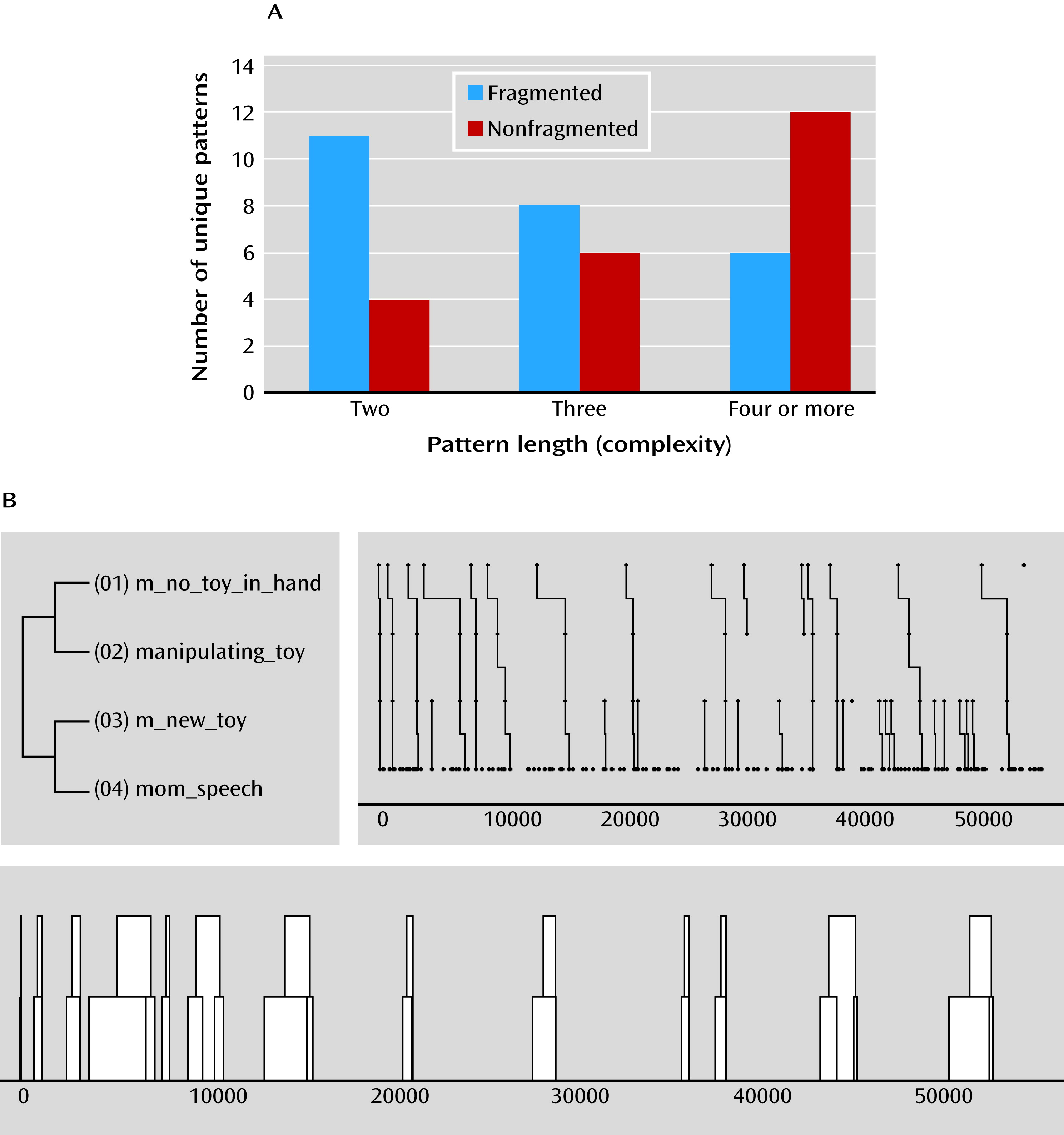
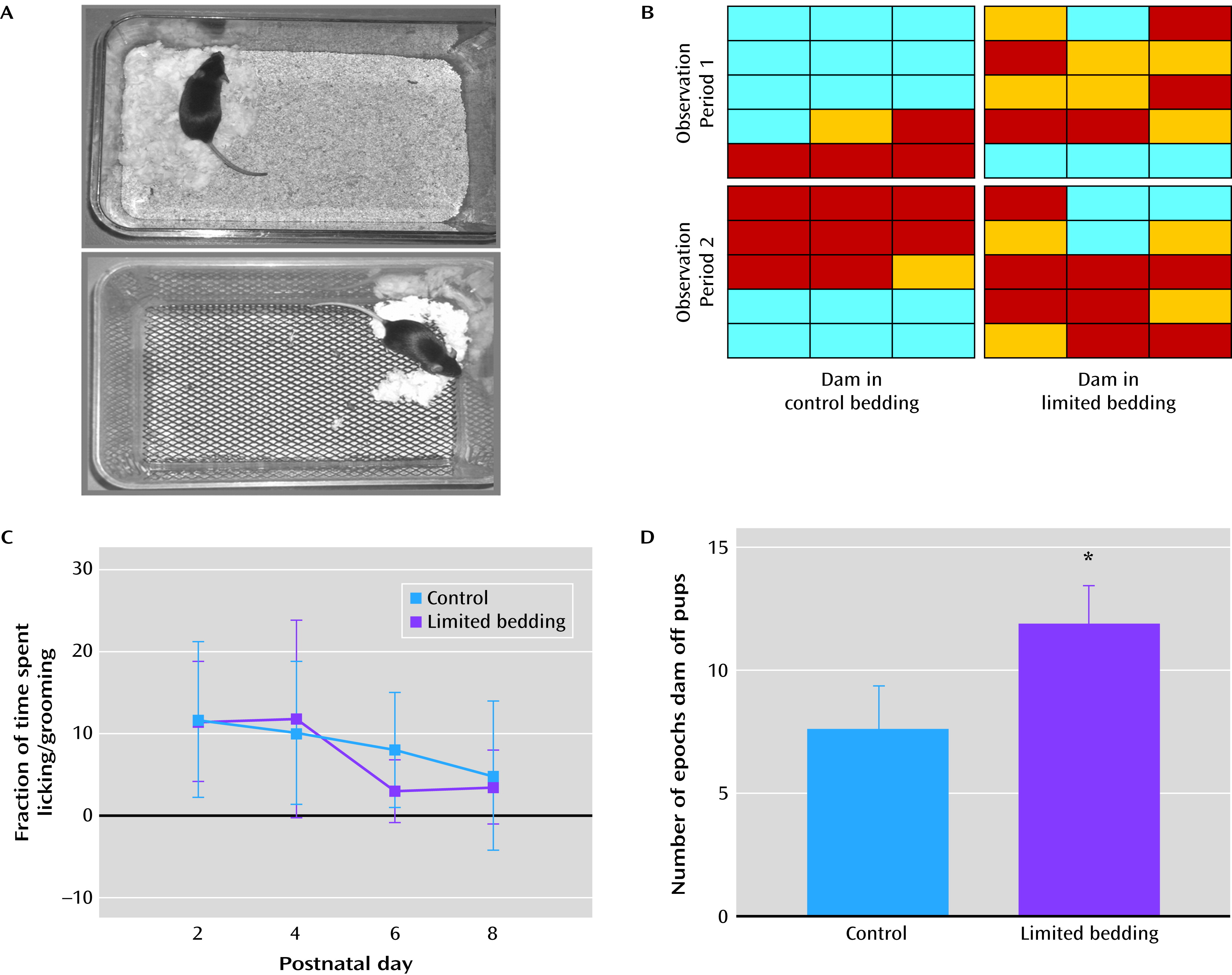
In human maternal behaviors, fragmentation refers to a behavior consisting of many sequences of simple patterns (Figure 1). In rodents, fragmentation refers to the degree to which a caring behavior occurs in numerous short episodes, rather than a small number of long episodes, under conditions when the total amount of the behavior remains relatively constant (Figure 2). Unpredictable maternal behavior in both humans and rodents refers to patterns involving more than one behavior and measures the occurrence of consistent versus inconsistent sequences or patterns of behavior (Figure 1) (66). Thus, in considering unpredictability, we ask, for example, whether smiling always follows eye contact (in humans) or grooming always follows nursing (in rodents). This measure is independent of the sensitivity of the mother-to-infant cues; rather, it assesses the observed regularities and irregularities in a sequence of behaviors. These may be quantified by the proportion of time one behavior follows another or by measuring the entropy (randomness [67]) of the conditional probability distribution following a given behavior.
FIGURE 1. Analyses of the Fragmentation and Unpredictability of Maternal Behavior in a Naturalistic Setting in Humansa
a The bar graph in panel A depicts the behavioral profile of a mother with fragmented care behavior compared with the behavioral profile of a mother with a more complex behavioral pattern. Fragmented care is defined as a large number of simple, two-component behaviors (e.g., smiling and then holding up a toy), compared with complex behavioral patterns of three or more components. A mother with nonfragmented behavior exhibits a large number of complex (four or more components) behavioral sequences. Panel B depicts single temporal patterns from a mother interacting with her 12-month-old child while exhibiting nonfragmented care. The left top subpanels show that the temporal pattern is initiated by the behavior “no toy in hand,” has four behavioral elements (e.g., look at infant, coo at infant, touch infant, pick up toy), and is repeated 13 times. This pattern consists of two hierarchical levels (smaller patterns connected to form larger patterns). The smaller pattern, “new toy speech,” is repeated 15 times without being part of the larger pattern. Both the length and level are considered measures of complexity. The frequency and temporal ordering of the events evolving into patterns are depicted in the upper right subpanel, and the sequence of events is depicted in the bottom subpanel. (Unpublished data, available upon request from the authors.)
FIGURE 2. Fragmentation of Rodent Maternal Behavior Provoked by Adversitya
a The fragmentation resulted in little change in total care, and the provoked adversity was the limiting of the bedding and nesting materials in the cage. Panel A depicts a typical cage and a cage with limited bedding, which leads to maternal stress and fragmented nurturing behaviors. The grid charts in Panel B represent the activity of a dam in a typical cage (control condition) and in the limited bedding cage during two matched observation periods. Each grid depicts one dam’s activity during 15 1-minute epochs (starting at the top left corner); individual blocks are color-coded to represent the dam’s activity during each epoch (blue=nursing; red=licking/grooming; yellow=more than one behavior). The grid demonstrates the consecutive epochs of the same behavior (consistent) in the control condition and the short epochs of each behavior type (fragmentation) in the limited nesting cage. Also apparent is the rapid alteration among nurturing behavior types (inconsistency, unpredictability) in the limited bedding cage. Panel C depicts fragmented behavior not associated with reduced time of individual caring behaviors; the total licking/grooming duration among dams in the limited bedding cages was comparable to that of dams in the control condition. The bar graph in Panel D depicts the augmented number of epochs when the dam was away from the nest, an additional measure of fragmentation. Differences between the two conditions reached statistical significance (p<0.05). Adapted from Brunson et al. (52). Copyright © 2005. Society for Neuroscience. Used with permission. Adapted from Rice et al. (57). Copyright © 2008 Endocrinology. Used with permission.

Fragmented and Unpredictable Maternal Signals in Pre- and Postnatal Humans
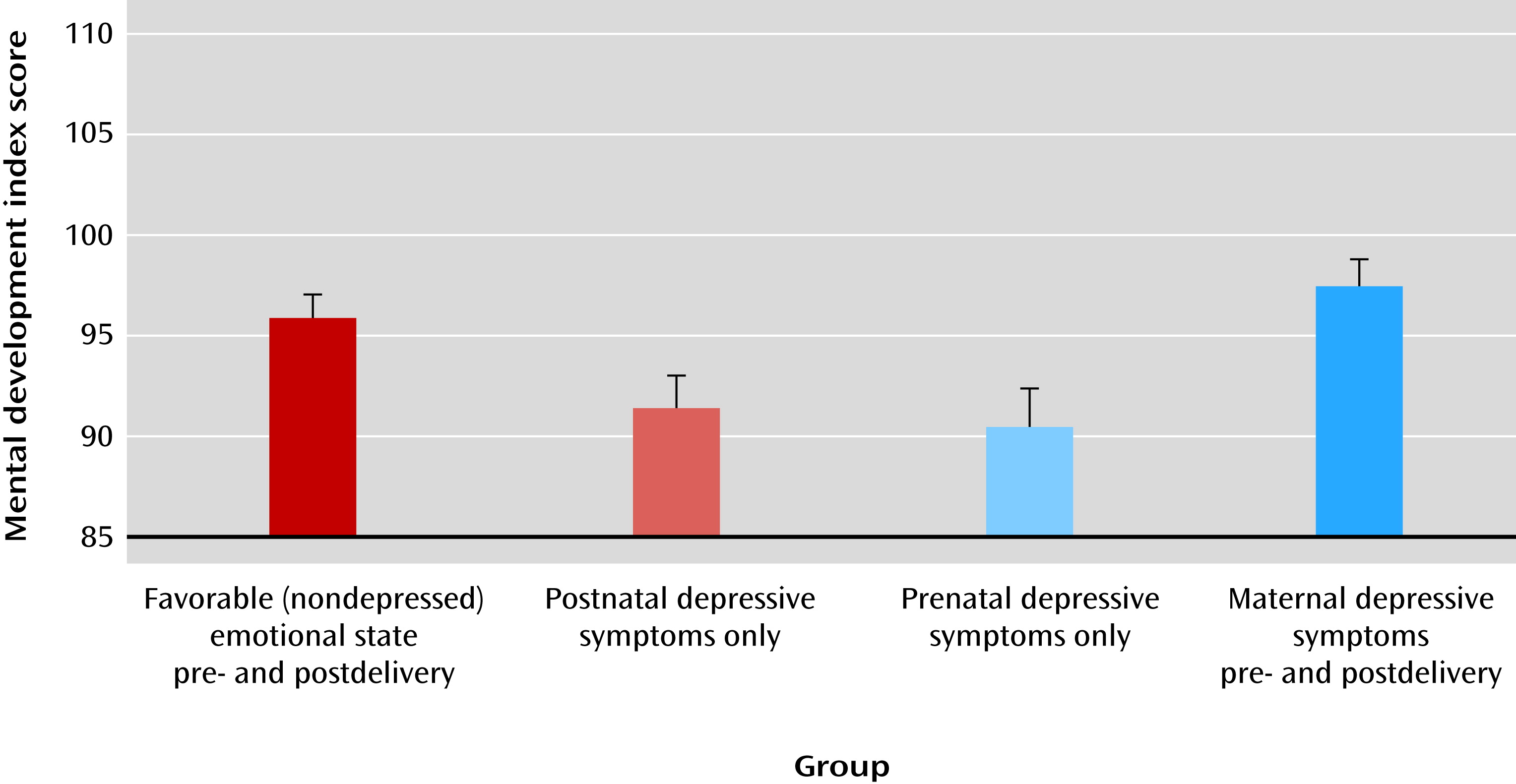
A potential contribution of fragmented maternally derived signals to adverse cognitive and emotional outcomes has emerged in studies of the human fetus and of early postnatal life. The nature of the sensory input conveyed by the mother to the fetus is not fully understood, yet maternal emotional states during gestation have been found to contribute significantly to neuropsychiatric outcome (6870). Our group recently began to explore the importance of consistency relative to fragmentation of maternal emotional states in influencing neuropsychiatric outcomes in the infant and child. We observed that mental development was greater in 1-year-old children whose mothers experienced consistent emotional states before and after delivery (71). This was true even when the emotional state was of depressive symptoms and contrasted with outcomes for offspring of mothers with inconsistent emotional states before and after delivery (Figure 3).
FIGURE 3. Effect of Consistency and Fragmentation of Pre- and Postnatal Maternal Emotional States on Neuropsychiatric Outcomea
a Mean scores on the Mental Development Index of the Bayley Scales of Infant Development for 186 1-year-old children were divided into four groups according to maternal emotional states. Presence of depressive symptoms (71) was assessed both before and after delivery in the mothers. Significant difference between groups, p<0.001 (two-way analysis of covariance). Adapted from Sandman et al. (71). Copyright © 2012 Psychological Science. Used with permission.
While these findings are remarkable, it remains unclear how these maternal emotional states are transmitted to the fetus. An intriguing possibility is that the mother conveys emotional information to her fetus via physiological parameters, such as hormone levels or patterns of respiration or heart rate (69, 70). For example, the fetal auditory system is capable of detecting and responding to maternal heartbeats by 25 weeks’ gestation (72, 73), and magnetoencephalography studies have reported that the human fetal brain responds to maternal heartbeats and external sounds (72). Unpublished data from our group suggest that maternal heartbeat patterns can also be categorized as consistent or fragmented and that there are wide individual differences in unpredictable and fragmented maternal heartbeat patterns in healthy populations of pregnant women. It is not yet known whether maternal heart rate patterns join stress hormones in providing potential biomarkers of maternal emotional states that are known to alter offspring outcomes (6971).
Regarding postnatal maternal signals (i.e., maternal care), the importance of patterns of care is evident from the work of Bowlby (15) and the large body of subsequent literature. In essence, this work strongly supports the notion that patterns of maternal behavior during neonatal and infant life may influence the infant brain and contribute to the emotional profile and neurocognitive development of offspring. The question that arises concerns whether it is possible to quantify and then categorize maternal behavioral patterns by their unpredictability and fragmentation, as distinct from maternal sensitivity to the infant. A key follow-up question is whether such analyses would provide important new information about the nature of maternal behavioral effects on emotional and cognitive outcomes. The answers to these questions are emerging. We have used video recordings to identify and code specific sensory signals from the mother to the infant, which were analyzed in several ways to infer fragmentation and unpredictability (Figure 1). Within a population of mothers without diagnoses of mental disorders, unpredictable maternal behavior (i.e., low conditional probabilities of faithful occurrence of a given pair of nurturing behaviors) varied widely and was normally distributed. Ongoing studies are examining whether this unpredictability of maternal care, independent of total care duration, influences aspects of infant behavior and developmental parameters that predict mental and cognitive outcomes.

Fragmented and Unpredictable Maternal Signals in Rodent Models
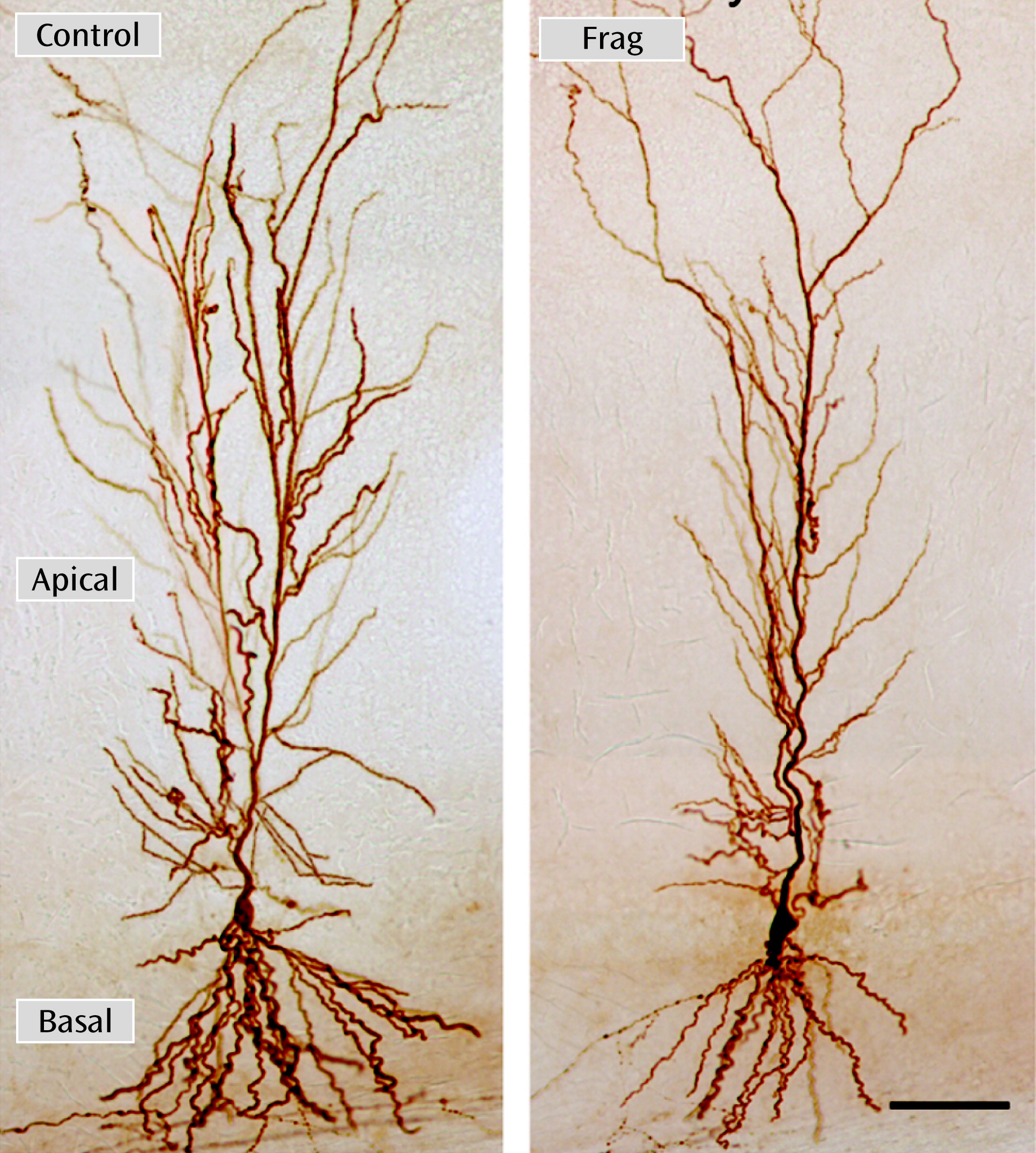
In rodents, the qualitative and quantitative aspects of maternal care (74) were evaluated by our group using a model of simulated poverty that consisted of reduced bedding and nesting materials in the home cage (52, 56, 57, 75). Surprisingly, fragmentation of maternal care into short epochs of individual nurturing behaviors, with preserved total duration of each behavior (Figure 2), provoked significant cognitive and emotional problems in the pups, which became apparent later in life and persisted throughout middle age (52, 55). Learning defects were associated with (and perhaps a result of) loss of hippocampal synapses and dendritic spines and branches (52) (Figure 4). Emotional problems resulting from fragmented maternal signals included augmented anxiety-like behavior in weanling rats and increased vulnerability to depressive-like behavior. In contrast, consistent and predictable maternal care led to effects that were opposite those generated by fragmented care; daily bouts of maternally derived licking and grooming were increased experimentally by returning rat pups to their mothers after a brief separation. Receiving recurrent, consistent, and predictable care for even 1 week resulted in a phenotype of resilience to subsequent stress and to depressive-like behaviors associated with reduced levels of CRH expression in the hypothalamus (45, 76, 77).
FIGURE 4. Fragmented Maternal Care During Early Postnatal Life Leading to Impoverished Dendritic Trees in Hippocampal Area CA1 Neuronsa
a A biocytin-filled cell from a middle-aged rat with fragmented maternal care (Frag) during postnatal days 2–9 (right) is depicted. Fragmentation of maternal care was elicited as described in Figure 2. The comparison with a neuron from a matched control section (left) illustrates the reduction in total dendritic length and in dendritic arborization following fragmented care. Scale bar=80 microns. Adapted from Brunson et al. (52). Copyright © 2005. Society for Neuroscience. Used with permission.
The mechanisms through which distinct patterns of maternally derived signals lead to these opposing molecular and functional/behavioral phenotypes are not fully understood. Recurrent, predictable episodes of maternal care activate brain regions, including the thalamic paraventricular nucleus, that are not engaged by a single episode of care (43). The paraventricular nucleus has been considered a “stress memory” storage region (78), and it exerts inhibitory effects on limbic regions, including the central nucleus of the amygdala and the bed nucleus of the stria terminalis, that regulate stress responses and CRH expression within the hypothalamus (75, 79). Indeed, predictable and recurrent patterns of maternal care have been reported to reduce the number and function of excitatory synapses on CRH-expressing neurons within the hypothalamus (42, 47), essentially modulating neuronal “wiring.” These data suggest that maternally derived sensory signals may influence intercellular signaling in the developing brain. This in turn modulates intracellular processes, including gene expression, leading to enduring alterations of neuronal function (47, 80). Interestingly, reduced CRH expression was found to begin in infancy and to lead to diminished CRH release during stress. The resulting attenuation of plasma glucocorticoid levels during stress enhanced the expression levels of hippocampal glucocorticoid receptors (45, 76). Other studies reproduced the augmented glucocorticoid receptor expression by partial blocking of CRH receptors (77), suggesting that, in rodent pups, reduction of hypothalamic CRH expression and of hormonal responses to future stresses is an important mechanism for long-lasting resilience to stress-provoking signals (2, 6, 14, 31, 44, 58, 68, 76, 8183).
If predictable and consistent sensory input from the mother reduces excitation and gene expression in key neurons, it seems reasonable to speculate that fragmented or unpredictable sensory signals may have an opposite effect. In support of this possibility, CRH expression in the hippocampus is persistently enhanced in adult rats that experienced fragmented maternal care early in life, an aberrant hyperexpression that endures for 12 months or longer (55). However, much work is needed to identify the synaptic and intracellular processes through which specific patterns of maternally derived sensory input “program” gene expression and behavioral patterns in the long term.

Discussion: Challenges and Future Directions

We propose that fragmented and unpredictable patterns and sequences of maternally derived signals (distinct from maternal sensitivity to infant needs and overall quantity of maternal care) influence brain development in a manner that contributes to emotional and cognitive outcomes. This concept is not unexpected if one considers that sensory input in early life might govern neuronal activity (31, 70, 84) and that neuronal activity influences synapse development and brain organization (8588). An important link in this proposed chain of events is the demonstration that specific patterns of maternally derived sensory input influence the number and function of synaptic connections onto specific neurons. This type of study is feasible in animal models, in which it has been found that maternal signals travel from sensory regions to stress-related brain regions (70, 8991). Notably, the patterns of maternal care have been reported to modulate the number and function of synapses of stress-responsive neurons (42). These alterations in synaptic intercellular signaling are predicted to influence intracellular signaling, turning on epigenetic processes that program the expression of genes contributing to vulnerability or resilience to stress-related neuropsychiatric disorders.
The patterns—and especially the degree of fragmentation and unpredictability—of maternal signals are important both before and after birth, influencing the child’s neuropsychiatric outcome. Furthermore, these sequential epochs of development seem to interact in their combined effects on outcome, and the consistency of pre- and postnatal maternal signals may be as (or more) important than the nature of the signals themselves (71) (Figure 3).
In summary, the contribution of maternally derived signals to vulnerability and resilience to mental illnesses is well established. Less understood is the spectrum of maternal care patterns that influence the infant/fetal brain and the pathways and mechanisms through which these crucial influences take place. We propose the novel concept that fragmentation and predictability of maternal sensory signals influence the developing brain and contribute to future cognitive and emotional vulnerabilities. We need to study the inter- and intracellular mechanisms involved at both molecular/cellular and network levels. We also need to establish the trajectories of the effects and to define biomarkers that will enable early recognition and eventual intervention. Hopefully, the concept of fragmented and unpredictable early-life environment as a cardinal contributor to subsequent vulnerability to disorders, including depression, anxiety, and certain cognitive deficits, will provide a theoretical unifying framework for a large and important body of existing literature on the origin of mental and cognitive illnesses.

Footnote

The authors report no financial relationships with commercial interests.
Supported by NIH grants MH-73136 and NS28912 (Dr. Baram), MH-086062 (Dr. Davis), HD51852 and NS41298 (Dr. Sandman), NS54942 (Dr. Solodkin), DC03378 (Dr. Small), and UL1RR031985 (Dr. Stern); and by a National Science Foundation grant, AGS 1027354 (Dr. Stern).
The authors thank Mrs. Barbara Cartwright for editorial assistance.

References

1.
Kessler RC, Demler O, Frank RG, Olfson M, Pincus HA, Walters EE, Wang P, Wells KB, Zaslavsky AM: Prevalence and treatment of mental disorders, 1990 to 2003. N Engl J Med 2005; 352:2515–2523
2.
Transformative neurodevelopmental research in mental illness, Results of the NIMH Workgroup, 2009 NIH. http://www.nimh.nih.gov/about/advisory-boards-and-groups/namhc/neurodevelopment_workgroup_report.pdf
3.
Huttenlocher PR, Dabholkar AS: Regional differences in synaptogenesis in human cerebral cortex. J Comp Neurol 1997; 387:167–178
4.
Levitt P: Structural and functional maturation of the developing primate brain. J Pediatr 2003; 143(Suppl):S35–S45
5.
Caspi A, Sugden K, Moffitt TE, Taylor A, Craig IW, Harrington H, McClay J, Mill J, Martin J, Braithwaite A, Poulton R: Influence of life stress on depression: moderation by a polymorphism in the 5-HTT gene. Science 2003; 301:386–389
6.
Martin EI, Ressler KJ, Binder E, Nemeroff CB: The neurobiology of anxiety disorders: brain imaging, genetics, and psychoneuroendocrinology. Psychiatr Clin North Am 2009; 32:549–575
7.
Gluckman PD, Hanson MA, Cooper C, Thornburg KL: Effect of in utero and early-life conditions on adult health and disease. N Engl J Med 2008; 359:61–73
8.
Krishnan V, Nestler EJ: Linking molecules to mood: new insight into the biology of depression. Am J Psychiatry 2010; 167:1305–1320
9.
Amir RE, Van den Veyver IB, Wan M, Tran CQ, Francke U, Zoghbi HY: Rett syndrome is caused by mutations in X-linked MECP2, encoding methyl-CpG-binding protein 2. Nat Genet 1999; 23:185–188
10.
Morrow EM, Yoo SY, Flavell SW, Kim TK, Lin Y, Hill RS, Mukaddes NM, Balkhy S, Gascon G, Hashmi A, Al-Saad S, Ware J, Joseph RM, Greenblatt R, Gleason D, Ertelt JA, Apse KA, Bodell A, Partlow JN, Barry B, Yao H, Markianos K, Ferland RJ, Greenberg ME, Walsh CA: Identifying autism loci and genes by tracing recent shared ancestry. Science 2008; 321:218–223
11.
Everson-Rose SA, Mendes de Leon CF, Bienias JL, Wilson RS, Evans DA: Early life conditions and cognitive functioning in later life. Am J Epidemiol 2003; 158:1083–1089
12.
Freedman R, Goldowitz D: Studies on the hippocampal formation: From basic development to clinical applications: Studies on schizophrenia. Prog Neurobiol 2010; 90:263–275
13.
van Os J, Kenis G, Rutten BP: The environment and schizophrenia. Nature 2010; 468:203–212
14.
Wilson RS, Scherr PA, Bienias JL, Mendes de Leon CF, Everson-Rose SA, Bennett DA, Evans DA: Socioeconomic characteristics of the community in childhood and cognition in old age. Exp Aging Res 2005; 31:393–407
15.
Bowlby J: Research into the origins of delinquent behaviour. BMJ 1950; 1:570–573
16.
Als H, Duffy FH, McAnulty GB, Rivkin MJ, Vajapeyam S, Mulkern RV, Warfield SK, Huppi PS, Butler SC, Conneman N, Fischer C, Eichenwald EC: Early experience alters brain function and structure. Pediatrics 2004; 113:846–857
17.
Network NECCR: Affect dysregulation in the mother-child relationship in the toddler years: antecedents and consequences. Dev Psychopathol 2004; 16:43–68
18.
Paavola L, Kemppinen K, Kumpulainen K, Moilanen I, Ebeling H: Maternal sensitivity, infant co-operation and early linguistic development: some predictive relations. Eur J Dev Psychol 2006; 3:13–30
19.
Masur EF, Flynn V, Eichorst DL: Maternal responsive and directive behaviours and utterances as predictors of children’s lexical development. J Child Lang 2005; 32:63–91
20.
McElwain NL, Volling BL: Attachment security and parental sensitivity during infancy: associations with friendship quality and false-belief understanding at age 4. J Soc Pers Relat 2004; 21:639–667
21.
Belsky J, Fearon RMP: Early attachment security, subsequent maternal sensitivity, and later child development: does continuity in development depend upon continuity of caregiving? Attach Hum Dev 2002; 4:361–387
22.
Network NECCR and NICHD Early Care Research Network: Infant-mother attachment classification: risk and protection in relation to changing maternal caregiving quality. Dev Psychol 2006; 42:38–58
23.
Ainsworth MDS, Blehar MC, Waters E, Wall S: Patterns of Attachment. A Psychological Study of the Strange Situation. Hillsdale, NJ, Lawrence Erlbaum Associates, 1978
24.
De Wolff MS, van Ijzendoorn MH: Sensitivity and attachment: a meta-analysis on parental antecedents of infant attachment. Child Dev 1997; 68:571–591
25.
Egeland B, Farber EA: Infant-mother attachment: factors related to its development and changes over time. Child Dev 1984; 55:753–771
26.
Sroufe LA: Attachment and development: a prospective, longitudinal study from birth to adulthood. Attach Hum Dev 2005; 7:349–367
27.
Belsky J, Fearon RM: Infant-mother attachment security, contextual risk, and early development: a moderational analysis. Dev Psychopathol 2002; 14:293–310
28.
Egeland B, Sroufe LA, Erickson M: The developmental consequence of different patterns of maltreatment. Child Abuse Negl 1983; 7:459–469
29.
Hane AA, Fox NA: Ordinary variations in maternal caregiving influence human infants’ stress reactivity. Psychol Sci 2006; 17:550–556
30.
Nelson CA, Carver LJ: The effects of stress and trauma on brain and memory: a view from developmental cognitive neuroscience. Dev Psychopathol 1998; 10:793–809
31.
Gunnar MR: Reversing the effects of early deprivation after infancy: giving children families may not be enough. Frontiers in Neuroscience 2010; 4:170
32.
Nachmias M, Gunnar MR, Mangelsdorf S, Parritz RH, Buss K: Behavioral inhibition and stress reactivity: the moderating role of attachment security. Child Dev 1996; 67:508–522
33.
Albers EM, Riksen-Walraven JM, Sweep FC, de Weerth C: Maternal behavior predicts infant cortisol recovery from a mild everyday stressor. J Child Psychol Psychiatry 2008; 49:97–103
34.
Parker KJ, Buckmaster CL, Sundlass K, Schatzberg AF, Lyons DM: Maternal mediation, stress inoculation, and the development of neuroendocrine stress resistance in primates. Proc Natl Acad Sci USA 2006; 103:3000–3005
35.
Sabatini MJ, Ebert P, Lewis DA, Levitt P, Cameron JL, Mirnics K: Amygdala gene expression correlates of social behavior in monkeys experiencing maternal separation. J Neurosci 2007; 27:3295–3304
36.
Suchecki D, Rosenfeld P, Levine S: Maternal regulation of the hypothalamic-pituitary-adrenal axis in the infant rat: the roles of feeding and stroking. Brain Res Dev Brain Res 1993; 75:185–192
37.
Suchecki D, Nelson DY, Van Oers H, Levine S: Activation and inhibition of the hypothalamic-pituitary-adrenal axis of the neonatal rat: effects of maternal deprivation. Psychoneuroendocrinology 1995; 20:169–182
38.
Korosi A, Baram TZ: Plasticity of the stress response early in life: mechanisms and significance. Dev Psychobiol 2010; 52:661–670
39.
Liu D, Diorio J, Tannenbaum B, Caldji C, Francis D, Freedman A, Sharma S, Pearson D, Plotsky PM, Meaney MJ: Maternal care, hippocampal glucocorticoid receptors, and hypothalamic-pituitary-adrenal responses to stress. Science 1997; 277:1659–1662
40.
Meaney MJ, Szyf M: Maternal care as a model for experience-dependent chromatin plasticity? Trends Neurosci 2005; 28:456–463
41.
Eghbal-Ahmadi M, Avishai-Eliner S, Hatalski CG, Baram TZ: Differential regulation of the expression of corticotropin-releasing factor receptor type 2 (CRF2) in hypothalamus and amygdala of the immature rat by sensory input and food intake. J Neurosci 1999; 19:3982–3991
42.
Korosi A, Shanabrough M, McClelland S, Liu ZW, Borok E, Gao XB, Horvath TL, Baram TZ: Early-life experience reduces excitation to stress-responsive hypothalamic neurons and reprograms the expression of corticotropin-releasing hormone. J Neurosci 2010; 30:703–713
43.
Fenoglio KA, Chen Y, Baram TZ: Neuroplasticity of the hypothalamic-pituitary-adrenal axis early in life requires recurrent recruitment of stress-regulating brain regions. J Neurosci 2006; 26:2434–2442
44.
Plotsky PM, Meaney MJ: Early, postnatal experience alters hypothalamic corticotropin-releasing factor (CRF) mRNA, median eminence CRF content and stress-induced release in adult rats. Brain Res Mol Brain Res 1993; 18:195–200
45.
Avishai-Eliner S, Eghbal-Ahmadi M, Tabachnik E, Brunson KL, Baram TZ: Down-regulation of hypothalamic corticotropin-releasing hormone messenger ribonucleic acid (mRNA) precedes early-life experience-induced changes in hippocampal glucocorticoid receptor mRNA. Endocrinology 2001; 142:89–97
46.
Weaver IC, Cervoni N, Champagne FA, D’Alessio AC, Sharma S, Seckl JR, Dymov S, Szyf M, Meaney MJ: Epigenetic programming by maternal behavior. Nat Neurosci 2004; 7:847–854
47.
McClelland S, Korosi A, Cope J, Ivy A, Baram TZ: Emerging roles of epigenetic mechanisms in the enduring effects of early-life stress and experience on learning and memory. Neurobiol Learn Mem 2011; 96:79–88
48.
van Hasselt FN, Cornelisse S, Zhang TY, Meaney MJ, Velzing EH, Krugers HJ, Joëls M: Adult hippocampal glucocorticoid receptor expression and dentate synaptic plasticity correlate with maternal care received by individuals early in life. Hippocampus 2012; 22:255–266
49.
Walker CD: Maternal touch and feed as critical regulators of behavioral and stress responses in the offspring. Dev Psychobiol 2010; 52:638–650
50.
Bale TL, Baram TZ, Brown AS, Goldstein JM, Insel TR, McCarthy MM, Nemeroff CB, Reyes TM, Simerly RB, Susser ES, Nestler EJ: Early life programming and neurodevelopmental disorders. Biol Psychiatry 2010; 68:314–319
51.
Mueller BR, Bale TL: Sex-specific programming of offspring emotionality after stress early in pregnancy. J Neurosci 2008; 28:9055–9065
52.
Brunson KL, Kramár E, Lin B, Chen Y, Colgin LL, Yanagihara TK, Lynch G, Baram TZ: Mechanisms of late-onset cognitive decline after early-life stress. J Neurosci 2005; 25:9328–9338
53.
Roth TL, Sullivan RM: Memory of early maltreatment: neonatal behavioral and neural correlates of maternal maltreatment within the context of classical conditioning. Biol Psychiatry 2005; 57:823–831
54.
Moriceau S, Roth TL, Sullivan RM: Rodent model of infant attachment learning and stress. Dev Psychobiol 2010; 52:651–660
55.
Ivy AS, Rex CS, Chen Y, Dubé C, Maras PM, Grigoriadis DE, Gall CM, Lynch G, Baram TZ: Hippocampal dysfunction and cognitive impairments provoked by chronic early-life stress involve excessive activation of CRH receptors. J Neurosci 2010; 30:13005–13015
56.
Ivy AS, Brunson KL, Sandman C, Baram TZ: Dysfunctional nurturing behavior in rat dams with limited access to nesting material: a clinically relevant model for early-life stress. Neuroscience 2008; 154:1132–1142
57.
Rice CJ, Sandman CA, Lenjavi MR, Baram TZ: A novel mouse model for acute and long-lasting consequences of early life stress. Endocrinology 2008; 149:4892–4900
58.
McEwen BS: Early life influences on life-long patterns of behavior and health. Ment Retard Dev Disabil Res Rev 2003; 9:149–154
59.
Fox NA, Almas AN, Degnan KA, Nelson CA, Zeanah CH: The effects of severe psychosocial deprivation and foster care intervention on cognitive development at 8 years of age: findings from the Bucharest Early Intervention Project. J Child Psychol Psychiatry 2011; 52:919–928
60.
Nelson CA, Zeanah CH, Fox NA, Marshall PJ, Smyke AT, Guthrie D: Cognitive recovery in socially deprived young children: the Bucharest Early Intervention Project. Science 2007; 318:1937–1940
61.
Suomi SJ: Surrogate rehabilitation of monkeys reared in total social isolation. J Child Psychol Psychiatry 1973; 14:71–77
62.
Meyer JS, Novak MA, Bowman RE, Harlow HF: Behavioral and hormonal effects of attachment object separation in surrogate-peer-reared and mother-reared infant rhesus monkeys. Dev Psychobiol 1975; 8:425–435
63.
Mendoza SP, Smotherman WP, Miner MT, Kaplan J, Levine S: Pituitary-adrenal response to separation in mother and infant squirrel monkeys. Dev Psychobiol 1978; 11:169–175
64.
Champagne DL, Bagot RC, van Hasselt F, Ramakers G, Meaney MJ, de Kloet ER, Joëls M, Krugers H: Maternal care and hippocampal plasticity: evidence for experience-dependent structural plasticity, altered synaptic functioning, and differential responsiveness to glucocorticoids and stress. J Neurosci 2008; 28:6037–6045
65.
Lupien SJ, McEwen BS, Gunnar MR, Heim C: Effects of stress throughout the lifespan on the brain, behaviour and cognition. Nat Rev Neurosci 2009; 10:434–445
66.
Magnusson MS: Discovering hidden time patterns in behavior: T-patterns and their detection. Behav Res Methods Instrum Comput 2000; 32:93–110
67.
Thomas JA: Elements of Information Theory, 2nd ed. New York, Wiley-Interscience, 2006
68.
Avishai-Eliner S, Brunson KL, Sandman CA, Baram TZ: Stressed-out, or in (utero)? Trends Neurosci 2002; 25:518–524
69.
Sandman CA, Davis EP, Buss C, Glynn LM: Exposure to prenatal psychobiological stress exerts programming influences on the mother and her fetus. Neuroendocrinology (Epub ahead of print, April 15, 2011)
70.
Dipietro JA: Psychological and psychophysiological considerations regarding the maternal-fetal relationship. Infant Child Dev 2010; 19:27–38
71.
Sandman CA, Davis EP, Glynn LM: Prescient human fetuses thrive. Psychol Sci 2012; 23:93–100
72.
Porcaro C, Zappasodi F, Barbati G, Salustri C, Pizzella V, Rossini PM, Tecchio F: Fetal auditory responses to external sounds and mother’s heart beat: detection improved by Independent Component Analysis. Brain Res 2006; 1101:51–58
73.
Pujol R, Lavigne-Rebillard M, Uziel A: Physiological correlates of development of the human cochlea. Semin Perinatol 1990; 14:275–280
74.
Hofer MA, Zmitrovich A, Shair HN: Nursing interaction of Wistar rats is modified by prior experience of altered nursing bout length. Dev Psychobiol 1989; 22:321–345
75.
Avishai-Eliner S, Gilles EE, Eghbal-Ahmadi M, Bar-El Y, Baram TZ: Altered regulation of gene and protein expression of hypothalamic-pituitary-adrenal axis components in an immature rat model of chronic stress. J Neuroendocrinol 2001; 13:799–807
76.
Korosi A, Baram TZ: The pathways from mother’s love to baby’s future. Frontiers in Behavioral Neuroscience 2009; 3:27
77.
Fenoglio KA, Brunson KL, Avishai-Eliner S, Stone BA, Kapadia BJ, Baram TZ: Enduring, handling-evoked enhancement of hippocampal memory function and glucocorticoid receptor expression involves activation of the corticotropin-releasing factor type-1 receptor. Endocrinology 2005; 146:4090–4096
78.
Bhatnagar S, Dallman M: Neuroanatomical basis for facilitation of hypothalamic-pituitary-adrenal responses to a novel stressor after chronic stress. Neuroscience 1998; 84:1025–1039
79.
Herman JP, Figueiredo H, Mueller NK, Ulrich-Lai Y, Ostrander MM, Choi DC, Cullinan WE: Central mechanisms of stress integration: hierarchical circuitry controlling hypothalamo-pituitary-adrenocortical responsiveness. Front Neuroendocrinol 2003; 24:151–180
80.
Brunson KL, Chen Y, Avishai-Eliner S, Baram TZ: Stress and the developing hippocampus: a double-edged sword? Mol Neurobiol 2003; 27:121–136
81.
Insel TR: Translating scientific opportunity into public health impact: a strategic plan for research on mental illness. Arch Gen Psychiatry 2009; 66:128–133
82.
Chen ZY, Jing D, Bath KG, Ieraci A, Khan T, Siao CJ, Herrera DG, Toth M, Yang C, McEwen BS, Hempstead BL, Lee FS: Genetic variant BDNF (Val66Met) polymorphism alters anxiety-related behavior. Science 2006; 314:140–143
83.
Nestler EJ, Hyman SE: Animal models of neuropsychiatric disorders. Nat Neurosci 2010; 13:1161–1169
84.
Khazipov R, Sirota A, Leinekugel X, Holmes GL, Ben-Ari Y, Buzsáki G: Early motor activity drives spindle bursts in the developing somatosensory cortex. Nature 2004; 432:758–761
85.
Grossman AW, Aldridge GM, Weiler IJ, Greenough WT: Local protein synthesis and spine morphogenesis: Fragile X syndrome and beyond. J Neurosci 2006; 26:7151–7155
86.
Lynch G, Rex CS, Chen LY, Gall CM: The substrates of memory: defects, treatments, and enhancement. Eur J Pharmacol 2008; 585:2–13
87.
Zoghbi HY: Postnatal neurodevelopmental disorders: meeting at the synapse? Science 2003; 302:826–830
88.
Ben-Ari Y, Spitzer NC: Phenotypic checkpoints regulate neuronal development. Trends Neurosci 2010; 33:485–492
89.
Fenoglio KA, Brunson KL, Baram TZ: Hippocampal neuroplasticity induced by early-life stress: functional and molecular aspects. Front Neuroendocrinol 2006; 27:180–192
90.
Davis EP, Sandman CA: The timing of prenatal exposure to maternal cortisol and psychosocial stress is associated with human infant cognitive development. Child Dev 2010; 81:131–148
91.
Hare TA, Tottenham N, Galvan A, Voss HU, Glover GH, Casey BJ: Biological substrates of emotional reactivity and regulation in adolescence during an emotional go-nogo task. Biol Psychiatry 2008; 63:927–934

Information & Authors

Information

Published In

Go to American Journal of Psychiatry
American Journal of Psychiatry
Pages: 907 - 915
PubMed: 22885631

History

Received: 7 September 2011
Revision received: 21 December 2011
Revision received: 8 March 2012
Revision received: 2 April 2012
Accepted: 9 April 2012
Published online: 1 September 2012
Published in print: September 2012

Authors

Details

Tallie Z. Baram, M.D., Ph.D.
From the Departments of Pediatrics, Anatomy/Neurobiology, Neurology, and Psychiatry, School of Medicine, and the Bren School of Information and Computer Sciences, University of California, Irvine; and the Department of Pediatrics, School of Medicine, Loma Linda University, Loma Linda, Calif.
Elysia P. Davis, Ph.D.
From the Departments of Pediatrics, Anatomy/Neurobiology, Neurology, and Psychiatry, School of Medicine, and the Bren School of Information and Computer Sciences, University of California, Irvine; and the Department of Pediatrics, School of Medicine, Loma Linda University, Loma Linda, Calif.
Andre Obenaus, Ph.D.
From the Departments of Pediatrics, Anatomy/Neurobiology, Neurology, and Psychiatry, School of Medicine, and the Bren School of Information and Computer Sciences, University of California, Irvine; and the Department of Pediatrics, School of Medicine, Loma Linda University, Loma Linda, Calif.
Curt A. Sandman, Ph.D.
From the Departments of Pediatrics, Anatomy/Neurobiology, Neurology, and Psychiatry, School of Medicine, and the Bren School of Information and Computer Sciences, University of California, Irvine; and the Department of Pediatrics, School of Medicine, Loma Linda University, Loma Linda, Calif.
Steven L. Small, M.D., Ph.D.
From the Departments of Pediatrics, Anatomy/Neurobiology, Neurology, and Psychiatry, School of Medicine, and the Bren School of Information and Computer Sciences, University of California, Irvine; and the Department of Pediatrics, School of Medicine, Loma Linda University, Loma Linda, Calif.
Ana Solodkin, Ph.D.
From the Departments of Pediatrics, Anatomy/Neurobiology, Neurology, and Psychiatry, School of Medicine, and the Bren School of Information and Computer Sciences, University of California, Irvine; and the Department of Pediatrics, School of Medicine, Loma Linda University, Loma Linda, Calif.
Hal Stern, Ph.D.
From the Departments of Pediatrics, Anatomy/Neurobiology, Neurology, and Psychiatry, School of Medicine, and the Bren School of Information and Computer Sciences, University of California, Irvine; and the Department of Pediatrics, School of Medicine, Loma Linda University, Loma Linda, Calif.

Notes

Address correspondence to Dr. Baram ([email protected]).

Metrics & Citations

Metrics

Citations

Export Citations

If you have the appropriate software installed, you can download article citation data to the citation manager of your choice. Simply select your manager software from the list below and click Download.

For more information or tips please see 'Downloading to a citation manager' in the Help menu.

Format
Citation style
Style
Copy to clipboard

View Options

View options

PDF/EPUB

View PDF/EPUB

Login options

Already a subscriber? Access your subscription through your login credentials or your institution for full access to this article.

Personal login Institutional Login Open Athens login
Purchase Options

Purchase this article to access the full text.

PPV Articles - American Journal of Psychiatry

PPV Articles - American Journal of Psychiatry

Not a subscriber?

Subscribe Now / Learn More

PsychiatryOnline subscription options offer access to the DSM-5-TR® library, books, journals, CME, and patient resources. This all-in-one virtual library provides psychiatrists and mental health professionals with key resources for diagnosis, treatment, research, and professional development.

Need more help? PsychiatryOnline Customer Service may be reached by emailing [email protected] or by calling 800-368-5777 (in the U.S.) or 703-907-7322 (outside the U.S.).

Media

Figures

Other

Tables

Share

Share

Share article link

Share