Full access
Communications and Updates
Published Online: 1 January 2013

Altered Functional and Structural Connectivity in a Schizophrenia Patient With Complete Agenesis of the Corpus Callosum

To the Editor: We incidentally detected complete agenesis of the corpus callosum (1, 2) in a 34-year-old married African American man with schizophrenia who was referred for a resting-state functional MRI (fMRI) and diffusion tensor imaging (DTI) research study. The patient had a 5-year history of schizophrenia that was treated with risperidone, 4 mg/day, and he was mildly ill at the time of the study (Positive and Negative Syndrome Scale subscores of 7 [positive], 10 [negative], and 20 [general psychopathology]). We compared his images from the 3-T scanner with those of an age-, gender-, and ethnicity-matched comparison subject.
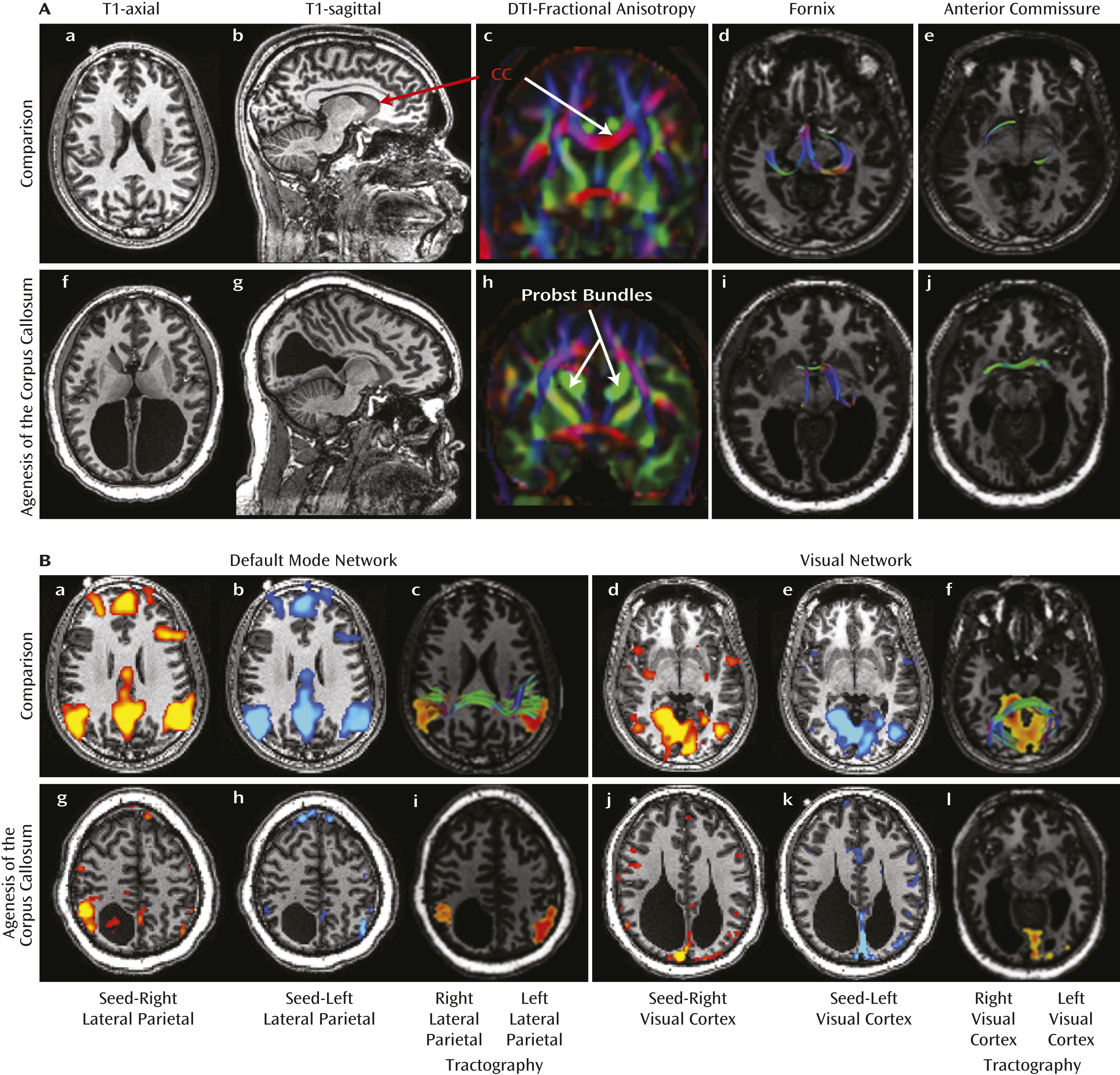
The images in Figure 1A show the absent corpus callosum in the patient and prominent Probst bundles running parallel to the interhemispheric fissure. The fornix, although skewed in orientation, was intact, and the anterior commissure was more prominent in our patient than in the comparison subject. 
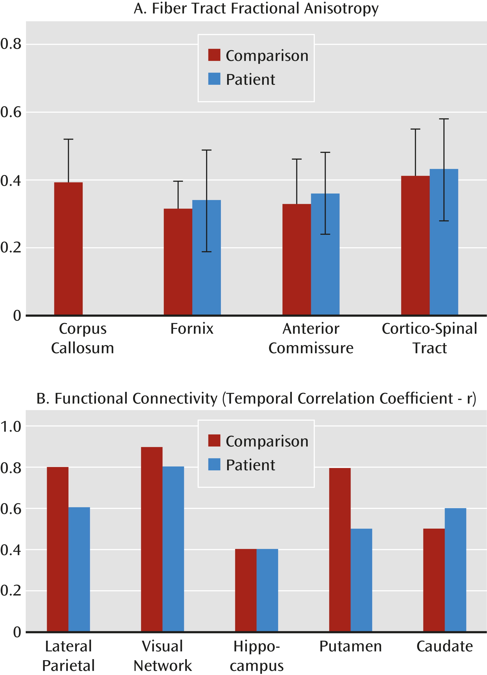
Figure 2A shows that the patient had slightly higher fractional anisotropy values in the fornix and anterior commissure.
FIGURE 1. Brain Imaging From a Study of Complete Agenesis of the Corpus Callosuma
a Panel A depicts the anatomical and DTI images from a healthy comparison subject (a–e) and the patient with agenesis of the corpus callosum (CC) (f–j). The corpus callosum is completely absent in the patient, leading to enlarged ventricles (colcocephaly) and prominent Probst bundles (green in panel h). In contrast, the fornix (d, i) is preserved, and the anterior commissure is prominent (e, j). Panel B depicts resting blood-oxygen-level-dependent images (a, b, d, e, g, h, j, k) and DTI tractography images (c, f, i, l) from a healthy comparison subject (a–f) and the patient with agenesis of the corpus callosum (g–l). Regions of interest were placed in the left and right lateral parietal and visual cortex to study the default mode (a, b) and visual networks (d, e). Relative to the healthy comparison subject, default mode network (g, h) and visual network (g, h) connectivity were markedly reduced in the patient (3<z<7). The callosal fibers connecting the parietal (c) and visual (f) cortex region of interest in the healthy comparison subject are missing in the patient with agenesis of the corpus callosum (i, l).
FIGURE 2. Fractional Anisotropy Values and Bilateral Functional Connectivity in a Study of Complete Agenesis of the Corpus Callosuma
a Panel A shows that the fractional anisotropy values of interhemispheric pathways (fornix and anterior commissure) and the corticospinal tract are higher in the patient with agenesis of the corpus callosum. In Panel B, correlation coefficients (r) for temporal synchrony between left and right brain hemispheres were reduced in cortical regions and the putamen but normal in the hippocampus and caudate.
We used independent component analysis in FMRIB’s Software Library to identify resting-state activity involving the lateral parietal region in the default mode network and involving the primary visual cortex in the visual network. These functionally defined regions of interest in the lateral parietal and primary visual cortex were then used for two-region DTI tractography using MedINRIA (3) to evaluate interhemispheric connections via the corpus callosum as well as noncallosal connections via the cortico-spinal tract, fornix, and anterior commissure.
The patient showed dramatically reduced interhemispheric but strong ipsilateral functional connectivity in the default mode network and the primary visual cortex (Figure 1B). DTI tractography showed that connections between the left and right lateral parietal and primary visual cortex regions, mediated via the corpus callosum, were absent in the patient (panels i and l of Figure 1B). We calculated correlation coefficients between the left and right brain regions (Figure 2B) and found reduced temporal synchrony in the lateral parietal and primary visual cortex (connected via the corpus callosum) but normal synchrony in the hippocampus (connected via the fornix and anterior commissure) and the caudate nuclei (possibly from long-term compensatory or robustly developed indirect pathways).
Consistent with the view that functional connectivity demonstrated by resting-state blood-oxygen-level-dependent fMRI is related to anatomical connectivity, we observed highly lateralized and reduced interhemispheric connectivity in regions dependent on an intact corpus callosum in our patient. Nonetheless, functional and structural connectivity across the two brain hemispheres via other interhemispheric fiber tracts were preserved. This case report provides a broad understanding of the morphological and functional modifications that occur in agenesis of the corpus callosum. Altered brain function in a patient with complete agenesis of the corpus callosum was successfully illustrated using fMRI. Furthermore, these changes also conformed to the altered anatomical connectivity that was observed with DTI.

References

1.
Lungu O, Stip E: Agenesis of corpus callosum and emotional information processing in schizophrenia. Front Psychiatry 2012; 3:1–8
2.
Johnston JM, Vaishnavi SN, Smyth MD, Zhang D, He BJ, Zempel JM, Shimony JS, Snyder AZ, Raichle ME: Loss of resting interhemispheric functional connectivity after complete section of the corpus callosum. J Neurosci 2008; 28:6453–6458
3.
Toussaint N, Souplet JC, Fillard P: MedINRIA: Medical Image Navigation and Research Tool by INRIA, in Proceedings of MICCAI Workshop on Interaction in Medical Image Analysis and Visualization. Brisbane, Australia, 2007. Available at http://www-sop.inria.fr/asclepios/software/MedINRIA.

Information & Authors

Information

Published In

Go to American Journal of Psychiatry
American Journal of Psychiatry
Pages: 122 - 123
PubMed: 23288389

History

Accepted: October 2012
Published online: 1 January 2013
Published in print: January 2013

Authors

Affiliations

Swati Rane, Ph.D.
From the Institute of Imaging Science, Department of Radiology and Radiological Sciences, and the Department of Psychiatry, Vanderbilt University, Nashville.
Samet Kose, M.D., Ph.D.
From the Institute of Imaging Science, Department of Radiology and Radiological Sciences, and the Department of Psychiatry, Vanderbilt University, Nashville.
John C. Gore, Ph.D.
From the Institute of Imaging Science, Department of Radiology and Radiological Sciences, and the Department of Psychiatry, Vanderbilt University, Nashville.
Stephan Heckers, M.D.
From the Institute of Imaging Science, Department of Radiology and Radiological Sciences, and the Department of Psychiatry, Vanderbilt University, Nashville.

Competing Interests

The authors report no financial relationships with commercial interests.

Metrics & Citations

Metrics

Citations

Export Citations

If you have the appropriate software installed, you can download article citation data to the citation manager of your choice. Simply select your manager software from the list below and click Download.

For more information or tips please see 'Downloading to a citation manager' in the Help menu.

Format
Citation style
Style
Copy to clipboard

There are no citations for this item

View Options

View options

PDF/ePub

View PDF/ePub

Get Access

Login options

Already a subscriber? Access your subscription through your login credentials or your institution for full access to this article.

Personal login Institutional Login Open Athens login
Purchase Options

Purchase this article to access the full text.

PPV Articles - American Journal of Psychiatry

PPV Articles - American Journal of Psychiatry

Not a subscriber?

Subscribe Now / Learn More

PsychiatryOnline subscription options offer access to the DSM-5-TR® library, books, journals, CME, and patient resources. This all-in-one virtual library provides psychiatrists and mental health professionals with key resources for diagnosis, treatment, research, and professional development.

Need more help? PsychiatryOnline Customer Service may be reached by emailing [email protected] or by calling 800-368-5777 (in the U.S.) or 703-907-7322 (outside the U.S.).

Media

Figures

Other

Tables

Share

Share

Share article link

Share