Abstract

Objective:

In this conceptual review, the authors propose a novel mechanistic candidate in the etiology of depression with onset in the menopause transition (“perimenopausal depression”) involving alterations in stress-responsive pathways, induced by ovarian hormone fluctuation.

Method:

The relevant literature in perimenopausal depression, including prevalence, predictors, and treatment with estrogen therapy, was reviewed. Subsequently, the growing evidence from animal models and clinical research in other reproductive mood disorders was synthesized to describe a heuristic model of perimenopausal depression development.

Results:

The rate of major depressive disorder and clinically meaningful elevations in depressive symptoms increases two- to threefold during the menopause transition. While the mechanisms by which ovarian hormone fluctuation might impact mood are poorly understood, growing evidence from basic and clinical research suggests that fluctuations in ovarian hormones and derived neurosteroids result in alterations in regulation of the HPA axis by γ-aminobutyric acid (GABA). The authors’ heuristic model suggests that for some women, failure of the GABAA receptor to regulate overall GABA-ergic tone in the face of shifting levels of these neurosteroids may induce HPA axis dysfunction, thereby increasing sensitivity to stress and generating greater vulnerability to depression.

Conclusions:

The proposed model provides a basis for understanding the mechanisms by which the changing hormonal environment of the menopause transition may interact with the psychosocial environment of midlife to contribute to perimenopausal depression risk. Future research investigating this model may inform the development of novel pharmacological treatments for perimenopausal depression and related disorders, such as postpartum depression and premenstrual dysphoric disorder.
The rate of major depressive disorder in women of reproductive age is double that of men’s. Depressive disorders tied to reproductive events may partially account for this higher risk. Premenstrual dysphoric disorder (PMDD) and postpartum depression are two such disorders for which pathophysiological mechanisms include an increased vulnerability to fluctuations in ovarian-derived hormones as well as hypothalamic-pituitary-adrenal (HPA) axis dysregulation. Substantially less research has been conducted on depressive disorders tied to the menopause transition.
The neuroendocrine mechanisms by which the complex hormonal milieu of the menopause transition might trigger depressive symptoms also remain poorly understood, though multiple candidate mechanisms are under investigation. These include, but are not limited to, withdrawal from the anti-inflammatory (1) and neuroprotective (2) effects of estradiol as well as its modulation of the limbic processing (3) and memory (4, 5) of emotionally relevant information. The primary goal of the current review is to set forth an additional mechanistic hypothesis involving interactions among reproductive steroids, neurosteroids mediated by γ-aminobutyric acid (GABA), and HPA axis function, which can serve as the basis for further investigation. We will discuss the literature implicating ovarian hormone variability in the development of depression with onset in the menopause transition (a.k.a. perimenopausal depression) and describe a paradigm in which changes in GABA-ergic neurosteroids derived from progesterone may induce dysfunction of the GABA-ergic system and, in turn, the HPA axis. To first provide a context in which to discuss this potential mechanism, we briefly describe the following: 1) the endocrine environment characterizing the menopause transition, 2) the prevalence of perimenopausal depression, 3) risk factors for perimenopausal depression, and 4) the evidence for the use of estrogen therapy as a treatment for perimenopausal depression.

Endocrine Environment of The Menopause Transition

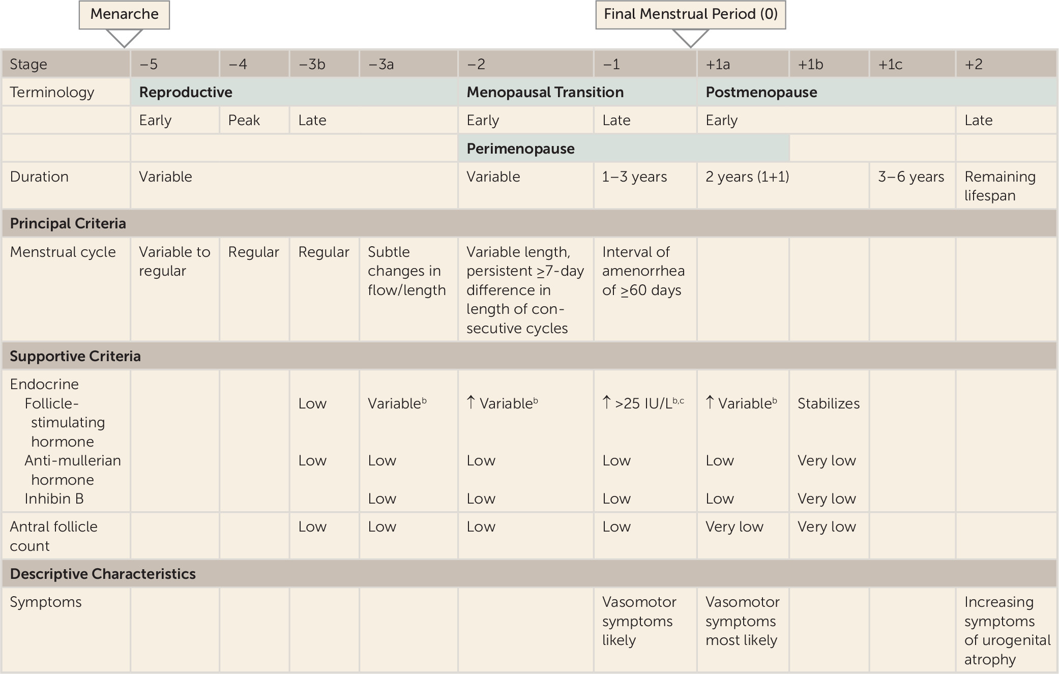
The menopause transition, triggered by a woman’s diminishing supply of ovarian follicles, represents the reproductive stage transitioning from reproductively capable ovulatory cycles through the loss of ovulatory function and to the cessation of menses. The latter marks the onset of the menopause. In premenopausal women, antral ovarian follicles produce inhibin B, a protein complex that inhibits follicle-stimulating hormone (FSH) release, which stimulates the recruitment and growth of ovarian follicles. As women approach the end of their reproductive years and fewer antral ovarian follicles are available to produce inhibin B, FSH concentrations gradually rise. While FSH levels have historically been used as an endocrine marker of postmenopausal status, FSH is less useful for reproductive staging in the menopause transition because of variability in FSH concentrations at this time. Consequently, standard criteria for reproductive staging are based primarily on menstrual bleeding patterns, which can be corroborated with endocrine data. The Stages of Reproductive Aging Workshop system (STRAW), first developed in 2001 and revised in 2011 (6), is among the most commonly used staging systems and divides a woman’s reproductive lifespan into stages, using the final menstrual period as the anchor (Figure 1).
FIGURE 1. Phases of Women’s Reproductive Lives as Represented in the Stages of Reproductive Aging Workshop System (STRAW+10)a
a Reprinted with permission of Informa Healthcare from the work of Harlow et al. (6).
b Blood draw on cycle days 2–5. ↑, elevated.
c Approximate expected level based on assays using current international pituitary standard.
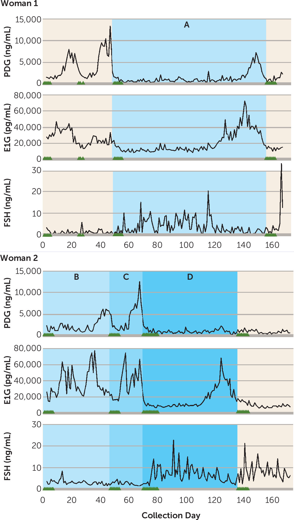
While there are substantial individual differences in the hormonal trajectory through the menopause transition (7, 8), most women experience the following changes (reviewed in references 9 and 10), beginning in the early menopause transition and progressing into the late stage. First, menstrual cycle length becomes increasingly variable, with long cycles becoming more common as the transition progresses. This is illustrated in Figure 2, showing daily concentrations of urinary FSH and the urinary metabolites of estradiol and progesterone (proxies for estradiol and progesterone concentrations) in two women in the menopause transition. While short cycles are due to early follicular recruitment by intermittently high FSH levels, long cycles can be due to anovulation (Figure 2, woman 2, cycle D) or a delayed ovarian response to FSH stimulation, resulting in an extended (low-estradiol) early follicular phase (Figure 2, woman 1, cycle A). Second, luteal progesterone also decreases, thought to result from declining dominant follicle quality (Figure 2, woman 2, cycle B). Third, appearing in the early and continuing into the late menopause transition are cycles in which concentrations of estradiol are higher than premenopausal concentrations, also thought to result from elevated FSH. In addition to these changes in ovulatory cycles, the late menopause transition is marked by an increasing frequency of anovulatory cycles (60%−70% of cycles), which are characterized by low progesterone and erratic estradiol concentrations. Eventually, estradiol and progesterone production ceases, though recent evidence suggests that the early postmenopausal period is characterized by more ovarian activity than previously believed (8). In summary, most women undergoing a natural transition to menopause are exposed to highly erratic hormonal flux. While an increasing proportion of anovulatory cycles translates to fewer luteal-phase increases in progesterone, variable FSH concentrations cause periods of both hypo- and hyper-estrogenism. This exposure to erratic ovarian hormone concentrations may extend over 5 years (12).
FIGURE 2. Daily Urinary Levels of FSH and Metabolites of Estradiol (E1G) and Progesterone (PDG) in Two Perimenopausal Womena,b
a Adapted with permission of Wolters Kluwer Health from the work of O’Connor et al. (11).
b A, a long ovulatory cycle; B, a long ovulatory cycle with low luteal progesterone; C, a normal ovulatory cycle; and D, a long anovulatory cycle. Triangles on the x-axis represent days of menstrual bleeding. PDG, pregnanediol-glucuronide; E1G, estrone-glucuronide; FSH, follicle-stimulating hormone.

Prevalence of Perimenopausal Depression

Though the existence of menopause-associated depression has long been debated (13) and several cross-sectional studies find no relationship between the prevalence of major depressive disorder and menopausal status (14, 15), longitudinal studies have been more consistent in finding that the menopause transition is associated with a substantial increase in vulnerability to clinically significant depressive symptoms, with odds ratios ranging from 1.33 to 1.79 (1618); this increased vulnerability is also observed in longitudinal studies in women with no history of major depression (17, 19, 20). Furthermore, studies using the Structured Clinical Interview for DSM-IV to examine the incidence of syndromal depression in the menopause transition echo the above findings in mixed samples of women with or without a history of major depressive disorder (21). For example, in a subanalysis of 221 initially premenopausal participants from the Study of Women’s Health Across the Nation (SWAN), which included women with or without a history of depression who were followed for 10 years, the rate of syndromal depression was doubled during the menopause transition and tripled in the early postmenopausal period (22). In contrast, two longitudinal studies (19, 23) suggest that the risk of first-onset syndromal major depressive disorder may not be increased in the menopause transition, though additional research to confirm this is warranted.
In all, there is strong evidence that the menopause transition and early postmenopausal period are a time of increased vulnerability to dysphoric mood, though the risk of a major depressive episode may be elevated only in the context of recurrent major depressive disorder.

Risk Factors for Perimenopausal Depression

Multiple longitudinal studies have provided insight regarding the factors that are most predictive of the development of depressive symptoms in the menopause transition. These predictors fall into two broad categories, the first relating to traditional psychosocial factors and the second relating to an increased sensitivity to ovarian hormone fluctuations and menopausal symptoms.
A history of major depressive disorder is the strongest predictor of both elevated depressive symptoms and syndromal depression in the menopause transition, with odds ratios of 4–6 (17, 22, 24). Psychosocial stress, including unemployment (17), financial strain (16), lack of social support (16), and stressful life events proximate to the menopause transition (16, 22), also predict increased risk for both depressive symptoms and syndromal major depressive disorder. Finally, poor sleep during the menopause transition, independent of night sweats, has been associated with an increased risk of perimenopausal depression (17).
In addition to any role for psychosocial factors in perimenopausal depression, there is evidence to suggest that women vulnerable to perimenopausal depression exhibit a greater “hormonal sensitivity” to the endocrine profile of the menopause transition (25). First, both a history of PMDD and a history of postpartum depression—two disorders for which reproductive hormonal flux may be pathophysiologically relevant (26, 27)—are each strong predictors of perimenopausal depression (17, 18, 20). Second, vasomotor symptoms in the menopause transition are also associated with an increased risk of elevated depressive symptoms (1719). While the relationship between vasomotor symptoms and depressive symptoms is multifactorial, some evidence suggests that increasingly erratic ovarian hormone fluctuation may represent a shared mediator of risk for both vasomotor symptoms and perimenopausal depression (28). Third, there are data suggesting that a longer duration of the menopause transition, and therefore a longer exposure to these fluctuations in hormones, is positively associated with perimenopausal depression risk (24). A recent report from the Penn Ovarian Aging Study (POAS) (29) found that a more rapid rise in FSH prior to the final menstrual period predicted a decreased risk of elevated depressive symptoms after the final menstrual period, suggesting that a shorter menopause transition may protect against perimenopausal depression.

Estrogen Therapy for Perimenopausal Depression

Studies demonstrating the efficacy of estradiol to treat perimenopausal depression provide additional support that fluctuating estradiol during the menopause transition may be etiologically relevant to perimenopausal depression. While randomized controlled trials evaluating the efficacy of oral conjugated estrogen for the treatment of perimenopausal depression have been inconsistent in demonstrating a beneficial effect (reviewed in reference 30), studies using transdermal estradiol have proven promising. To date, three small randomized controlled trials have examined the efficacy of transdermal estradiol as a treatment for perimenopausal depression. In two, remission rates of 68% and 80% were observed, compared with rates of approximately 20% and 22%, respectively, in the placebo groups (30, 31). Interestingly, the study obtaining the remission rate of 80% (31) included only patients reporting that the onset of their depressive symptoms coincided with the onset of menstrual irregularity. A third randomized controlled trial (32) of depressed peri- and postmenopausal women that did not require depression onset to coincide with menstrual irregularity failed to find any differences between transdermal estradiol, the hypnotic zolpidem, and placebo in effects on mood. However, this study did find that increases in serum estradiol predicted symptom improvement among depressed perimenopausal, but not postmenopausal, women, suggesting specificity of mood effects of increasing estradiol levels for perimenopausal women. Also of relevance is an open-label study that examined the efficacy of transdermal estradiol to treat depression in peri- versus postmenopausal women. The authors reported that a greater proportion of perimenopausal women achieved remission compared with postmenopausal women (33). These results are consistent with the findings of Morrison et al., who found that transdermal estradiol was an ineffective treatment for depression among postmenopausal women (34).
It is well documented that estradiol “beneficially” modulates pathways implicated in the pathophysiology of depression, including serotonin (35, 36) and norepinephrine pathways, and exerts strong antidepressant effects in animal models (reviewed in reference 37). In addition, transdermal estradiol, which can impair ovulation (38) and restore early to mid follicular phase levels of FSH (39) and estradiol (40), may reduce the degree of ovarian hormone variability to which a perimenopausal woman is exposed. Although the evidence is limited, the efficacy of transdermal estradiol in depression treatment for peri- but not postmenopausal women therefore suggests that it is stabilization of estradiol during the menopause transition that is effective for mood and, indirectly, that estradiol fluctuation may act as a trigger for perimenopausal depression (32). Clearly there is a need for larger randomized controlled trials, including a direct comparison of estradiol therapy and antidepressant medication, as there is an insufficient evidence base with which to inform clinical decisions about treating perimenopausal depression. Treatment studies distinguishing between midlife women with the onset of major depressive disorder in the menopause transition and those with onset before the menopause transition would inform clinical practice and have implications for understanding the pathophysiology of perimenopausal depression.

Mechanisms

While the etiology of perimenopausal depression is not well understood, most studies suggest it is not simply due to low basal hormone concentrations. It has been hypothesized (for instance, in references 41 and 42) that the ovarian hormone fluctuations that characterize the menopause transition trigger mood disturbances in vulnerable women. To our knowledge, five studies have evaluated the hormone variability hypothesis by examining naturally occurring fluctuations in ovarian hormones in relation to mood among women in the menopause transition (reviewed in reference 41). The first of these, the Massachusetts Women’s Health Study (43) measured serum estradiol annually for 3 years in 309 women ranging from premenopausal to postmenopausal (STRAW stages –3 to +1) and found no association between estradiol variability and score on the Center for Epidemiologic Studies Depression Scale (CES-D). In a subset of participants in the Seattle Midlife Women’s Health Study, CES-D score was not associated with a urinary metabolite of estradiol, FSH, or testosterone in 131 women in STRAW stage –3 or –2 at baseline (18). In that study, the CES-D was administered annually for 8 years and hormone concentrations were measured monthly for 4 years and then quarterly for 4 years. However, only 714 observations were collected in total, suggesting that, on average, participants provided five samples over the course of the 8-year study. Bromberger et al. (22) also reported that among 3,302 SWAN participants, estradiol or FSH variability, calculated across eight annual measurements, was not associated with depressive symptoms. However, independent of menopausal status, testosterone levels and the change (increase) in testosterone from baseline were positively associated with CES-D score. While, overall, the above studies do not support a relationship between estradiol or FSH variability and mood, the absence of a positive finding may be related to the infrequent hormone sampling. Given the considerable hormonal variation occurring in the menopause transition, such infrequent measurements may be limited in their ability to capture the dynamics of the hormonal environment characterizing the menopause transition.
In contrast, Freeman et al. (20) found that over 8 years in the Penn Ovarian Aging Study, clinical elevations in depressive symptoms and syndromal major depressive disorder were more likely to occur at times when estradiol variability was highest. Ten estradiol and FSH assessment periods occurred over the 8 years, each consisting of two blood draws taken 1 month apart. Estradiol variability at each assessment was calculated as the standard deviation across the two estradiol levels obtained during each assessment period. In cycling women, these measurements were taken in the early follicular phase. An important finding was that the relationship between estradiol variability and depressive symptoms continued to be significant after adjustment for increases in poor sleep, which may also accompany periods of increased hormonal flux. This study has several strengths that may explain its ability to detect a relationship between estradiol variability and perimenopausal depression. First, unlike the Massachusetts Women’s Health Study and the Seattle Midlife Women’s Health Study, the Penn study included only euthymic participants at baseline, ensuring that they were examining depression with onset in the menopause transition rather than major depressive disorder that began prior to, and continued into, the menopause transition. Second, the Penn study used more frequent hormonal assessments (twice annually, 1 month apart) than the aforementioned studies. A study by Daly et al. (44), which also employed more frequent hormone sampling, assessed FSH weekly in 110 women with documented onset of depression in the menopause transition and found that the women who experienced a 50% drop in FSH over 6 weeks, indicating a return to premenopausal ovarian function, experienced a significant decline in depressive symptoms. Future research using more frequent assessments of depressed mood and ovarian hormone concentrations and isolating depression with onset during the menopause transition may therefore more definitively implicate the involvement of hormonal variability in the etiology of perimenopausal depression.
To the extent that variability in reproductive steroid hormones may play a role in the etiology of perimenopausal depression, by what mechanism(s) would it do so? On the basis of the evidence that estradiol modulates serotonergic and noradrenergic function (see references 37 and 45 for reviews), combined with the documentation that SSRIs and SNRIs are effective in some women with perimenopausal depression, it seems possible that estradiol fluctuation in the menopause transition may adversely impact the serotonergic and noradrenergic systems (37, 45, 46), though this has never been directly tested, to our knowledge.
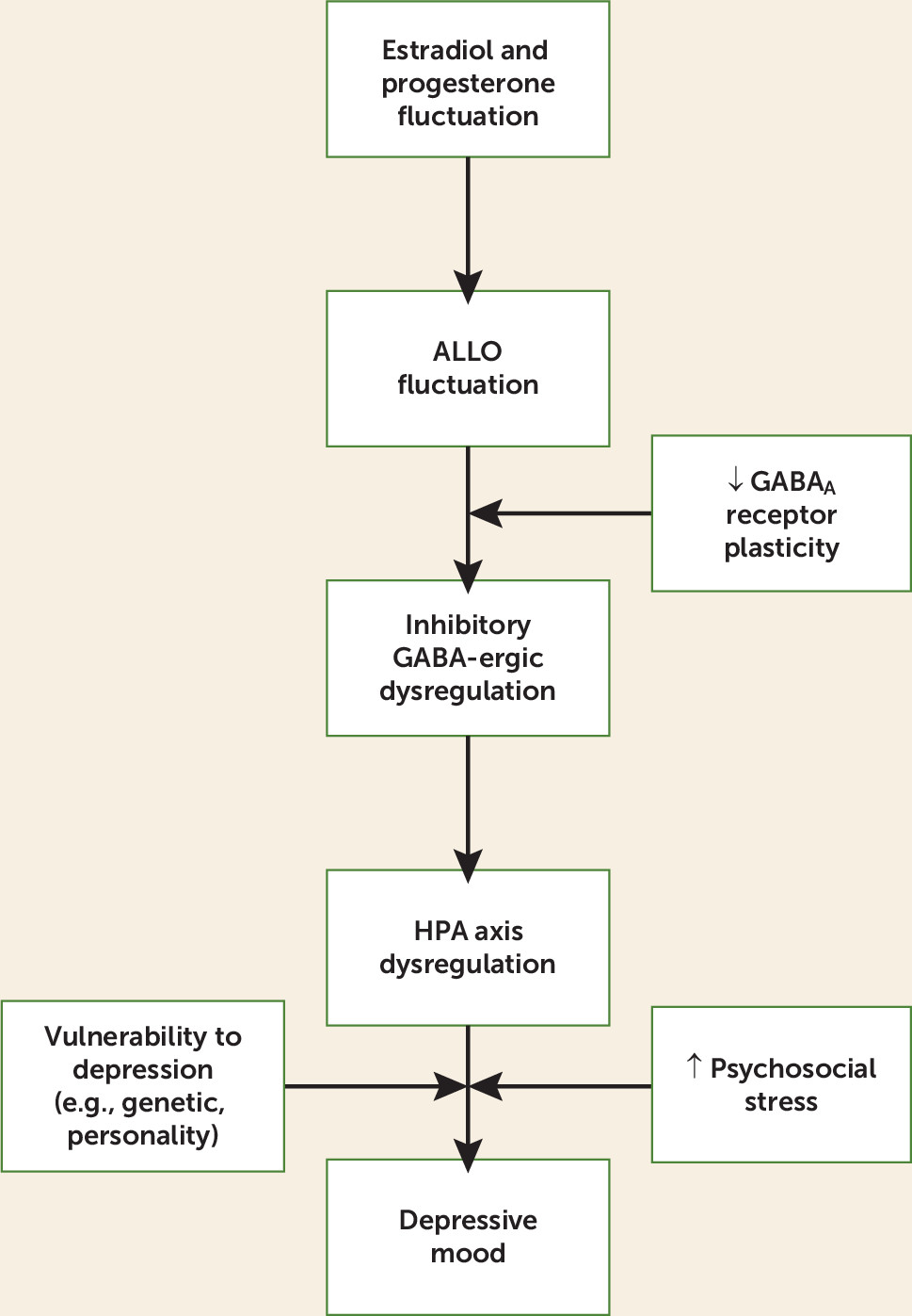
In this review, we discuss the available evidence to support another plausible mechanism: reproductive steroid modulation of GABA-ergic regulation of the HPA axis. More specifically, we present evidence for the role of progesterone-derived neurosteroids, including allopregnanolone (3α-hydroxy-5α-pregnan-20-one; ALLO), in altering GABA-ergic modulation of the HPA axis and discuss how these alterations may sensitize perimenopausal women to stress and, consequently, to the development of perimenopausal depression. When this increased stress sensitivity is combined with stressful life events and/or a genetic or dispositional vulnerability to depression, clinical elevations in depressed mood may ensue. This mechanistic candidate is illustrated in Figure 3 and will be expanded on in the following sections.
FIGURE 3. Proposed Etiologic Model of Depression With Onset in the Menopause Transitiona
a ALLO, allopregnanolone; ↑, elevated; ↓ decreased.

GABA-Ergic Neurosteroids in the Menopause Transition

While most studies have focused on the role of estradiol in the risk of perimenopausal depression, evidence from animal models suggests that ovarian hormone fluctuation increases the risk of perimenopausal depression, at least in part, because of the concurrent changes in progesterone-derived neurosteroids. Among the most studied progesterone-derived neurosteroids in humans is ALLO, an A-ring-reduced metabolite of progesterone. ALLO is stress responsive in animals and humans (for a review, see reference 47) and serves as a potent, positive allosteric modulator of GABAA receptors through dose-dependent enhancement of GABA-induced chloride-ion channels (48). GABA is the chief inhibitory neurotransmitter in the mammalian central nervous system. The role of GABA in regulating the HPA axis in response to stress by limiting the extent and duration of the HPA axis stress response is well established (49). In part, it is through ALLO’s modulation of the GABAA receptor to increase GABA-ergic transmission that ALLO not only negatively modulates the HPA axis to return it to homeostasis following stress (50) but also exerts profound anxiolytic (51) and antidepressant (52) actions. However, ALLO’s anxiolytic properties may also be partially mediated through its effects on the bed nucleus of the stria terminalis, the “relay center” linking stress-responsive pathways such as the HPA axis and limbic structures such as the amygdala (53). Two major sources of ALLO in women of reproductive age are the adrenal glands and the corpus luteum, where ALLO is converted from progesterone (54). Because of ovarian ALLO contributions, ALLO concentrations in premenopausal women are highest in the luteal phase and lowest in the follicular phase (54). However, in postmenopausal women, the adrenal glands become the exclusive source of peripheral ALLO (54). It is important to note that peripherally derived ALLO freely crosses the blood-brain barrier (55) and contributes significantly to CNS concentrations (56).
As mentioned earlier, an increasing proportion of anovulatory cycles results in less frequent luteal phases and therefore overall lower levels of progesterone. Although the availability of progesterone is an important determinant of ALLO, estradiol is also likely to positively influence ALLO production through its modulation of the enzymes involved in the conversion of progesterone to ALLO, 5α-reductase and 3α-hydroxysteroid dehydrogenase (57). This is supported by both basic and clinical research. For example, while ovariectomy in animals has been shown to decrease ALLO concentrations in the hippocampus, hypothalamus, pituitary, and plasma, transdermal estradiol administration restores preovariectomy brain and plasma ALLO concentrations (see reference 57 for a review). Similarly, transdermal estradiol increases plasma ALLO in postmenopausal women (58, 59). Thus, even in the absence of ovulation and the consequent production of progesterone, intermittent endogenous production of estradiol during the menopause transition may cause fluctuations in the synthesis and release of ALLO.
ALLO fluctuation may have important implications at the GABAA receptor, which is composed of a combination of five (out of 19 existing) subunits. Which subunits a receptor contains will greatly influence its sensitivity to neurosteroids (see reference 60 for a review). For example, the δ subunit has been shown to greatly increase receptor sensitivity to very low concentrations of neurosteroids; mice lacking the δ subunit therefore exhibit greatly reduced neurosteroid sensitivity (61). In contrast, GABAA receptors containing the ε subunit are relatively insensitive to neurosteroids like ALLO. Furthermore, the subunit composition of GABAA receptors is extremely plastic and influenced by neurosteroid levels; for example, during pregnancy, when levels of progesterone, and thus progesterone-derived neurosteroids, are extremely elevated, the expression of δ subunits is down-regulated in multiple areas of the brain, thus reducing receptor sensitivity to elevated ALLO levels (62).
This ability of the GABAA receptor to change its composition in response to ALLO concentrations is likely to be especially important in times of considerable ALLO flux. Failure of the GABAA receptor to match its composition to an ever-changing hormonal environment could result in either too high or too low GABA-ergic inhibitory tone. In addition, ovarian hormone fluctuation may actually trigger maladaptive changes in GABAA receptor configuration. Evidence supporting this comes from animal models of puberty, in which ovarian hormone fluctuations have been shown to promote the expression of GABAA receptors containing α4, β2, and δ subunits in rodents, the combination of which has been found to transform ALLO’s effects from excitatory to inhibitory at the GABAA receptor (63). Thus, rather than positively modulating the GABAA receptor, ALLO inhibits it. In turn, there is an overall decline in GABA-ergic inhibitory tone. Furthermore, this reduction in GABA-ergic tone during puberty is also accompanied by an increase in anxiety, as indicated by less time spent in the open arms of the elevated plus maze and more anxiety behavior following a restraint stress (63). Hormone fluctuation across the estrous cycle (64) or progesterone (and therefore ALLO) withdrawal induced in the laboratory (65) have also been shown to result in similar changes in GABAA receptor subunit expression.
Failure of the GABAA receptor to demonstrate adaptive homeostatic plasticity in the context of steroid hormone fluctuations is theorized to be involved in the development of PMDD and postpartum depression (see references 60, 62, and 66 for reviews). This may be one mechanism contributing to decreased saccadic eye velocity, an indirect measure of GABAA receptor sensitivity, in women with PMDD during their symptomatic phase (67). In light of this, we propose that insufficient plasticity of the GABAA receptor in the menopause transition, or maladaptive changes to the GABAA receptor, may contribute to mood disturbance during the menopause transition, when concentrations of both estradiol and progesterone become erratic and unpredictable. How GABAA receptors “respond” to fluctuating ALLO concentrations in the menopause transition would be critical in determining overall GABA-ergic tone, mood, and, theoretically, regulation of the HPA axis. This process, in which hormonal fluctuation can trigger GABAA subunit changes such that ALLO’s effects become paradoxical (inhibitory rather than excitatory at the GABAA receptor; anxiogenic rather than anxiolytic), may shed light on the results of a study by Andréen et al. (68). This study of 36 women in late perimenopause or early postmenopause who were treated with progesterone supplementation showed that women with resultant medium ALLO concentrations reported significantly more negative mood when compared with women with low ALLO levels. The possibility exists that the context of the perimenopausal fluctuating hormonal environment to which these women were exposed had contributed to GABAA receptor subunit changes with consequent alterations in ALLO’s effects at the receptor, and in turn, its effects on mood (69).
To the extent that GABA-ergic dysregulation is involved in perimenopausal depression, genes coding for GABAA receptor subunits may be implicated in predisposing some individuals to respond maladaptively to ALLO fluctuations and thus be at increased risk for perimenopausal depression. GABAA receptor subunit gene polymorphisms are differentially associated with risk for other mental disorders, such as alcohol dependence (70), major depressive disorder (71), bipolar disorder (71), and schizophrenia (71). In animal models, mice genetically designed to lack the δ subunit do not exhibit ALLO’s paradoxical effects at the GABAA receptor during puberty that are seen in wild-type mice (63). An investigation of which GABAA receptor subunit gene polymorphisms are associated with an increased risk for perimenopausal depression may be warranted. Large-scale epidemiologic studies such as SWAN may provide appropriate specimens for such genotyping.
Although ALLO’s effects on the GABA-ergic system are the best characterized and are thus the primary focus of this review, alterations in overall GABA-ergic tone are likely to impact the release of other neurotransmitters relevant to the development of psychopathology. For example, recent in vitro studies of rodent hippocampal neurons suggest that ALLO, via presynaptic GABAA receptors, modulates glutamate release (72). ALLO’s effects on glutamate may be particularly relevant to the study of perimenopausal depression in light of the recent recognition that glutamate transmission is likely a key player in the etiology of major depressive disorder and other psychopathologies (73).

HPA Axis Dysregulation in the Menopause Transition

GABA plays a critical role in regulating the HPA axis and limiting HPA activation following exposure to stress (49). As such, any failure of the GABAA receptor to adapt appropriately to the hormonal environment can be expected to have direct consequences for HPA axis activity. For example, if ALLO were to negatively modulate the GABAA receptor in response to hormonal fluctuation (as opposed to serving as a positive allosteric modulator of the GABAA receptor), as is seen in animal models of puberty, this would contribute to an overall increase in HPA axis activity since the GABA-ergic inhibition of the HPA axis is removed. Under these conditions, because the ALLO concentration increases following stress, this could potentiate HPA axis reactivity and prolong recovery in response to stress. In this way, we speculate that menopause-transition-related ovarian hormone fluctuation could trigger HPA axis dysregulation.
Dysregulation of the HPA axis in major depression has frequently been described as one of the most consistent findings in psychiatry (74). However, only in the last decade have we begun to view altered HPA axis activity as a risk factor increasing one’s susceptibility to depression rather than a consequence or epiphenomenon of depression (75). This view is supported not only by prospective studies identifying an elevated cortisol level as a precursor to the onset of first-episode major depression (76), as well as relapse (77), but also by studies observing increased HPA axis activation among the euthymic relatives of individuals with a history of depression, compared with control subjects with no family history of depression (78). Research on postpartum depression (79, 80) and PMDD (see reference 81 for a review) also suggest that HPA axis dysregulation has pathophysiological relevance to reproductive mood disorders.
To date, little is known about HPA axis activity in the menopause transition. Komesaroff et al. (82) examined cortisol reactivity to the Trier Social Stress Test, a speech and arithmetic stressor battery, following 8 weeks of oral estradiol or placebo in women during the menopause transition. It was found that estradiol therapy resulted in an attenuated cortisol response to the stressor. Two additional studies have examined basal cortisol levels in the menopause transition. In the Seattle Midlife Women’s Health Study, 91 women provided monthly first-morning urine specimens for cortisol measurement as they transitioned across menopausal stages (early to middle, middle to late, and late menopause transition to postmenopause) (83). It was found that 68% of the 22 women transitioning from the middle to late menopause transition exhibited an increase in cortisol, the magnitude of which has previously been associated with a decrement in memory performance in older women (84) and may therefore have clinical significance. While the increase in cortisol during the late menopause transition was not associated with depressive symptoms in this community sample, this study was limited in that it examined basal cortisol and not cortisol reactivity to stress. A study by Schmidt et al. (85) found no difference in the basal cortisol levels of 24 women with perimenopausal depression when compared with 26 asymptomatic control subjects. However, again, this study did not examine cortisol reactivity; it also did not account for STRAW stage. Together, these studies suggest that perimenopausal depression may not be associated with alterations in basal HPA axis hormone concentrations but that ovarian hormones do regulate HPA axis responses to stress in women during the menopause transition. To our knowledge, there have been no studies examining HPA axis activation in response to stress in women with perimenopausal depression.
Conclusions and Future Directions

On the basis of emerging evidence from both animal and clinical research, we propose a heuristic model of perimenopausal depression whereby failure of the GABAA receptor to adapt to fluctuations in ALLO over the course of the menopause transition increases the risk of perimenopausal depression in vulnerable women. Specifically (see Figure 3), in the context of the menopause transition, characterized by fluctuations in ALLO that are consequent to estradiol and progesterone fluctuations, an inability of the GABAA receptor to demonstrate the plasticity necessary to maintain GABA-ergic homeostatic control might exacerbate the response of the HPA axis to stress. Combined with an increased vulnerability to major depressive disorder due to personality or genetic factors (e.g., in women with a history of major depression) and/or stressful life events proximate to the menopause transition, the endocrine profile of the menopause transition sets the stage for depressive symptoms. While speculative, this model is consistent with studies linking both HPA axis dysregulation (81) and altered GABAA receptor sensitivity (67) to other reproductive mood disorders. Future research investigating this model has the potential to inform the development of novel pharmacological treatments for perimenopausal depression.
While novel, our model remains speculative as there is virtually no research examining these pathways in the menopause transition. However, the risk factors predictive of perimenopausal depression, including sensitivity to hormonal fluctuations and greater psychosocial stress and/or increased sensitivity to stress, are consistent with this model. Furthermore, evidence from other reproductive mood disorders indirectly suggests that neurosteroid and HPA axis dysregulation may be involved in the etiology of perimenopausal depression. However, we wish to acknowledge that our model is by no means comprehensive. There are likely multiple complex downstream effects of GABA-ergic and HPA axis dysregulation as well as entirely separate mechanisms involving serotonin, dopamine, and norepinephrine that contribute to the etiology of perimenopausal depression and warrant further investigation.
Our intent is that this model will foster research in perimenopausal depression and its etiological mechanisms. Based on our review of the existing literature, we offer several recommendations for future research, First, studies seeking to clarify the mechanisms involved in perimenopausal depression should confirm that the onset of depressive symptoms coincides with the menopause transition, since differing etiological mechanisms may be involved in perimenopausal depression and major depressive disorder with onset at other life stages. Second, research aimed at detecting an effect of ovarian hormones on perimenopausal depression and/or its underlying mechanisms should measure hormone concentrations frequently, as once-yearly assessments of hormones may be insufficient to capture the erratic changes in ovarian hormones occurring in the menopause transition. Third, examination of ovarian hormone and ALLO variability in relation to mood disturbance before and after the treatment of perimenopausal depression with estradiol therapy will help to advance the proposed etiologic model of perimenopausal depression. To the extent that the etiologic model proposed here is predictive of perimenopausal depression, estradiol and ALLO stabilization with estradiol therapy would be expected to predict clinical outcomes. Couched within a placebo-controlled randomized trial comparing estradiol therapy and an antidepressant medication, such research would further inform clinical decision making in the treatment of perimenopausal depression.

References

1.
Vegeto E, Benedusi V, Maggi A: Estrogen anti-inflammatory activity in brain: a therapeutic opportunity for menopause and neurodegenerative diseases. Front Neuroendocrinol 2008; 29:507–519
2.
Bredemann TM, McMahon LL: 17β Estradiol increases resilience and improves hippocampal synaptic function in helpless ovariectomized rats. Psychoneuroendocrinology 2014; 42:77–88
3.
Epperson CN, Amin Z, Ruparel K, et al: Interactive effects of estrogen and serotonin on brain activation during working memory and affective processing in menopausal women. Psychoneuroendocrinology 2012; 37:372–382
4.
Barha CK, Dalton GL, Galea LA: Low doses of 17alpha-estradiol and 17beta-estradiol facilitate, whereas higher doses of estrone and 17alpha- and 17beta-estradiol impair, contextual fear conditioning in adult female rats. Neuropsychopharmacology 2010; 35:547–559
5.
Milad MR, Zeidan MA, Contero A, et al: The influence of gonadal hormones on conditioned fear extinction in healthy humans. Neuroscience 2010; 168:652–658
6.
Harlow SD, Gass M, Hall JE, et al: Executive summary of the Stages of Reproductive Aging Workshop +10: addressing the unfinished agenda of staging reproductive aging. Climacteric 2012; 15:105–114
7.
Santoro N, Lasley B, McConnell D, et al: Body size and ethnicity are associated with menstrual cycle alterations in women in the early menopausal transition: the Study of Women’s Health across the Nation (SWAN) Daily Hormone Study. J Clin Endocrinol Metab 2004; 89:2622–2631
8.
Tepper PG, Randolph JF Jr, McConnell DS, et al: Trajectory clustering of estradiol and follicle-stimulating hormone during the menopausal transition among women in the Study of Women’s Health across the Nation (SWAN). J Clin Endocrinol Metab 2012; 97:2872–2880
9.
Hale GE, Robertson DM, Burger HG: The perimenopausal woman: endocrinology and management. J Steroid Biochem Mol Biol 2014; 142:121–131
10.
Santoro N, Randolph JF Jr: Reproductive hormones and the menopause transition. Obstet Gynecol Clin North Am 2011; 38:455–466
11.
O’Connor KA, Ferrell R, Brindle E, et al: Progesterone and ovulation across stages of the transition to menopause. Menopause 2009; 16:1178–1187
12.
McKinlay SM, Brambilla DJ, Posner JG: The normal menopause transition. Maturitas 1992; 14:103–115
13.
Weissman MM: The myth of involutional melancholia. JAMA 1979; 242:742–744
14.
Kaufert PA, Gilbert P, Tate R: The Manitoba Project: a re-examination of the link between menopause and depression. Maturitas 1992; 14:143–155
15.
McKinlay JB, McKinlay SM, Brambilla D: The relative contributions of endocrine changes and social circumstances to depression in mid-aged women. J Health Soc Behav 1987; 28:345–363
16.
Bromberger JT, Matthews KA, Schott LL, et al: Depressive symptoms during the menopausal transition: the Study of Women’s Health Across the Nation (SWAN). J Affect Disord 2007; 103:267–272
17.
Freeman EW, Sammel MD, Liu L, et al: Hormones and menopausal status as predictors of depression in women in transition to menopause. Arch Gen Psychiatry 2004; 61:62–70
18.
Woods NF, Smith-DiJulio K, Percival DB, et al: Depressed mood during the menopausal transition and early postmenopause: observations from the Seattle Midlife Women’s Health Study. Menopause 2008; 15:223–232
19.
Cohen LS, Soares CN, Vitonis AF, et al: Risk for new onset of depression during the menopausal transition: the Harvard study of moods and cycles. Arch Gen Psychiatry 2006; 63:385–390
20.
Freeman EW, Sammel MD, Lin H, et al: Associations of hormones and menopausal status with depressed mood in women with no history of depression. Arch Gen Psychiatry 2006; 63:375–382
21.
Schmidt PJ, Haq N, Rubinow DR: A longitudinal evaluation of the relationship between reproductive status and mood in perimenopausal women. Am J Psychiatry 2004; 161:2238–2244
22.
Bromberger JT, Kravitz HM, Chang Y-F, et al: Major depression during and after the menopausal transition: Study of Women’s Health Across the Nation (SWAN). Psychol Med 2011; 41:1879–1888
23.
Bromberger JT, Kravitz HM, Matthews K, et al: Predictors of first lifetime episodes of major depression in midlife women. Psychol Med 2009; 39:55–64
24.
Avis NE, Brambilla D, McKinlay SM, et al: A longitudinal analysis of the association between menopause and depression: results from the Massachusetts Women’s Health Study. Ann Epidemiol 1994; 4:214–220
25.
Payne JL, Palmer JT, Joffe H: A reproductive subtype of depression: conceptualizing models and moving toward etiology. Harv Rev Psychiatry 2009; 17:72–86
26.
Bloch M, Schmidt PJ, Danaceau M, et al: Effects of gonadal steroids in women with a history of postpartum depression. Am J Psychiatry 2000; 157:924–930
27.
Schmidt PJ, Nieman LK, Danaceau MA, et al: Differential behavioral effects of gonadal steroids in women with and in those without premenstrual syndrome. N Engl J Med 1998; 338:209–216
28.
Freeman EW, Sammel MD, Lin H, et al: Symptoms associated with menopausal transition and reproductive hormones in midlife women. Obstet Gynecol 2007; 110:230–240
29.
Freeman EW, Sammel MD, Boorman DW, et al: Longitudinal pattern of depressive symptoms around natural menopause. JAMA Psychiatry 2014; 71:36–43
30.
Soares CN, Almeida OP, Joffe H, et al: Efficacy of estradiol for the treatment of depressive disorders in perimenopausal women: a double-blind, randomized, placebo-controlled trial. Arch Gen Psychiatry 2001; 58:529–534
31.
Schmidt PJ, Nieman L, Danaceau MA, et al: Estrogen replacement in perimenopause-related depression: a preliminary report. Am J Obstet Gynecol 2000; 183:414–420
32.
Joffe H, Petrillo LF, Koukopoulos A, et al: Increased estradiol and improved sleep, but not hot flashes, predict enhanced mood during the menopausal transition. J Clin Endocrinol Metab 2011; 96:E1044–E1054
33.
Cohen LS, Soares CN, Poitras JR, et al: Short-term use of estradiol for depression in perimenopausal and postmenopausal women: a preliminary report. Am J Psychiatry 2003; 160:1519–1522
34.
Morrison MF, Kallan MJ, Ten Have T, et al: Lack of efficacy of estradiol for depression in postmenopausal women: a randomized, controlled trial. Biol Psychiatry 2004; 55:406–412
35.
Biegon A, Fischette CT, Rainbow TC, et al: Serotonin receptor modulation by estrogen in discrete brain nuclei. Neuroendocrinology 1982; 35:287–291
36.
Biegon A, Reches A, Snyder L, et al: Serotonergic and noradrenergic receptors in the rat brain: modulation by chronic exposure to ovarian hormones. Life Sci 1983; 32:2015–2021
37.
Deecher D, Andree TH, Sloan D, et al: From menarche to menopause: exploring the underlying biology of depression in women experiencing hormonal changes. Psychoneuroendocrinology 2008; 33:3–17
38.
De Leo V, Lanzetta D, Morgante G, et al: Inhibition of ovulation with transdermal estradiol and oral progestogens in perimenopausal women. Contraception 1997; 55:239–243
39.
De Leo V, Lanzetta D, D’Antona D, et al: Transdermal estrogen replacement therapy in normal perimenopausal women: effects on pituitary-ovarian function. Gynecol Endocrinol 1996; 10:49–53
40.
Girdler SS, Hinderliter AL, Wells EC, et al: Transdermal versus oral estrogen therapy in postmenopausal smokers: hemodynamic and endothelial effects. Obstet Gynecol 2004; 103:169–180
41.
Freeman EW: Associations of depression with the transition to menopause. Menopause 2010; 17:823–827
42.
Schmidt PJ, Rubinow DR: Sex hormones and mood in the perimenopause. Ann N Y Acad Sci 2009; 1179:70–85
43.
Avis NE, Crawford S, Stellato R, et al: Longitudinal study of hormone levels and depression among women transitioning through menopause. Climacteric 2001; 4:243–249
44.
Daly RC, Danaceau MA, Rubinow DR, et al: Concordant restoration of ovarian function and mood in perimenopausal depression. Am J Psychiatry 2003; 160:1842–1846
45.
Rubinow DR, Schmidt PJ, Roca CA: Estrogen-serotonin interactions: implications for affective regulation. Biol Psychiatry 1998; 44:839–850
46.
Joffe H, Cohen LS: Estrogen, serotonin, and mood disturbance: where is the therapeutic bridge? Biol Psychiatry 1998; 44:798–811
47.
Crowley SK, Girdler SS: Neurosteroid, GABAergic and hypothalamic pituitary adrenal (HPA) axis regulation: what is the current state of knowledge in humans? Psychopharmacology (Berl) 2014; 231:3619–3634
48.
Morrow AL, Suzdak PD, Paul SM: Steroid hormone metabolites potentiate GABA receptor-mediated chloride ion flux with nanomolar potency. Eur J Pharmacol 1987; 142:483–485
49.
Cullinan WE, Ziegler DR, Herman JP: Functional role of local GABAergic influences on the HPA axis. Brain Struct Funct 2008; 213:63–72
50.
Guo AL, Petraglia F, Criscuolo M, et al: Evidence for a role of neurosteroids in modulation of diurnal changes and acute stress-induced corticosterone secretion in rats. Gynecol Endocrinol 1995; 9:1–7
51.
Bitran D, Shiekh M, McLeod M: Anxiolytic effect of progesterone is mediated by the neurosteroid allopregnanolone at brain GABAA receptors. J Neuroendocrinol 1995; 7:171–177
52.
Rodrìguez-Landa JF, Contreras CM, Bernal-Morales B, et al: Allopregnanolone reduces immobility in the forced swimming test and increases the firing rate of lateral septal neurons through actions on the GABAA receptor in the rat. J Psychopharmacol 2007; 21:76–84
53.
Toufexis DJ, Davis C, Hammond A, et al: Progesterone attenuates corticotropin-releasing factor-enhanced but not fear-potentiated startle via the activity of its neuroactive metabolite, allopregnanolone. J Neurosci 2004; 24:10280–10287
54.
Genazzani AR, Petraglia F, Bernardi F, et al: Circulating levels of allopregnanolone in humans: gender, age, and endocrine influences. J Clin Endocrinol Metab 1998; 83:2099–2103
55.
Paul SM, Purdy RH: Neuroactive steroids. FASEB J 1992; 6:2311–2322
56.
Purdy RH, Morrow AL, Moore PH Jr, et al: Stress-induced elevations of gamma-aminobutyric acid type A receptor-active steroids in the rat brain. Proc Natl Acad Sci USA 1991; 88:4553–4557
57.
Pluchino N, Cubeddu A, Giannini A, et al: Progestogens and brain: an update. Maturitas 2009; 62:349–355
58.
Pluchino N, Genazzani AD, Bernardi F, et al: Tibolone, transdermal estradiol or oral estrogen-progestin therapies: effects on circulating allopregnanolone, cortisol and dehydroepiandrosterone levels. Gynecol Endocrinol 2005; 20:144–149
59.
Bernardi F, Pieri M, Stomati M, et al: Effect of different hormonal replacement therapies on circulating allopregnanolone and dehydroepiandrosterone levels in postmenopausal women. Gynecol Endocrinol 2003; 17:65–77
60.
MacKenzie G, Maguire J: The role of ovarian hormone-derived neurosteroids on the regulation of GABAA receptors in affective disorders. Psychopharmacology (Berl) 2014; 231:3333–3342
61.
Mihalek RM, Banerjee PK, Korpi ER, et al: Attenuated sensitivity to neuroactive steroids in γ-aminobutyrate type A receptor delta subunit knockout mice. Proc Natl Acad Sci USA 1999; 96:12905–12910
62.
Maguire J, Mody I: GABA(A)R plasticity during pregnancy: relevance to postpartum depression. Neuron 2008; 59:207–213
63.
Shen H, Gong QH, Aoki C, et al: Reversal of neurosteroid effects at α4β2δ GABAA receptors triggers anxiety at puberty. Nat Neurosci 2007; 10:469–477
64.
Lovick TA, Griffiths JL, Dunn SM, et al: Changes in GABA(A) receptor subunit expression in the midbrain during the oestrous cycle in Wistar rats. Neuroscience 2005; 131:397–405
65.
Griffiths J, Lovick T: Withdrawal from progesterone increases expression of α4, β1, and δ GABA(A) receptor subunits in neurons in the periaqueductal gray matter in female Wistar rats. J Comp Neurol 2005; 486:89–97
66.
Maguire J, Mody I: Steroid hormone fluctuations and GABA(A)R plasticity. Psychoneuroendocrinology 2009; 34(suppl 1):S84–S90
67.
Sundström I, Bäckström T: Patients with premenstrual syndrome have decreased saccadic eye velocity compared to control subjects. Biol Psychiatry 1998; 44:755–764
68.
Andréen L, Sundström-Poromaa I, Bixo M, et al: Relationship between allopregnanolone and negative mood in postmenopausal women taking sequential hormone replacement therapy with vaginal progesterone. Psychoneuroendocrinology 2005; 30:212–224
69.
Andréen L, Nyberg S, Turkmen S, et al: Sex steroid induced negative mood may be explained by the paradoxical effect mediated by GABAA modulators. Psychoneuroendocrinology 2009; 34:1121–1132
70.
Soyka M, Preuss UW, Hesselbrock V, et al: GABA-A2 receptor subunit gene (GABRA2) polymorphisms and risk for alcohol dependence. J Psychiatr Res 2008; 42:184–191
71.
Fatemi SH, Folsom TD, Rooney RJ, et al: Expression of GABAA α2-, β1- and ε-receptors are altered significantly in the lateral cerebellum of subjects with schizophrenia, major depression and bipolar disorder. Transl Psychiatr 2013; 3:e303
72.
Iwata S, Wakita M, Shin M-C, et al: Modulation of allopregnanolone on excitatory transmitters release from single glutamatergic terminal. Brain Res Bull 2013; 93:39–46
73.
Sanacora G, Treccani G, Popoli M: Towards a glutamate hypothesis of depression: an emerging frontier of neuropsychopharmacology for mood disorders. Neuropharmacology 2012; 62:63–77
74.
Parker KJ, Schatzberg AF, Lyons DM: Neuroendocrine aspects of hypercortisolism in major depression. Horm Behav 2003; 43:60–66
75.
Pariante CM, Lightman SL: The HPA axis in major depression: classical theories and new developments. Trends Neurosci 2008; 31:464–468
76.
Goodyer IM, Herbert J, Tamplin A, et al: Recent life events, cortisol, dehydroepiandrosterone and the onset of major depression in high-risk adolescents. Br J Psychiatry 2000; 177:499–504
77.
Appelhof BC, Huyser J, Verweij M, et al: Glucocorticoids and relapse of major depression (dexamethasone/corticotropin-releasing hormone test in relation to relapse of major depression). Biol Psychiatry 2006; 59:696–701
78.
Mannie ZN, Harmer CJ, Cowen PJ: Increased waking salivary cortisol levels in young people at familial risk of depression. Am J Psychiatry 2007; 164:617–621
79.
Magiakou MA, Mastorakos G, Rabin D, et al: Hypothalamic corticotropin-releasing hormone suppression during the postpartum period: implications for the increase in psychiatric manifestations at this time. J Clin Endocrinol Metab 1996; 81:1912–1917
80.
Bloch M, Rubinow DR, Schmidt PJ, et al: Cortisol response to ovine corticotropin-releasing hormone in a model of pregnancy and parturition in euthymic women with and without a history of postpartum depression. J Clin Endocrinol Metab 2005; 90:695–699
81.
Girdler SS, Klatzkin R: Neurosteroids in the context of stress: implications for depressive disorders. Pharmacol Ther 2007; 116:125–139
82.
Komesaroff PA, Esler MD, Sudhir K: Estrogen supplementation attenuates glucocorticoid and catecholamine responses to mental stress in perimenopausal women. J Clin Endocrinol Metab 1999; 84:606–610
83.
Woods NF, Carr MC, Tao EY, et al: Increased urinary cortisol levels during the menopausal transition. Menopause 2006; 13:212–221
84.
Seeman TE, McEwen BS, Singer BH, et al: Increase in urinary cortisol excretion and memory declines: MacArthur studies of successful aging. J Clin Endocrinol Metab 1997; 82:2458–2465
85.
Schmidt PJ, Murphy JH, Haq N, et al: Basal plasma hormone levels in depressed perimenopausal women. Psychoneuroendocrinology 2002; 27:907–920

Information & Authors

Information

Published In

Go to American Journal of Psychiatry
American Journal of Psychiatry
Pages: 227 - 236
PubMed: 25585035

History

Received: 24 July 2014
Revision received: 1 October 2014
Accepted: 14 October 2014
Published online: 13 January 2015
Published in print: March 01, 2015

Authors

Details

Jennifer L. Gordon, Ph.D.
From the Department of Psychiatry, University of North Carolina at Chapel Hill; the Department of Obstetrics/Gynecology and the Department of Psychiatry and Behavioral Sciences, Northwestern University, Chicago; the Department of Psychiatry, University of Pittsburgh School of Medicine; the Department of Obstetrics and Gynecology, Allegheny Health Network, Pittsburgh; and the Department of Psychiatry, Massachusetts General Hospital, Harvard Medical School, Boston.
Susan S. Girdler, Ph.D.
From the Department of Psychiatry, University of North Carolina at Chapel Hill; the Department of Obstetrics/Gynecology and the Department of Psychiatry and Behavioral Sciences, Northwestern University, Chicago; the Department of Psychiatry, University of Pittsburgh School of Medicine; the Department of Obstetrics and Gynecology, Allegheny Health Network, Pittsburgh; and the Department of Psychiatry, Massachusetts General Hospital, Harvard Medical School, Boston.
Samantha E. Meltzer-Brody, M.D.
From the Department of Psychiatry, University of North Carolina at Chapel Hill; the Department of Obstetrics/Gynecology and the Department of Psychiatry and Behavioral Sciences, Northwestern University, Chicago; the Department of Psychiatry, University of Pittsburgh School of Medicine; the Department of Obstetrics and Gynecology, Allegheny Health Network, Pittsburgh; and the Department of Psychiatry, Massachusetts General Hospital, Harvard Medical School, Boston.
Catherine S. Stika, M.D.
From the Department of Psychiatry, University of North Carolina at Chapel Hill; the Department of Obstetrics/Gynecology and the Department of Psychiatry and Behavioral Sciences, Northwestern University, Chicago; the Department of Psychiatry, University of Pittsburgh School of Medicine; the Department of Obstetrics and Gynecology, Allegheny Health Network, Pittsburgh; and the Department of Psychiatry, Massachusetts General Hospital, Harvard Medical School, Boston.
Rebecca C. Thurston, Ph.D.
From the Department of Psychiatry, University of North Carolina at Chapel Hill; the Department of Obstetrics/Gynecology and the Department of Psychiatry and Behavioral Sciences, Northwestern University, Chicago; the Department of Psychiatry, University of Pittsburgh School of Medicine; the Department of Obstetrics and Gynecology, Allegheny Health Network, Pittsburgh; and the Department of Psychiatry, Massachusetts General Hospital, Harvard Medical School, Boston.
Crystal T. Clark, M.D., M.Sc.
From the Department of Psychiatry, University of North Carolina at Chapel Hill; the Department of Obstetrics/Gynecology and the Department of Psychiatry and Behavioral Sciences, Northwestern University, Chicago; the Department of Psychiatry, University of Pittsburgh School of Medicine; the Department of Obstetrics and Gynecology, Allegheny Health Network, Pittsburgh; and the Department of Psychiatry, Massachusetts General Hospital, Harvard Medical School, Boston.
Beth A. Prairie, M.D.
From the Department of Psychiatry, University of North Carolina at Chapel Hill; the Department of Obstetrics/Gynecology and the Department of Psychiatry and Behavioral Sciences, Northwestern University, Chicago; the Department of Psychiatry, University of Pittsburgh School of Medicine; the Department of Obstetrics and Gynecology, Allegheny Health Network, Pittsburgh; and the Department of Psychiatry, Massachusetts General Hospital, Harvard Medical School, Boston.
Eydie Moses-Kolko, Ph.D.
From the Department of Psychiatry, University of North Carolina at Chapel Hill; the Department of Obstetrics/Gynecology and the Department of Psychiatry and Behavioral Sciences, Northwestern University, Chicago; the Department of Psychiatry, University of Pittsburgh School of Medicine; the Department of Obstetrics and Gynecology, Allegheny Health Network, Pittsburgh; and the Department of Psychiatry, Massachusetts General Hospital, Harvard Medical School, Boston.
Hadine Joffe, M.D., M.Sc.
From the Department of Psychiatry, University of North Carolina at Chapel Hill; the Department of Obstetrics/Gynecology and the Department of Psychiatry and Behavioral Sciences, Northwestern University, Chicago; the Department of Psychiatry, University of Pittsburgh School of Medicine; the Department of Obstetrics and Gynecology, Allegheny Health Network, Pittsburgh; and the Department of Psychiatry, Massachusetts General Hospital, Harvard Medical School, Boston.
Katherine L. Wisner, M.D., M.S.
From the Department of Psychiatry, University of North Carolina at Chapel Hill; the Department of Obstetrics/Gynecology and the Department of Psychiatry and Behavioral Sciences, Northwestern University, Chicago; the Department of Psychiatry, University of Pittsburgh School of Medicine; the Department of Obstetrics and Gynecology, Allegheny Health Network, Pittsburgh; and the Department of Psychiatry, Massachusetts General Hospital, Harvard Medical School, Boston.

Notes

Address correspondence to Dr. Girdler ([email protected]).

Author Contributions

Dr. Joffe and Dr. Wisner share last authorship.

Funding Information

Eunice Kennedy Shriver National Institute of Child Health & Human Development: K12 HD055884
National Institute of Mental Health10.13039/100000025: R01-MH087619
Fonds de Recherche du Québec - Santé10.13039/501100000156:
Supported by NIMH grant RO1 MH-087619. Dr. Gordon is the recipient of a postdoctoral fellowship of the Fonds de la Recherche du Québec–Santé (FRQS). Dr. Clark is supported in part by grant K12 HD-055884 from the Eunice Kennedy Shriver National Institute of Child Health and Human Development.Dr. Gordon reports receiving a Pilot Project Grant from the North Carolina Translational and Clinical Sciences Institute; previously she was the recipient of a Canadian Institutes of Health Research (CIHR) Vanier Canada Graduate Scholarship. Dr. Meltzer-Brody has received grant funding from AstraZeneca, the Foundation of Hope, and Sage Pharmaceuticals. Dr. Moses-Kolko has received support from grant HD-067185 from the National Institute of Child Health and Human Development. Dr. Joffe receives research support from Cephalon/Teva, serves as consultant to Noven and Merck, and participates in an advisory board for Merck. Dr. Wisner serves as consultant to Quinn Emanuel Urquhart & Sullivan, L.L.P., which legally represents Pfizer Pharmaceutical Company; the Department of Psychiatry at Northwestern University receives contractual fees for Dr. Wisner's consultation to Quinn Emanuel Urquhart & Sullivan. The other authors report no financial relationships with commercial interests.

Metrics & Citations

Metrics

Citations

Export Citations

If you have the appropriate software installed, you can download article citation data to the citation manager of your choice. Simply select your manager software from the list below and click Download.

For more information or tips please see 'Downloading to a citation manager' in the Help menu.

Format
Citation style
Style
Copy to clipboard

View Options

View options

PDF/EPUB

View PDF/EPUB

Get Access

Login options

Already a subscriber? Access your subscription through your login credentials or your institution for full access to this article.

Personal login Institutional Login Open Athens login
Purchase Options

Purchase this article to access the full text.

PPV Articles - American Journal of Psychiatry

PPV Articles - American Journal of Psychiatry

Not a subscriber?

Subscribe Now / Learn More

PsychiatryOnline subscription options offer access to the DSM-5-TR® library, books, journals, CME, and patient resources. This all-in-one virtual library provides psychiatrists and mental health professionals with key resources for diagnosis, treatment, research, and professional development.

Need more help? PsychiatryOnline Customer Service may be reached by emailing [email protected] or by calling 800-368-5777 (in the U.S.) or 703-907-7322 (outside the U.S.).

Media

Figures

Other

Tables

Share

Share

Share article link

Share