Full access
Articles
Published Online: 24 July 2015

CHRNA7 and CHRFAM7A mRNAs: Co-Localized and Their Expression Levels Altered in the Postmortem Dorsolateral Prefrontal Cortex in Major Psychiatric Disorders

Abstract

Objective:

CHRNA7, coding α-7 nicotinic acetylcholine receptor (α7 nAChR), is involved in cognition through interneuron modulation of dopamine and glutamate signaling. CHRNA7 and its partially duplicated chimeric gene CHRFAM7A have been implicated in schizophrenia through linkage and association studies.

Method:

Expression of CHRNA7 and CHRFAM7A mRNA was measured in the postmortem prefrontal cortex in more than 700 subjects, including patients with schizophrenia, bipolar disorder, major depression, and normal comparison subjects. The effects of antipsychotics and nicotine, as well as associations of CHRNA7 SNPs with gene expression, were explored. Fluorescent in-situ hybridization was used to examine coexpression of both transcripts in the human cortex.

Results:

CHRFAM7A expression and CHRFAM7A/CHRNA7 ratios were higher in fetal compared with postnatal life, whereas CHRNA7 expression was relatively stable. CHRFAM7A expression was significantly elevated in all diagnostic groups, while CHRNA7 expression was reduced in the schizophrenia group and increased in the major depression group compared with the comparison group. CHRFAM7A/CHRNA7 ratios were significantly increased in the schizophrenia and bipolar disorder groups compared with the comparison group. There was no effect of nicotine or antipsychotics and no association of SNPs in CHRNA7 with expression. CHRNA7 and CHRFAM7A mRNAs were expressed in the same neuronal nuclei of the human neocortex.

Conclusions:

These data show preferential fetal CHRFAM7A expression in the human prefrontal cortex and suggest abnormalities in the CHRFAM7A/CHRNA7 ratios in schizophrenia and bipolar disorder, due mainly to overexpression of CHRFAM7A. Given that these transcripts are coexpressed in a subset of human cortical neurons and can interact to alter function of nAChRs, these results support the concept of aberrant function of nAChRs in mental illness.
Alpha7 nicotinic acetylcholine receptor (α7 nAChR) is encoded by CHRNA7 located at 15q13-q14. This region has been reported to be associated with schizophrenia and a deficit in the P50 waveform of the auditory event-related potential (1, 2). Homomeric α7 nAChR, which consists of five α7 subunits, has low affinity for nicotine and its endogenous ligand, acetylcholine (3). α7 nAChRs are widely distributed in the brain (4, 5) and especially highly expressed in the prefrontal cortex and hippocampus (6, 7), two regions involved in a number of cognitive functions. α7 nAChRs mainly act presynaptically in the brain (8), although they exist at both pre- and postsynaptic sites (9, 10). At presynaptic sites, activation of α7 nAChRs leads to increased permeability to cations, including Ca(2+), and facilitates neurotransmitter release, including the release of noradrenaline, serotonin, GABA, glutamate, and dopamine (3, 11). Postsynaptically, α7 nAChRs are implicated in phosphorylation and regulation of gene expression (10, 12). CHRNA7 has a partial duplication that constitutes the α7-like nicotinic receptor gene, CHRFAM7A (see Figure S1 in the data supplement accompanying the online version of this article), which is unique to humans (13) and expressed in human brains at approximately 10-fold lower levels than CHRNA7 (11). This chimeric gene, CHRFAM7A, is considered to be a dominant negative regulator of function when coexpressed with the CHRNA7 gene product, α7 nAChR (11), because it lacks the majority of the neurotransmitter-gated ion-channel ligand binding domain but retains the transmembrane region that forms the ion channel.
Genetic studies of CHRNA7 and CHRFAM7A identified several schizophrenia-related single-nucleotide polymorphisms (SNPs), haplotypes, and mutations (2, 1417). There were also reports of associations of single SNPs and haplotypes with bipolar disorder and major depression (18, 19). A number of postmortem brain studies of α7 nAChRs in schizophrenia, using [125I]- α-bungarotoxin, for which α7 nAChRs have high affinity, indicated that subjects with schizophrenia have decreased binding of [125I]- α-bungarotoxin in various brain regions, including the frontal cortex and hippocampus (20, 21). In bipolar disorder, bungarotoxin binding in the CA1 and perirhinal cortex was increased (22). In contrast to these rather consistent results reflecting α7 nAChR binding abilities in schizophrenia, the results on CHRNA7 transcript levels were fewer and less clear (23). The mRNA levels of CHRNA7 and CHRFAM7A in the dorsolateral prefrontal cortex from subjects with schizophrenia and bipolar disorder were not significantly different from comparison subjects (24), and in the hippocampus, CHRNA7 mRNA levels were significantly decreased in nonsmokers with schizophrenia compared with comparison nonsmokers (25). In addition, two postmortem studies examined the effect of smoking on expression of CHRNA7 (25, 26). In the hippocampus of patients with schizophrenia, both CHRNA7 mRNA and protein were elevated in smokers compared with nonsmokers (25), and in the temporal cortex of comparison subjects, CHRNA7 protein levels were also higher in smokers than in nonsmokers (26). The incidence of smoking in schizophrenia is very high (more than 80% of schizophrenia patients smoke compared with 25% of the general population [27]), and thus smoking constitutes a potential confounding factor in the studies of schizophrenia.
In the present study, we investigated the expression of CHRNA7 and CHRFAM7A in the dorsolateral prefrontal cortex in a large cohort of patients with schizophrenia, bipolar disorder, and major depression, as well as comparison subjects, across the lifespan (N=700) using quantitative real-time polymerase chain reaction (PCR) and additionally examined the effects of smoking history and nicotine toxicology results obtained postmortem on the mRNA expression levels of these genes. Moreover, we explored the associations between a number of CHRNA7 and CHRFAM7A SNPs and mRNA expression levels of CHRNA7 and CHRFAM7A to examine potential contributions of common genetic variants to the pathophysiology of these disorders. Our data show that the expression of CHRNA7 and CHRFAM7A is significantly altered in patients with schizophrenia and affective disorders. However, because we were not able to identify the genetic basis for these changes or show that they are due to obvious confounding factors such as nicotine or medication use, the neurobiological mechanisms underlying altered expression of CHRNA7 and CHRFAM7A remain elusive.

Method

Human Postmortem Brain Tissue Collection

Postmortem brains were collected at the Clinical Brain Disorders Branch, National Institute of Mental Health (NIMH), with informed consent from the legal next of kin according to the National Institutes of Health Institutional Review Board and ethical guidelines under NIMH protocol (90-M-0142), and at the Brain and Tissue Bank for Developmental Disorders of the National Institute of Child Health and Human Development (under contracts NO1-HD-4–3368 and NO1-HD-4–3383). Clinical characterization, neuropathological screening, toxicological analyses, and dissection of the dorsolateral prefrontal cortex were performed as previously described (28) (see the online data supplement). Briefly, all patients met DSM-IV criteria for a lifetime axis I diagnosis of schizophrenia or schizoaffective disorder (N=176), bipolar disorder (N=61), or major depression (N=138), and comparison subjects (N=326) were defined as those individuals with no history of significant psychological problems or psychological care, psychiatric admissions, or drug detoxification and with no known history of psychiatric symptoms or substance abuse, as determined by both telephone screening and medical examiner documentation, as well as negative toxicology results. A total of 148 postmortem human brain specimens were collected through the Stanley Medical Research Institute. There was no significant effect of the source of tissue on expression data of CHRFAM7A and CHRNA7 (p>0.5). The Stanley subgroup consisted of 18 comparison subjects and 57 schizophrenia, 51 major depression, and 22 bipolar disorder patients. A vast majority of these cases were Caucasians (N=134). A total of 709 dorsolateral prefrontal cortex samples from postmortem brains were used for this study. Comparison samples included 326 nonpsychiatric subjects (male, N=220; female, N=106), ranging from fetal weeks 14–20 (fetal subjects, N=43; male, N=22; female, N=21) and early postnatal age to 85 years of age. Diagnostic studies were carried out using 176 schizophrenia subjects (male, N=111; female, N=65), 61 bipolar disorder subjects (male, N=36; female, N=25), 138 major depression subjects (male, N=79; female, N=59), and comparison subjects (male, N=171; female, N=73; >13 years old). Demographic data for these samples are summarized in Table 1.
TABLE 1. Demographic Characteristics of Postmortem Brain Sample
CharacteristicComparison GroupSchizophrenia Group (N=176)Bipolar Disorder Group (N=61)Major Depression Group (N=138)
Fetal (N=43)Postnatal (N=283)
 N%N%N%N%N%
Male2251198701116336597957
Race          
 Caucasian512132479655518411986
 African American37861394973416101410
 MeanSDMeanSDMeanSDMeanSDMeanSD
Age (years)a 35.020.850.015.044.814.245.014.2
Postmortem interval (hours)2.62.129.014.438.624.132.918.437.925.3
Brain pHNANA6.50.36.40.36.40.36.40.3
RNA integrity number8.81.38.20.87.81.08.00.98.00.9
a
Gestational weeks 14–20.

Antemortem Medication

Postmortem toxicology screening was performed by the medical examiner on every sample to test for illicit drug use. Prescription drug use at the time of death, including antipsychotic drugs, antidepressant medications, and nicotine was also measured in postmortem blood and/or cerebellar tissue by the medical examiner and/or by National Medical Services (www.nmslabs.com) to determine which prescribed medications were being used at the time of death. Positive results for antipsychotic drugs were seen in 62.5% (N=110) of the schizophrenia patients and 29.8% (N=17) of the bipolar disorder patients. Lithium medication records were obtained from the subjects’ medical charts. Nicotine exposure levels were measured using toxicology testing services of National Medical Services. Nicotine exposure was defined by the level of nicotine (present if ≥5ng/ml) and/or cotinine (present if ≥10 ng/ml) in postmortem blood or cerebellar tissue and used as a factor in the analysis.

DNA Collection and Genotyping

DNA was extracted from cerebellar tissues (Qiagen, Venlo, the Netherlands). All brain samples were genotyped using Illumina Human 1M-Duo BeadChips (Illumina, San Diego) according to the manufacturer’s instructions. Genotypes were called using Illumina BeadExpress software. All samples had >99% of SNPs called. In this study, we examined 81 SNPs located in or near the CHRNA7 gene (see Table S1 in the online data supplement). We assessed the frequency of hemideletion of CHRFAM7A utilizing genome-wide genotyping data as described previously (29). RNA sequencing data for a subset of subjects were used to assess two-base pair (2-bp) deletion in exon 6 of CHRFAM7A, as described previously (30) (also see the data supplement).

Quantitative Real-Time PCR

Total RNA was extracted from the dorsolateral prefrontal cortex (see the data supplement). The expression levels of CHRNA7 and CHRFAM7A were measured in the postmortem dorsolateral prefrontal cortex samples using quantitative real-time PCR on the ABI Prism 7900 sequence detection system with 384-well format (Applied Biosystems, Carlsbad, Calif.) by a standard curve method using Taqman assays (Hs01063372_m1, which measures CHRNA7 as it spans exons 3–4 of NM_000746.5 and NM_001190455.2, and Hs00415199_m1, which is specific for two existing variants of the hybrid gene CHRFAM7A as it spans exons 1–2 of NM_139320.1 and NM_148911.1) (see Figure S1 in the data supplement). The expression data were normalized to a geometric mean of three housekeeping genes, beta 2-microglobulin (B2M), beta glucuronidase (GUSB), and beta actin (ACTB) expression. Amplification efficiencies for CHRNA7 and CHRFAM7A assays were 97% and 90%, respectively, and the correlation coefficients (R-squared) for the standard curves were >0.99. The average number of cycles to threshold for adult subjects was 29.1 for CHRNA7 and 29.3 for CHRFAM7A, respectively.

Fluorescent In-Situ Hybridization

Sections (14 μm in thickness) through the dorsolateral prefrontal cortex and anterior cingulate cortex obtained from the left hemisphere of the brain from one subject were prepared using a cryostat and mounted on Superfrost Plus slides. Digoxigenin- or fluorescein-labeled riboprobes were generated from a commercial cDNA source using in vitro transcription kits (T7-riboprobe transcription kit; Promega, Madison, Wisc.) and RNA labeling mixes (Roche Products, Indianapolis, Ind.). These riboprobes were directed to the transcript regions used in quantitative real-time PCR analysis. Single- or double-labeled fluorescent in-situ hybridization was performed using a method similar to that in a previous study (31). CHRNA7 and CHRFAM7A sense probes were used to confirm the specificity of the labeling. For double-labeled fluorescent in-situ hybridization, digoxigenin-labeled CHRNA7 probe and fluorescein-labeled CHRFAM7A probe were added together during the hybridization step. The digoxigenin-labeled CHRNA7 was first detected with antidigoxigenin-horseradish peroxidase (Roche Products, Indianapolis, Ind.) and a cyanine-3 substrate kit (TSA Plus Cyanine-3 System; PerkinElmer, Waltham, Mass.). After the wash step, the slides were treated with 1% H2O2 to quench the residual horseradish peroxidase. Fluorescein-labeled CHRFAM7A probe was detected with antifluorescein-horseradish peroxidase (Roche Products, Indianapolis, Ind.) and a fluorescein substrate kit (TSA Plus Fluorescein Kit; PerkinElmer, Waltham, Mass.). Nuclei were counterstained with 4',6-diamidino−2-phenylindole (DAPI, Vector Laboratories, Burlingame, Calif.). The confocal microscopy (FV1000; Olympus, Tokyo) with three excitation lasers (405 nm, 488 nm, and 543 nm) was used to image the slides. Images were collected with an Olympus 40×/0.80NA water-immersion lens. The laser power, detector gain, and offset were kept constant between antisense probe-incubated slides and the corresponding sense probe control slides. The double-fluorescent in-situ hybridization images were acquired using the same setting as single-fluorescent in-situ hybridization images.

Statistical Analyses

Comparisons between groups were made by analyses of covariance (ANCOVAs) for each normalized expression level of mRNA with diagnosis, antemortem medication, and genotype as independent variables and sex, age at death, race, smoking status at death, and RNA quality (RNA integrity number) as covariates using STATISTICA version 7.1 (StatSoft, Tulsa, Okla.). Fisher’s least significant difference post hoc comparisons were used to evaluate group differences. Bonferroni corrections (p<0.05) were applied in the analysis of the SNP data.

Results

Expression of CHRNA7 and CHRFAM7A in the Diagnostic Groups

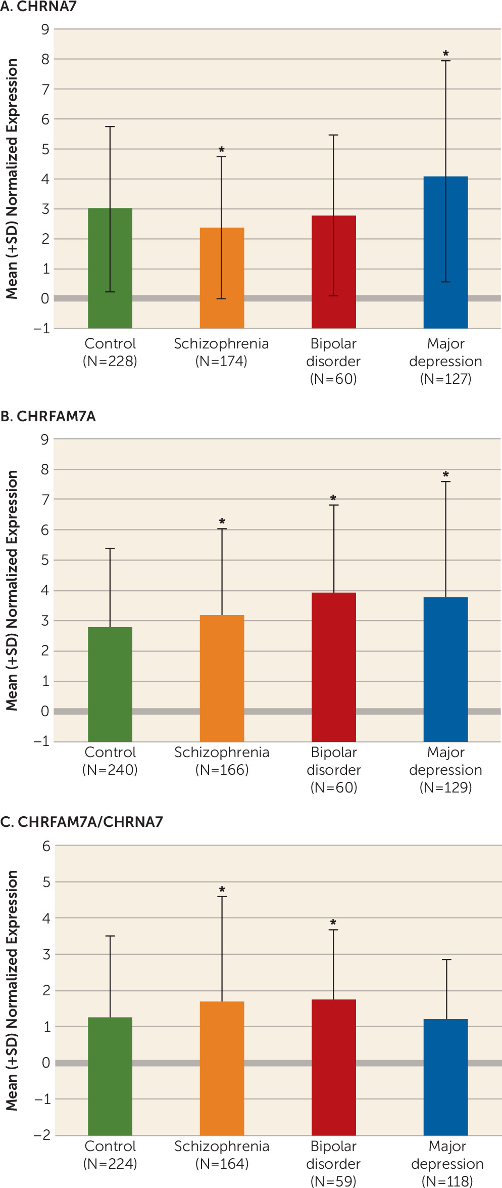
We found that the expression levels of CHRNA7 mRNA were significantly increased in major depression patients compared with all other groups (ANCOVA: F=31.5, df=3, 580, p<1.0E-17; post hoc Fisher’s least significant difference: p=8.1E-12 compared with comparison subjects, p<1.0E-17 compared with schizophrenia patients, p=2.3E-8 compared with bipolar disorder patients) (Figure 1A). In contrast, the expression of CHRNA7 in patients with schizophrenia was significantly lower than in all other groups (post hoc Fisher’s least significant difference: p=2.8E-5 compared with comparison subjects, p<1.0E-17 compared with major depression patients, p=0.048 compared with bipolar disorder patients) (Figure 1A).
FIGURE 1. Differences in the Expression of CHRNA7 and CHRFAM7A Between the Diagnostic Groupsa
a Mean mRNA expression of CHRNA7 (panel A), CHRFAM7A (panel B), and the ratios of CHRFAM7A/CHRNA7 (panel C) in the dorsolateral prefrontal cortex from comparison subjects and patients with schizophrenia, bipolar disorder, and major depression measured using quantitative real-time polymerase chain reaction. Expression data are normalized to a geometric mean of three housekeeping genes, beta 2-microglobulin (B2M), beta glucuronidase (GUSB), and beta actin (ACTB) expression. Asterisks indicate significant differences between comparison subjects and the diagnostic groups (p<0.05).
Expression of CHRFAM7A was significantly increased in the schizophrenia, bipolar disorder, and major depression groups compared with the comparison group (ANCOVA: F=11.9, df=3, 586, p=1.3E-7; post hoc Fisher’s least significant difference: p=0.025, p=3.4E-7, p=2.4E-9, respectively) (Figure 1B). Although significantly elevated compared with comparison subjects, the expression of CHRFAM7A in schizophrenia patients was significantly lower than in patients with bipolar disorder and major depression (post hoc Fisher’s least significant difference: p=6.3E-4, p=2.3E-4, respectively) (Figure 1B). Importantly, the ratios of CHRFAM7A/CHRNA7 levels were significantly different between the diagnostic groups (main effect of diagnosis by ANCOVA: F=3.63, df=3, 556, p<0.01) (Figure 1C); in particular, for schizophrenia and bipolar disorder patients these ratios were significantly higher than for comparison subjects and major depression patients (post hoc p values<0.0001 and<0.01, respectively), whereas major depression patients did not differ from comparison subjects (p>0.9).
There were no significant effects of antipsychotic medication, antidepressants, or lithium on the expression levels of CHRNA7 and CHRFAM7A transcripts in patients with schizophrenia and major depression (Table 2). Only for patients with bipolar disorder, the expression of CHRNA7 in the subjects positive for antipsychotic medication at the time of death was significantly higher than in subjects negative for antipsychotics (F=8.1, df=1, 31, p=0.0079], and the expression of CHRFAM7A in the subjects treated with lithium in their lifetime was significantly lower than in subjects without lithium treatment (F=4.7, df=1, 32, p=0.037). None of these differences, however, explain the diagnostic effects described above.
TABLE 2. Effects of Antemortem Medication on Expression of CHRNA7 and CHRFAM7A in Patient Groupsa
Group and TranscriptToxicology Test for NeurolepticsToxicology Test for AntidepressantsLifetime History of Lithium
Schizophrenia   
 CHRNA70.830.83NA
 CHRFAM7A0.430.75NA
Bipolar disorder   
 CHRNA70.0079**0.460.77
 CHRFAM7A0.150.270.037*
Major depression   
 CHRNA70.940.58NA
 CHRFAM7A0.930.23NA
a
The numbers indicate p values from analyses of covariance performed separately for each diagnostic group without correction for multiple testing.
*
p<0.05; **p<0.01.

Effects of Smoking Status and Nicotine on CHRNA7 and CHRFAM7A Expression

Given the previous reports on the involvement of CHRNA7 in smoking, we next examined the effects of smoking status (history of smoking) and nicotine status (negative or positive results of toxicological tests at death) on the mRNA expression levels of CHRNA7 and CHRFAM7A in the same cohort. In a two-way ANCOVA with smoking and diagnosis as main factors, there were no significant effects of smoking on either CHNRNA7 or CHRFAM7A expression (F=0.1, df=1, 531, p=0.74 and F=2.4, df=1, 536, p=0.12, respectively) and no significant diagnosis-by-smoking interaction for CHRFAM7A (F=0.57, df=3, 536, p=0.64). Since we detected a marginally significant smoking-by-diagnosis interaction for CHRNA7 (F=2.51, df=3, 531, p=0.058), we conducted post hoc tests and found that bipolar disorder patients with a history of smoking had higher levels of CHRNA7 than bipolar disorder patients who did not smoke (post hoc Fisher’s least significant difference: p=0.018), whereas major depression smokers had lower levels than major depression nonsmokers (post hoc Fisher’s least significant difference: p=0.046). There were no differences between smokers and nonsmokers among patients with schizophrenia or comparison subjects. Although these inconsistent results between the groups are difficult to interpret, they nevertheless suggest that smoking is not directly responsible for the observed diagnostic differences in the expression of CHRNA7 or CHRFAM7A in this study.

Expression of CHRNA7 and CHRFAM7A During the Lifespan

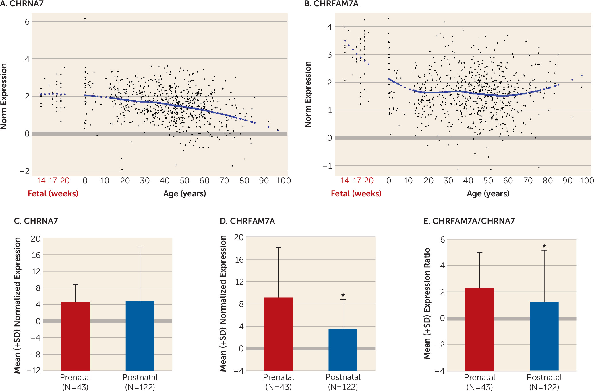
To investigate the developmental profile of expression of CHRNA7 and CHRFAM7A, we used samples from the dorsolateral prefrontal cortex of normal comparison subjects across the lifespan, from the 14th through 20th gestational week and from birth through old age (85 years old) (demographic characteristics are presented in Table 1). The expression of CHRNA7 was slightly higher during the prenatal period and decreased gradually throughout postnatal life until old age (Figure 2A). CHRFAM7A was expressed at much higher levels prenatally, decreased gradually from birth to young adulthood (approximately 30 years old), and then increased slightly with aging (Figure 2B). To statistically confirm our observation that CHRNA7 shows a different expression trajectory from CHRFAM7A, we examined the differences in CHRNA7 and CHRFAM7A expression between two age groups: fetal subjects and young individuals between 0 and 30 years old. Whereas there was no difference in the expression of CHRNA7 between the postnatal group and the fetal group (Figure 2C), CHRFAM7A expression was significantly lower in the postnatal group compared with the fetal group (Mann-Whitney U test: p=5.8E-14) (Figure 2D), Importantly, the ratios of CHRFAM7A/CHRNA7 were significantly higher in the fetal period compared with postnatal life (p=5.5E-12) (Figure 2E). Finally, we tested nicotine effects in these groups and found that CHRNA7 expression was marginally lower in fetal subjects positive for nicotine than in fetal subjects who were nicotine-negative (post hoc Fisher’s least significant difference: p=0.078).
FIGURE 2. Developmental Expression Patterns of CHRNA7 and CHRFAM7Aa
a Expression of CHRNA7 (panel A) and CHRFAM7A (panel B) in the dorsolateral prefrontal cortex of comparison subjects across the lifespan, from gestational week 14 through 20 and from birth (age 0) until old age (>80 years old) were measured using quantitative real-time polymerase chain reaction. Gene expression is displayed as a function of age (black dots represent individual subjects). A blue line represents a LOESS fit across the lifespan. Mean mRNA expression of CHRNA7 (panel C), CHRFAM7A (panel D), and the ratios of CHRFAM7A/CHRNA7 (panel E) from comparison subjects during prenatal (second trimester of gestation) and postnatal (0–30 years of age) periods are shown. Asterisks indicate significant differences between the groups (p<0.001).

Effects of CHRNA7 Genotype on CHRNA7 and CHRFAM7A Expression

To test whether genetic variants are associated with expression of these genes, we examined the effects of 81 CHRNA7 SNPs (see Table S1 in the online data supplement) on the mRNA expression levels of CHRNA7 and CHRFAM7A, covarying by diagnosis, sex, age at death, race, and RNA integrity number and Bonferroni-correcting for multiple comparisons. There were no significant associations between any SNPs examined in this study and the mRNA expression levels of CHRNA7 or CHRFAM7A and the ratio CHRFAM7A/CHRNA7 in all subjects or in any diagnostic group analyzed separately (all p values >0.05), including two SNPs (rs6494165 and rs3826029) previously examined but not associated with schizophrenia (16).
We also obtained the data for the 2-bp deletion in exon 6 through RNA sequencing of a subset of samples (comparison and schizophrenia groups, N=169). The 2-bp deletion was found in 74 subjects (comparison group, N=44; schizophrenia group, N=30), absent in 95 subjects (comparison group, N=42; schizophrenia group, N=53), and marginally less common in patients with schizophrenia than comparison subjects (36% compared with 51%, χ2=3.87, p=0.05). There was no significant difference in the frequency of 2-bp deletion between Caucasian and African American subjects. Interestingly, this 2-bp deletion was associated with decreased CHRFAM7A mRNA expression in all subjects (by approximately 26%, p=0.0009) and had no association with the expression of CHRNA7 mRNA (p=0.6). The 2-bp deletion had a similar effect in both diagnostic groups (not significant) and in both races (not significant) and did not account for the observation of increased expression of CHRFAM7A or decreased expression of CHRNA7 in schizophrenia.
Finally, we identified four subjects with hemideletion of CHRFAM7A (i.e., far fewer than previously reported by Sinkus et al. [17]: two patients with schizophrenia [out of 195], one comparison subject [out of 161], and one subject with major depressive disorder [out of 156]). There were no cases with the homozygous deletion of CHRFAM7A in our cohort. Moreover, the hemideletion did not appear to be associated with expression of CHRNA7 and CHRFAM7A (all p values >0.5).

Colocalization of CHRNA7 and CHRFAM7A Transcripts

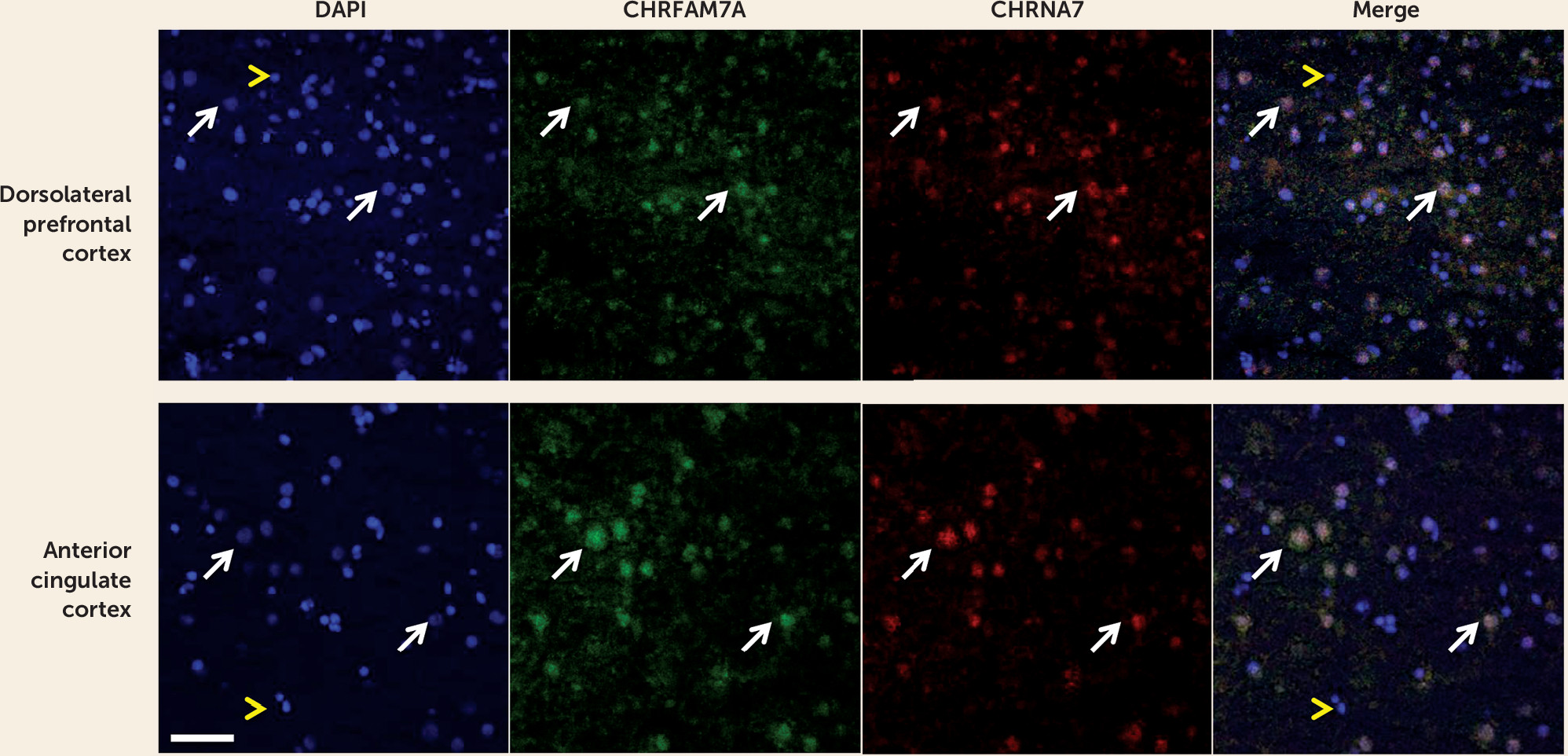
Using dual-label fluorescent in-situ hybridization and confocal microscopy, we examined whether CHRNA7 and CHRFAM7A mRNAs are expressed in the same cells of the human neocortex: the dorsolateral prefrontal cortex and anterior cingulate cortex. We chose the anterior cingulate cortex, in addition to the dorsolateral prefrontal cortex, because of the previous report by Allen Brain Atlas (http://www.brain-map.org/) of enhanced expression of CHRNA7 in this cortical region. We demonstrated that CHRNA7 and CHRFAM7A mRNAs are colocalized in a subset of putative neuronal nuclei (large DAPI-stained nuclei), both in the dorsolateral prefrontal cortex and anterior cingulate cortex (Figure 3). Sense probes produced no detectable signal. These findings support the view that the two gene products are colocalized and can function together.
FIGURE 3. Colocalization of CHRNA7 and CHRFAM7A Transcripts in a Subset of Neocortical Cellsa
a Fluorescently labeled CHRNA7 and CHRFAM7A riboprobes were imaged in representative sections of human dorsolateral prefrontal cortex and anterior cingulate cortex by confocal microscopy. The green signal (fluorescein-labeled) represents CHRFAM7A, the red signal (digoxigenin-labeled) represents CHRNA7, the blue signal results from counterstaining with 4',6-diamidino−2-phenylindole (DAPI) and indicates cell nuclei. The merged images show colocalization of CHRNA7 and CHRFAM7A riboprobes in putative neurons (large DAPI-stained nuclei, examples indicated by white arrows). Putative glial cells show DAPI staining over small nuclei and a lack of CHRNA7 or CHRFAM7A expression (examples indicated by yellow arrow heads). Scale bar: 50 μm.

Discussion

Our study is the first, to our knowledge, to show that CHRFAM7A is expressed in fetal human brain and that CHRFAM7A expression and CHRFAM7A/CHRNA7 expression ratios are markedly increased during the prenatal period compared with postnatal life. We are also the first to demonstrate coexpression of the two transcripts in a subset of neuronal cells in the human neocortex. Moreover, we found that CHRFAM7A/CHRNA7 expression ratios are significantly higher in the dorsolateral prefrontal cortex of patients with schizophrenia and bipolar disorder compared with normal comparison subjects due primarily to overexpression of CHRFAM7A in cases, a pattern of expression mimicking fetal life. Taken together, these findings suggest that the composition and function of α7 nACh receptors may be altered in the dorsolateral prefrontal cortex of patients with schizophrenia and bipolar disorder.
CHRFAM7A is considered to act as a dominant negative regulator of α7 nAChR function in humans (11). CHRFAM7A lacks CHRNA7 exons 1–4, and in consequence, CHRFAM7A protein is missing the signal peptide and part of the binding site for ligands. As a result, the receptor, which comprises the gene product of CHRFAM7A, would not function as effectively as α7 nAChRs containing only the gene products of CHRNA7. Indeed, in vitro oocyte-based studies have shown that the activity of α7 nAChRs is diminished (32) and [125I]-α-bungarotoxin binding is lower in cells coexpressing CHRNA7 with the duplicate gene product (11, 13). The mechanisms underlying these changes in receptor properties are not clear. It has been speculated that in addition to the formation of heteromeric receptors by these atypical duplicate subunits, they also interfere with oligomerization and assembly of α7 subunits in the endoplasmic reticulum, decreasing the number of mature α7 nAChRs able to migrate to the membrane (13). It has been suggested that regulating the levels of cell surface α7 nAChRs by the human-specific duplicate subunit may be important for immune responses of the cholinergic anti-inflammatory pathway in the periphery (3335). Furthermore, a recent report by Wang et al. (36) provided evidence that the duplicated subunits are colocalized with full-length-7 subunits in the transfected mouse neuroblastoma cells (Neuro2a), as well as rat hippocampal neurons, are assembled and transported to the cell membrane together with full-length-7 subunits, and alter the function of the nAChRs. These findings support the view that the two gene products are colocalized and can function together. Our results of coexpression of the two transcripts in the human neocortex provide additional support for this concept.
The results of altered balance between CHRNA7 and CHRFAM7A in schizophrenia and bipolar disorder are in agreement with the only other published study on this topic, by De Luca et al. (24), which showed nearly significant changes in the same direction in these disorders using the Stanley Foundation 35/35/35 postmortem dorsolateral prefrontal cortex collection (i.e., higher ratios of CHRFAM7A/CHRNA7 in case compared with comparison subjects). If indeed these altered CHRFAM7A/CHRNA7 ratios lead to functional changes, such as altered receptor assembly or receptor migration to the surface, they may perhaps explain reduced [125I]- α-bungarotoxin binding reported in schizophrenia, as it would be expected that cells expressing the duplicated chimeric gene have a smaller number of binding sites (11).
The results of our analysis of nicotine effects are not easy to interpret and reconcile with the existing literature. In contrast to our prediction based on the study by de Lucas-Cerrillo et al. (13), who exposed cultured cells to nicotine in vitro and found significantly reduced expression of CHRFAM7A, we did not detect changes in CHRFAM7A expression in individuals who smoked or who were positive for nicotine at the time of death (13). We also did not find an increase in CHRNA7 expression in smokers with schizophrenia as reported previously in a relatively small postmortem study of the hippocampus (37), but we used samples from a different brain region, and this may explain the discrepancy. On the other hand, we found contrasting changes between smokers and nonsmokers in two other diagnostic groups: an increase in CHRNA7 expression in bipolar patients and a decrease in major depression patients who smoked compared with nonsmokers. These effects were weak, however, and did not survive corrections for multiple testing and thus may be spurious. In fetal subjects, we showed that CHRNA7 expression tended to be decreased in subjects exposed to nicotine, but again these effects were weak and not statistically significant. Thus, although nicotine exposure during pregnancy has been associated with various obstetrical complications, such as spontaneous abortion, preterm birth, stillbirth, and low birth weight (38) and with adverse effects on brain development, it is not clear whether any of these effects are mediated even partly through α7 nAChRs.
We found that the developmental trajectory of expression in the dorsolateral prefrontal cortex differed markedly between CHRNA7 and CHRFAM7A. CHRFAM7A was relatively highly expressed during fetal life and then decreased dramatically from fetal age to young adulthood, whereas CHRNA7 was more stable during these periods and decreased gradually with aging (3941). This preferential fetal profile of expression of CHRFAM7A suggests that CHRFAM7A may play a yet unknown role in early brain development. Our findings of an altered balance between the fetal variant of the gene and the variant that constitutes a fully functional mature α7 nAChR are reminiscent of the recent findings suggesting delayed maturation of GABA signaling in patients with schizophrenia. In the hippocampal formation, GAD25/GAD67 and NKCC1/KCC2 ratios were increased in patients with schizophrenia compared with normal comparison subjects, reflecting a potentially immature GABA physiology (42).
In this study, we did not find significant associations of CHRNA7 and CHRFAM7A expression with any of the SNPs examined, although previous genetic linkage analysis indicated that CHRNA7 is associated with smoking in schizophrenia (43) and several CHRNA7 SNPs are associated with smoking in nonpsychiatric subjects (44). D15S1360 polymorphism of the CHRNA7 was also associated with susceptibility to nicotine dependence in subjects with schizophrenia (45). We could not, however, detect a molecular basis for these clinical associations.
It is important to point out potential limitations of this study. As in any postmortem human brain examination, there are confounding factors, such as antemortem medication (antipsychotic drugs, antidepressants, lithium) or alcohol and illicit drug use (substance abuse was not an exclusion criterion for psychiatric patients, but it was for comparison subjects). Thus, it is possible that these factors contributed to or were responsible for the observed changes in patients. Another limitation is a lack of evidence in the human brain that CHRFAM7A mRNA is translated and that the protein product assembles in humans with α7 nAChRs.
In conclusion, we showed expression changes of CHRNA7 and CHRFAM7A and altered proportions of CHRFAM7A/ CHRNA7 in schizophrenia and bipolar disorder. We also demonstrated that the two transcripts are expressed together in a subset of cortical neurons. Given the evidence that the duplicated subunits interact with α7 nAChRs as well as other nAChRs (α3 and α4) and alter their function in the transfected cells, our results support the concept of aberrant function of nAChRs in mental illness.
Acknowledgments

The authors thank Liqin Wang, Jonathan Sirovatka, and Vesna Imamovic for their technical expertise, Dr. Ran Tao for genotyping, Dr. Ningping Feng for the gene sequence examination, and Drs. Llewellyn Bigelow and Mary Herman for their diagnostic and neuropathological contributions, as well as Yoshiyuki Matsui for his contribution to the literature searches and analyses, Dr. Mizuki Hino for valuable advice and help in depiction of a gene chart, and the Offices of the Chief Medical Examiner of Washington, DC, and of Northern Virginia-Northern District. The authors also thank Drs. Ronald Zielke, Robert Johnson, and Robert Vigorito at the NICHD Brain and Tissue Bank for Developmental Disorders, University of Maryland School of Medicine, and Dr. Maree Webster at the Stanley Medical Research Institute for their contribution of brain tissues, and the families of the deceased for the donations of brain tissue and their time and effort devoted to the consent process and interviews.

Supplementary Material

File (appi.ajp.2015.14080978.ds001.pdf)

References

1.
Harrison PJ, Weinberger DR: Schizophrenia genes, gene expression, and neuropathology: on the matter of their convergence. Mol Psychiatry 2005; 10:40–68; image 45
2.
Stephens SH, Franks A, Berger R, et al: Multiple genes in the 15q13-q14 chromosomal region are associated with schizophrenia. Psychiatr Genet 2012; 22:1–14
3.
Hajós M, Rogers BN: Targeting alpha7 nicotinic acetylcholine receptors in the treatment of schizophrenia. Curr Pharm Des 2010; 16:538–554
4.
Marks MJ, Stitzel JA, Romm E, et al: Nicotinic binding sites in rat and mouse brain: comparison of acetylcholine, nicotine, and alpha-bungarotoxin. Mol Pharmacol 1986; 30:427–436
5.
Quik M, Choremis J, Komourian J, et al: Similarity between rat brain nicotinic alpha-bungarotoxin receptors and stably expressed alpha-bungarotoxin binding sites. J Neurochem 1996; 67:145–154
6.
Whiteaker P, Davies AR, Marks MJ, et al: An autoradiographic study of the distribution of binding sites for the novel alpha7-selective nicotinic radioligand [3H]-methyllycaconitine in the mouse brain. Eur J Neurosci 1999; 11:2689–2696
7.
Tribollet E, Bertrand D, Marguerat A, et al: Comparative distribution of nicotinic receptor subtypes during development, adulthood and aging: an autoradiographic study in the rat brain. Neuroscience 2004; 124:405–420
8.
Wonnacott S: Presynaptic nicotinic ACh receptors. Trends Neurosci 1997; 20:92–98
9.
Toyohara J, Sakata M, Wu J, et al: Preclinical and the first clinical studies on [11C]CHIBA-1001 for mapping alpha7 nicotinic receptors by positron emission tomography. Ann Nucl Med 2009; 23:301–309
10.
Conroy WG, Liu Z, Nai Q, et al: PDZ-containing proteins provide a functional postsynaptic scaffold for nicotinic receptors in neurons. Neuron 2003; 38:759–771
11.
Araud T, Graw S, Berger R, et al: The chimeric gene CHRFAM7A, a partial duplication of the CHRNA7 gene, is a dominant negative regulator of α7*nAChR function. Biochem Pharmacol 2011; 82:904–914
12.
Mexal S, Frank M, Berger R, et al: Differential modulation of gene expression in the NMDA postsynaptic density of schizophrenic and control smokers. Brain Res Mol Brain Res 2005; 139:317–332
13.
de Lucas-Cerrillo AM, Maldifassi MC, Arnalich F, et al: Function of partially duplicated human α77 nicotinic receptor subunit CHRFAM7A gene: potential implications for the cholinergic anti-inflammatory response. J Biol Chem 2011; 286:594–606
14.
Freedman R, Coon H, Myles-Worsley M, et al: Linkage of a neurophysiological deficit in schizophrenia to a chromosome 15 locus. Proc Natl Acad Sci USA 1997; 94:587–592
15.
Leonard S, Gault J, Hopkins J, et al: Association of promoter variants in the alpha7 nicotinic acetylcholine receptor subunit gene with an inhibitory deficit found in schizophrenia. Arch Gen Psychiatry 2002; 59:1085–1096
16.
Stephens SH, Logel J, Barton A, et al: Association of the 5′-upstream regulatory region of the alpha7 nicotinic acetylcholine receptor subunit gene (CHRNA7) with schizophrenia. Schizophr Res 2009; 109:102–112
17.
Sinkus ML, Lee MJ, Gault J, et al: A 2-base pair deletion polymorphism in the partial duplication of the alpha7 nicotinic acetylcholine gene (CHRFAM7A) on chromosome 15q14 is associated with schizophrenia. Brain Res 2009; 1291:1–11
18.
Joo EJ, Lee KY, Kim HS, et al: Genetic association study of the alpha 7 nicotinic receptor (CHRNA7) with the development of schizophrenia and bipolar disorder in Korean population. Psychiatry Investig 2010; 7:196–201
19.
Lai IC, Hong CJ, Tsai SJ: Association study of nicotinic-receptor variants and major depressive disorder. J Affect Disord 2001; 66:79–82
20.
Freedman R, Hall M, Adler LE, et al: Evidence in postmortem brain tissue for decreased numbers of hippocampal nicotinic receptors in schizophrenia. Biol Psychiatry 1995; 38:22–33
21.
Guan ZZ, Zhang X, Blennow K, et al: Decreased protein level of nicotinic receptor alpha7 subunit in the frontal cortex from schizophrenic brain. Neuroreport 1999; 10:1779–1782
22.
Thomsen MS, Weyn A, Mikkelsen JD: Hippocampal α7 nicotinic acetylcholine receptor levels in patients with schizophrenia, bipolar disorder, or major depressive disorder. Bipolar Disord 2011; 13:701–707
23.
Mathew SV, Law AJ, Lipska BK, et al: Alpha7 nicotinic acetylcholine receptor mRNA expression and binding in postmortem human brain are associated with genetic variation in neuregulin 1. Hum Mol Genet 2007; 16:2921–2932
24.
De Luca V, Likhodi O, Van Tol HH, et al: Regulation of alpha7-nicotinic receptor subunit and alpha7-like gene expression in the prefrontal cortex of patients with bipolar disorder and schizophrenia. Acta Psychiatr Scand 2006; 114:211–215
25.
Mexal S, Berger R, Logel J, et al: Differential regulation of alpha7 nicotinic receptor gene (CHRNA7) expression in schizophrenic smokers. J Mol Neurosci 2010; 40:185–195
26.
Mousavi M, Hellström-Lindahl E, Guan ZZ, et al: Protein and mRNA levels of nicotinic receptors in brain of tobacco using controls and patients with Alzheimer’s disease. Neuroscience 2003; 122:515–520
27.
de Leon J, Diaz FJ: A meta-analysis of worldwide studies demonstrates an association between schizophrenia and tobacco smoking behaviors. Schizophr Res 2005; 76:135–157
28.
Lipska BK, Deep-Soboslay A, Weickert CS, et al: Critical factors in gene expression in postmortem human brain: focus on studies in schizophrenia. Biol Psychiatry 2006; 60:650–658
29.
Ye T, Lipska BK, Tao R, et al: Analysis of copy number variations in brain DNA from patients with schizophrenia and other psychiatric disorders. Biol Psychiatry 2012; 72:651–654
30.
Tao R, Cousijn H, Jaffe AE, et al: Expression of ZNF804A in human brain and alterations in schizophrenia, bipolar disorder, and major depressive disorder: a novel transcript fetally regulated by the psychosis risk variant rs1344706. JAMA Psychiatry 2014; 71:1112–1120
31.
Vazdarjanova A, McNaughton BL, Barnes CA, et al: Experience-dependent coincident expression of the effector immediate-early genes arc and Homer 1a in hippocampal and neocortical neuronal networks. J Neurosci 2002; 22:10067–10071
32.
Criado M, Valor LM, Mulet J, et al: Expression and functional properties of α7 acetylcholine nicotinic receptors are modified in the presence of other receptor subunits. J Neurochem 2012; 123:504–514
33.
Villiger Y, Szanto I, Jaconi S, et al: Expression of an alpha7 duplicate nicotinic acetylcholine receptor-related protein in human leukocytes. J Neuroimmunol 2002; 126:86–98
34.
Wang H, Yu M, Ochani M, et al: Nicotinic acetylcholine receptor alpha7 subunit is an essential regulator of inflammation. Nature 2003; 421:384–388
35.
Benfante R, Antonini RA, De Pizzol M, et al: Expression of the α7 nAChR subunit duplicate form (CHRFAM7A) is down-regulated in the monocytic cell line THP-1 on treatment with LPS. J Neuroimmunol 2011; 230:74–84
36.
Wang Y, Xiao C, Indersmitten T, et al: The duplicated α7 subunits assemble and form functional nicotinic receptors with the full-length α7. J Biol Chem 2014; 289:26451–26463
37.
Hong CJ, Lai IC, Liou LL, et al: Association study of the human partially duplicated alpha7 nicotinic acetylcholine receptor genetic variant with bipolar disorder. Neurosci Lett 2004; 355:69–72
38.
Bruin JE, Gerstein HC, Holloway AC: Long-term consequences of fetal and neonatal nicotine exposure: a critical review. Toxicol Sci 2010; 116:364–374
39.
Utsugisawa K, Nagane Y, Tohgi H, et al: Changes with aging and ischemia in nicotinic acetylcholine receptor subunit alpha7 mRNA expression in postmortem human frontal cortex and putamen. Neurosci Lett 1999; 270:145–148
40.
Adams CE, Broide RS, Chen Y, et al: Development of the alpha7 nicotinic cholinergic receptor in rat hippocampal formation. Brain Res Dev Brain Res 2002; 139:175–187
41.
Falk L, Nordberg A, Seiger A, et al: Higher expression of alpha7 nicotinic acetylcholine receptors in human fetal compared to adult brain. Brain Res Dev Brain Res 2003; 142:151–160
42.
Hyde TM, Lipska BK, Ali T, et al: Expression of GABA signaling molecules KCC2, NKCC1, and GAD1 in cortical development and schizophrenia. J Neurosci 2011; 31:11088–11095
43.
Faraone SV, Su J, Taylor L, et al: A novel permutation testing method implicates sixteen nicotinic acetylcholine receptor genes as risk factors for smoking in schizophrenia families. Hum Hered 2004; 57:59–68
44.
Saccone NL, Schwantes-An TH, Wang JC, et al: Multiple cholinergic nicotinic receptor genes affect nicotine dependence risk in African and European Americans. Genes Brain Behav 2010; 9:741–750
45.
De Luca V, Wong AH, Muller DJ, et al: Evidence of association between smoking and alpha7 nicotinic receptor subunit gene in schizophrenia patients. Neuropsychopharmacology 2004; 29:1522–1526

Information & Authors

Information

Published In

Go to American Journal of Psychiatry
American Journal of Psychiatry
Pages: 1122 - 1130
PubMed: 26206074

History

Received: 6 August 2014
Revision received: 30 December 2014
Revision received: 24 February 2015
Accepted: 12 March 2015
Published online: 24 July 2015
Published in print: November 01, 2015

Authors

Details

Yasuto Kunii, M.D., Ph.D.
From the Human Brain Collection Core, Division of Intramural Research Programs, National Institute of Mental Health, National Institutes of Health, Bethesda, Md.; the Department of Neuropsychiatry, Fukushima Medical University School of Medicine, Hikarigaoka, Fukushima City, Fukushima, Japan; the Unit on Neural Circuits and Adaptive Behaviors, National Institute of Mental Health, National Institutes of Health, Bethesda, Md.; the Lieber Institute for Brain Development, Johns Hopkins Medical Campus, Baltimore; the Department of Psychiatry and Behavioral Sciences, John Hopkins University School of Medicine, Baltimore; and the Department of Neurology, Johns Hopkins University School of Medicine, Baltimore.
Wenyu Zhang, Ph.D.
From the Human Brain Collection Core, Division of Intramural Research Programs, National Institute of Mental Health, National Institutes of Health, Bethesda, Md.; the Department of Neuropsychiatry, Fukushima Medical University School of Medicine, Hikarigaoka, Fukushima City, Fukushima, Japan; the Unit on Neural Circuits and Adaptive Behaviors, National Institute of Mental Health, National Institutes of Health, Bethesda, Md.; the Lieber Institute for Brain Development, Johns Hopkins Medical Campus, Baltimore; the Department of Psychiatry and Behavioral Sciences, John Hopkins University School of Medicine, Baltimore; and the Department of Neurology, Johns Hopkins University School of Medicine, Baltimore.
Qing Xu
From the Human Brain Collection Core, Division of Intramural Research Programs, National Institute of Mental Health, National Institutes of Health, Bethesda, Md.; the Department of Neuropsychiatry, Fukushima Medical University School of Medicine, Hikarigaoka, Fukushima City, Fukushima, Japan; the Unit on Neural Circuits and Adaptive Behaviors, National Institute of Mental Health, National Institutes of Health, Bethesda, Md.; the Lieber Institute for Brain Development, Johns Hopkins Medical Campus, Baltimore; the Department of Psychiatry and Behavioral Sciences, John Hopkins University School of Medicine, Baltimore; and the Department of Neurology, Johns Hopkins University School of Medicine, Baltimore.
Thomas M. Hyde, M.D., Ph.D.
From the Human Brain Collection Core, Division of Intramural Research Programs, National Institute of Mental Health, National Institutes of Health, Bethesda, Md.; the Department of Neuropsychiatry, Fukushima Medical University School of Medicine, Hikarigaoka, Fukushima City, Fukushima, Japan; the Unit on Neural Circuits and Adaptive Behaviors, National Institute of Mental Health, National Institutes of Health, Bethesda, Md.; the Lieber Institute for Brain Development, Johns Hopkins Medical Campus, Baltimore; the Department of Psychiatry and Behavioral Sciences, John Hopkins University School of Medicine, Baltimore; and the Department of Neurology, Johns Hopkins University School of Medicine, Baltimore.
Whitney McFadden
From the Human Brain Collection Core, Division of Intramural Research Programs, National Institute of Mental Health, National Institutes of Health, Bethesda, Md.; the Department of Neuropsychiatry, Fukushima Medical University School of Medicine, Hikarigaoka, Fukushima City, Fukushima, Japan; the Unit on Neural Circuits and Adaptive Behaviors, National Institute of Mental Health, National Institutes of Health, Bethesda, Md.; the Lieber Institute for Brain Development, Johns Hopkins Medical Campus, Baltimore; the Department of Psychiatry and Behavioral Sciences, John Hopkins University School of Medicine, Baltimore; and the Department of Neurology, Johns Hopkins University School of Medicine, Baltimore.
Joo Heon Shin, Ph.D.
From the Human Brain Collection Core, Division of Intramural Research Programs, National Institute of Mental Health, National Institutes of Health, Bethesda, Md.; the Department of Neuropsychiatry, Fukushima Medical University School of Medicine, Hikarigaoka, Fukushima City, Fukushima, Japan; the Unit on Neural Circuits and Adaptive Behaviors, National Institute of Mental Health, National Institutes of Health, Bethesda, Md.; the Lieber Institute for Brain Development, Johns Hopkins Medical Campus, Baltimore; the Department of Psychiatry and Behavioral Sciences, John Hopkins University School of Medicine, Baltimore; and the Department of Neurology, Johns Hopkins University School of Medicine, Baltimore.
Amy Deep-Soboslay, M.Ed.
From the Human Brain Collection Core, Division of Intramural Research Programs, National Institute of Mental Health, National Institutes of Health, Bethesda, Md.; the Department of Neuropsychiatry, Fukushima Medical University School of Medicine, Hikarigaoka, Fukushima City, Fukushima, Japan; the Unit on Neural Circuits and Adaptive Behaviors, National Institute of Mental Health, National Institutes of Health, Bethesda, Md.; the Lieber Institute for Brain Development, Johns Hopkins Medical Campus, Baltimore; the Department of Psychiatry and Behavioral Sciences, John Hopkins University School of Medicine, Baltimore; and the Department of Neurology, Johns Hopkins University School of Medicine, Baltimore.
Tianzhang Ye
From the Human Brain Collection Core, Division of Intramural Research Programs, National Institute of Mental Health, National Institutes of Health, Bethesda, Md.; the Department of Neuropsychiatry, Fukushima Medical University School of Medicine, Hikarigaoka, Fukushima City, Fukushima, Japan; the Unit on Neural Circuits and Adaptive Behaviors, National Institute of Mental Health, National Institutes of Health, Bethesda, Md.; the Lieber Institute for Brain Development, Johns Hopkins Medical Campus, Baltimore; the Department of Psychiatry and Behavioral Sciences, John Hopkins University School of Medicine, Baltimore; and the Department of Neurology, Johns Hopkins University School of Medicine, Baltimore.
Chao Li, Ph.D.
From the Human Brain Collection Core, Division of Intramural Research Programs, National Institute of Mental Health, National Institutes of Health, Bethesda, Md.; the Department of Neuropsychiatry, Fukushima Medical University School of Medicine, Hikarigaoka, Fukushima City, Fukushima, Japan; the Unit on Neural Circuits and Adaptive Behaviors, National Institute of Mental Health, National Institutes of Health, Bethesda, Md.; the Lieber Institute for Brain Development, Johns Hopkins Medical Campus, Baltimore; the Department of Psychiatry and Behavioral Sciences, John Hopkins University School of Medicine, Baltimore; and the Department of Neurology, Johns Hopkins University School of Medicine, Baltimore.
Joel E. Kleinman, M.D., Ph.D.
From the Human Brain Collection Core, Division of Intramural Research Programs, National Institute of Mental Health, National Institutes of Health, Bethesda, Md.; the Department of Neuropsychiatry, Fukushima Medical University School of Medicine, Hikarigaoka, Fukushima City, Fukushima, Japan; the Unit on Neural Circuits and Adaptive Behaviors, National Institute of Mental Health, National Institutes of Health, Bethesda, Md.; the Lieber Institute for Brain Development, Johns Hopkins Medical Campus, Baltimore; the Department of Psychiatry and Behavioral Sciences, John Hopkins University School of Medicine, Baltimore; and the Department of Neurology, Johns Hopkins University School of Medicine, Baltimore.
Kuan Hong Wang, Ph.D.
From the Human Brain Collection Core, Division of Intramural Research Programs, National Institute of Mental Health, National Institutes of Health, Bethesda, Md.; the Department of Neuropsychiatry, Fukushima Medical University School of Medicine, Hikarigaoka, Fukushima City, Fukushima, Japan; the Unit on Neural Circuits and Adaptive Behaviors, National Institute of Mental Health, National Institutes of Health, Bethesda, Md.; the Lieber Institute for Brain Development, Johns Hopkins Medical Campus, Baltimore; the Department of Psychiatry and Behavioral Sciences, John Hopkins University School of Medicine, Baltimore; and the Department of Neurology, Johns Hopkins University School of Medicine, Baltimore.
Barbara K. Lipska, Ph.D.
From the Human Brain Collection Core, Division of Intramural Research Programs, National Institute of Mental Health, National Institutes of Health, Bethesda, Md.; the Department of Neuropsychiatry, Fukushima Medical University School of Medicine, Hikarigaoka, Fukushima City, Fukushima, Japan; the Unit on Neural Circuits and Adaptive Behaviors, National Institute of Mental Health, National Institutes of Health, Bethesda, Md.; the Lieber Institute for Brain Development, Johns Hopkins Medical Campus, Baltimore; the Department of Psychiatry and Behavioral Sciences, John Hopkins University School of Medicine, Baltimore; and the Department of Neurology, Johns Hopkins University School of Medicine, Baltimore.

Notes

Address correspondence to Dr. Lipska ([email protected]).
Previously presented in part at the 52nd American College of Neuropsychopharmacology Annual Meeting, Dec. 8–12, 2013, Hollywood, Fla.

Funding Information

Supported by the Intramural Research Program of the National Institute of Mental Health at the National Institutes of Health.The authors report no financial relationships with commercial interests.

Metrics & Citations

Metrics

Citations

Export Citations

If you have the appropriate software installed, you can download article citation data to the citation manager of your choice. Simply select your manager software from the list below and click Download.

For more information or tips please see 'Downloading to a citation manager' in the Help menu.

Format
Citation style
Style
Copy to clipboard

View Options

View options

PDF/EPUB

View PDF/EPUB

Login options

Already a subscriber? Access your subscription through your login credentials or your institution for full access to this article.

Personal login Institutional Login Open Athens login
Purchase Options

Purchase this article to access the full text.

PPV Articles - American Journal of Psychiatry

PPV Articles - American Journal of Psychiatry

Not a subscriber?

Subscribe Now / Learn More

PsychiatryOnline subscription options offer access to the DSM-5-TR® library, books, journals, CME, and patient resources. This all-in-one virtual library provides psychiatrists and mental health professionals with key resources for diagnosis, treatment, research, and professional development.

Need more help? PsychiatryOnline Customer Service may be reached by emailing [email protected] or by calling 800-368-5777 (in the U.S.) or 703-907-7322 (outside the U.S.).

Media

Figures

Other

Tables

Share

Share

Share article link

Share