Full access
Regular Articles
Published Online: 4 October 2018

Resting-State Functional Connectivity of the Habenula in Mood Disorder Patients With and Without Suicide-Related Behaviors

Publication: The Journal of Neuropsychiatry and Clinical Neurosciences

Abstract

The habenula is a small midbrain structure that is important for brain signaling and learning from negative events. Thus, the habenula is strongly connected to both the reward system and motor regions. Increasing evidence suggests a role for the habenula in the etiology of psychiatric disorders, including mood and substance use disorders. However, no studies to date have investigated habenular resting-state functional connectivity (rsFC) in suicide-related behaviors (SB). The authors enrolled 123 individuals with major depressive disorder (MDD) or bipolar disorder and a history of suicide-related behaviors (SB+), 74 individuals with MDD or bipolar disorder and a history of suicidal ideation but no history of SB (SB–), and 75 healthy control subjects (HC). A seed-based approach was used to identify regions showing different rsFC with the habenula followed by region of interest to region of interest post hoc comparisons. Compared with both the SB– and HC groups, the SB+ group showed higher connectivity between the left habenula and the left parahippocampal gyrus, the right amygdala, and the right precentral and postcentral gyri. Patients with mood disorders displayed higher rsFC between the left habenula and left middle temporal gyrus, the left angular gyrus, and the left posterior cingulate cortex, as well as lower rsFC between the right habenula and the left thalamus, when compared with HCs. These findings suggest that the habenula is involved in the neural circuitry of suicide. The higher habenular rsFC found in the SB+ group may mediate a dysfunction in the mechanism that links the habenula with motor activity and contextual associative processing.
Death by suicide is a tragic event that occurs at higher rates among individuals with a psychiatric disorder, particularly mood disorders.1 However, it remains unclear why not all depressed patients who have suicidal ideation attempt suicide. Theoretical frameworks of suicidal behavior, such as the stress-vulnerability model, indicate that suicidal behavior may result from an interaction between exposure to stressors and a vulnerability in individuals suffering from psychiatric disorders.2 Suicide attempts have been associated with anhedonia and deficits in reward processing in both elderly3 and adolescent4 attempters when compared with nonattempters.
Studies on the neurobiology of suicide suggest that interrelated neurobiological systems linked to dysfunction in the stress response systems are involved. The corticotropin-releasing hormone/hypothalamic-pituitary-adrenal axis and the locus coeruleus-based norepinephrine system, which interact with the serotonin and opioid systems, are likely to be involved.5 Although they are usually small and heterogeneous, neuroimaging findings report associations between suicidal ideation as well as suicide-related behaviors with abnormalities involving the frontal neural system and the serotonergic system, including the orbitofrontal cortex, the anterior cingulate cortex, and the amygdala.69 Taken together, these findings suggest that brain regions regulating the stress response, as well as the reward-dopaminergic and serotonergic systems, may be involved in the circuitry underlying suicidal ideation and suicide-related behaviors.
One likely locus of concerted control over multiple neurobiological systems is the habenula, a region that links the forebrain to the midbrain. Indeed, because of its unique anatomical and functional position, the habenula modulates downstream limbic midbrain dopamine and serotonin circuits by controlling activity in the monoaminergic nuclei, the substantia nigra compacta/ventral tegmental area, and the raphe nucleus.10 Thus, as a relay interface between the basal ganglia and the limbic system, the habenula is involved in both motivational and emotional control of behavior,11 playing a critical role in both the stress response and the behavioral responses induced by expected reward.12,13 Not surprisingly, increasing evidence suggests the involvement of the habenula in the pathophysiology of a variety of psychiatric disorders, including mood disorders, schizophrenia, and substance use disorder.14,15 Specifically, increased connectivity and activation of the habenula has been associated with depression16,17 as well as sleep disturbances.18 It is not known, however, whether the habenula is involved in suicidal ideation and suicide-related behaviors.
The aim of the current study was to compare habenula resting state functional connectivity (rsFC) in a sample of patients with mood disorder, with and without a history of suicide-related behavior and healthy control subjects. We hypothesized that higher connectivity of the habenula with limbic regions would be associated with suicidal ideation and suicide-related behaviors.

Methods

Participants

The Baylor College of Medicine Internal Review Board approved the study protocol and procedures. All participants provided informed consent.
Inpatients who met criteria for current major depressive episode and with a primary DSM-IV-TR diagnosis of either major depressive disorder, recurrent (MDD; N=165) or bipolar disorder type I or type II (N=32) were recruited for the study. They were culled from a sample of 316 inpatients who gave their consent to take part in the study after being admitted at The Menninger Clinic in Houston. Participants with comorbid substance use disorder diagnoses were included, given the pervasive comorbidity between mood and substance use disorders19 and the need to investigate ecologically valid samples in neuroimaging studies.20 Psychiatric diagnoses were made using the Structured Clinical Interview for DSM-IV Disorders.21,22 Tobacco, alcohol, and cannabis were the most prevalent substances of abuse in the sample, according to the World Health Organization Alcohol, Smoking, and Substance Involvement Screening Test. All patients were medicated at the time of the scanning.
Healthy control (HC) subjects were recruited from the general population to serve as comparators for the rsFC analyses. HCs were excluded if they had a self-reported history of psychiatric diagnosis or substance abuse. Additional exclusion criteria for both patients and controls were a previous history of traumatic brain injury with loss of consciousness of more than 10 minutes or any other MRI contraindication.

Clinical Measures

Participants completed a comprehensive diagnostic assessment. Suicidal ideation and suicide-related behaviors were assessed using the Columbia Scale for Rating of Suicide Severity (C-SSRS).23,24 The C-SSRS is a semistructured interview that assesses severity of suicidal ideation, intensity of ideation, suicidal behavior and associated lethality of suicide attempts. The Suicidal Ideation (SI) subscale consists of five items: a passive wish to be dead; nonspecific active thoughts of suicide; active suicidal ideation, including methods; suicidal intent without a specific plan; and active suicidal ideation with intent and a plan to act. The Intensity of Ideation subscale rates the frequency, duration, controllability, deterrents, and reasons for thoughts of suicide. The Suicidal Behavior (SB) scale assesses past actual attempts, past nonsuicidal self-injurious behavior, past interrupted attempts, and past aborted attempts. The lifetime SB subscale was used to stratify groups. Patients were categorized in the suicide-related behaviors group (SB+) if they responded affirmatively to any one of the following three items: actual attempt, interrupted attempt, or aborted attempt. They did not meet criteria for inclusion in the SB+ group if they responded affirmatively solely to the following items: self-injurious behavior and preparatory acts or behavior. This approach is consistent with previous studies.25,26 Patients were included in the non-suicide-related behaviors group (SB–) if they had positive response to any item of the SI but negative response to all items of the SB. The final sample groups included 123 patients categorized as SB+ and 74 as SB–.
Depression severity was assessed with the self-rated nine-item Patient Health Questionnaire (PHQ-9) depressive scale.27 The PHQ-9 is a reliable measure of depression severity that scores each of the nine diagnostic criteria of major depressive episodes over the past 2 weeks as “0” (not at all) to “3” (nearly every day).28

Neuroimaging Data Acquisition and Preprocessing

Participants underwent magnetic resonance imaging of the brain on a 3T Siemens Trio Magnetom system. A structural T1-MPRAGE (TR=1,200 ms, TE=2.66 ms, flip angle=12°, voxel size=1mm isotropic, field of view=245 mm, and total sequence time=4.5 minutes) was first acquired. A 5-minute resting-state echo-planar imaging scan (echo time=30 ms, repetition time=2,000 ms, flip angle=90°, voxel size=3.4×3.4×4 mm, field of view=220 mm) was subsequently acquired for each subject. During the resting-state sequences, a large X was displayed on the screen; participants were asked to keep their eyes open or closed and not fall asleep.
Preprocessing of the functional data, including functional realignment and unwarp, functional slice-timing correction, structural segmentation and normalization, functional normalization, ART-based functional outlier detection and scrubbing, and functional smoothing (8-mm Gaussian kernel), were done in Montreal Neurological Institute (MNI)-space using CONN-fMRI Functional Connectivity toolbox v15.b29 with SPM8 (www.fil.ion.ucl.ac.uk/spm/). The Artifact Detection Toolbox (ART; http://www.nitrc.org/projects/artifact_detect/) was used for outlier detection and scrubbing as implemented in CONN. Motion outliers were defined as any frame where the motion exceeded 2 mm. Realignment parameters were entered as first-level covariates in the toolbox. Realignment, scrubbing, white matter, and cerebrospinal fluid were entered as potential confounders in the subject-level general linear model. Using the anatomical component correction (aCompCor) method of flexibly removing physiological noise and movement confounds on a voxel-by-voxel level,30,31 each of these effects was regressed out of the blood-oxygen-level-dependent (BOLD) signal before connectivity measures were computed by the toolbox. Functional images were then temporally band-pass filtered (0.01<f <0.1 Hz) to investigate low frequency correlations. Since we were interested in the habenula, which is a very small region, we downsampled the images to 3×3×3 mm. In addition, the habenular seed in rsFC analysis was not smoothed, to avoid likely contamination with adjacent areas.

Functional Connectivity Analyses

We first examined the rsFC between each habenula region of interest and each voxel of the brain. The left and right habenula seed regions of interest were manually created for every subject by using the T1 image to visually identify the right and left habenula of each subject’s landmarks with SPM8 (www.fil.ion.ucl.ac.uk/spm/). In T1 images, the habenula is clearly visible as two small triangular structures pointing into the third ventricle. Each functional region of interest was a 3×3×3-mm cube placed around a central MNI coordinate in the habenula.
The mean signal time course from the seeds was extracted and the Pearson’s correlation coefficients with the time course of all other voxels of the brain were calculated. Correlation maps were calculated for each subject and correlation coefficients were then converted to normally distributed z-scores using the Fisher’s transformation in order to perform general linear model analyses. In order to compare rsFC across the three groups (SB+, SB–, HC), assuming as the null hypothesis that the three groups would show equal rsFC with the left and right habenula seeds, analysis of covariance (ANCOVA) was performed with age and gender included as covariates. A voxel statistical height threshold of p<0.001 with a cluster threshold of p<0.05 family-wise error corrected was used to identify connectivity differences between the three groups. Significant clusters identified in this analysis were then imported as target regions of interest in the post hoc region of interest to region of interest analysis. The connectivity values between the significant clusters and left and right habenula were extracted from each participant in order to examine the nature of the connectivity in each group and the directionality of each post hoc contrast. The results were considered significant if p<0.05, false discovery rate corrected. To further explore whether additional factors such as MDD/bipolar disorder diagnosis, substance use/abuse disorder, or medications might have affected the significant results in the between patient group comparison (SB+ compared with SB–), we subsequently included these covariates in the model in a post hoc manner to avoid reducing statistical power of the original ANCOVA. Thus, mood disorder diagnosis, alcohol and substance use/abuse disorder, and the effects of antidepressants, lithium, antiepileptics, anxiolytics, and antipsychotics were entered as dichotomous covariates (absent versus present).
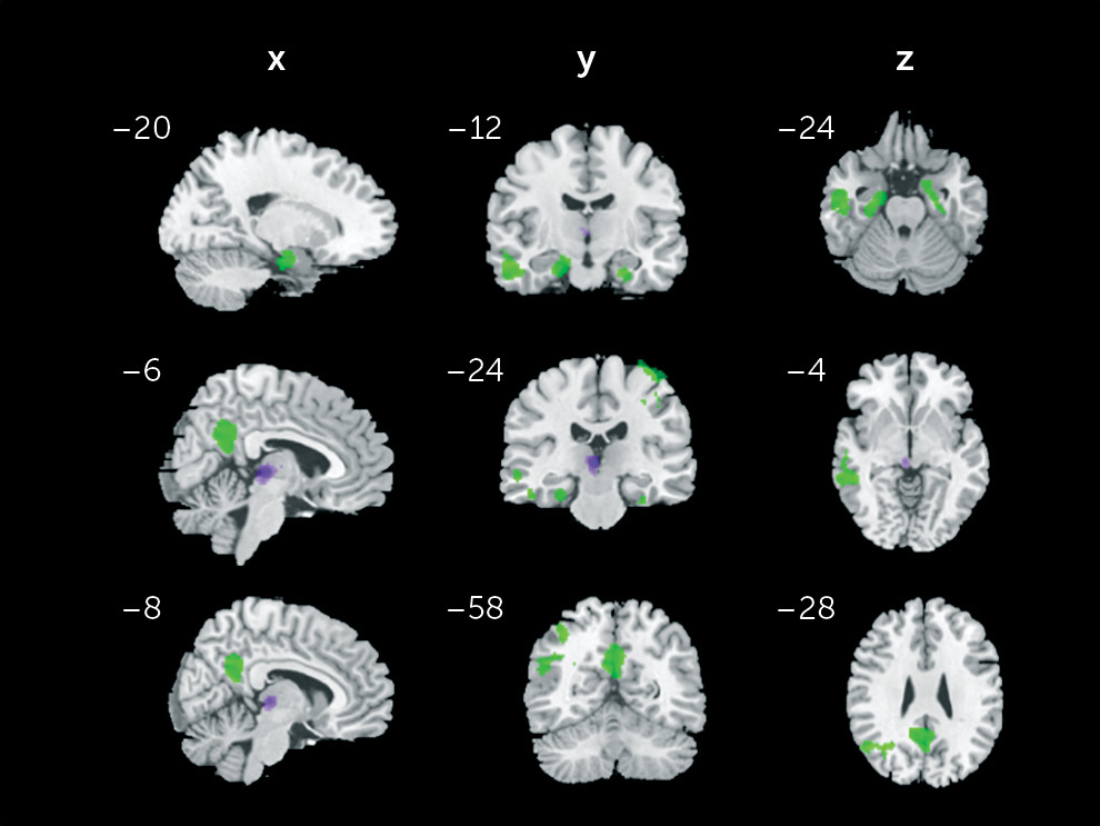
Additionally, to address whether the significant findings (clusters displayed in Table 1 would be specific to the habenula signal, we conducted an exploratory SB+ compared with SB– region of interest to region of interest comparison including thalamic regions as seeds. Specifically, the whole bilateral thalamus based on the automated anatomical labeling atlas32 as well as two small bilateral thalamic control regions of interest were chosen as seeds. The two small bilateral thalamic seeds were created by shifting the averaged habenula region of interest coordinates by 9 mm in the anterior direction (“thalamus medial,” MNI: right 4, –15, 2; left –3, –16, 2), and in the lateral direction (“thalamus lateral,” MNI: right 13, –24, 2; left –12, –25, 2). The results were considered significant if the p value was <0.05 (false discovery rate corrected).
TABLE 1. Seed-to-Voxel Analysis: Between-Group Differences Across the Three Study Groupsa
      Peak Voxel Montreal Neurological Institute Coordinates
RegionSideBrodmann’s AreaCluster SizeCluster p ValuePeak Z Valuexyz
Left habenula seed        
 Middle temporal gyrusLeft211,139<0.0014.71–52–10–24
 Angular gyrusLeft39817<0.0014.23–36–6250
 Posterior cingulateLeft31711<0.0014.9–2–5432
 Parahippocampal gyrusLeft3130.0024.54–30–22–22
 Postcentral gyrusRight12070.0054.3430–3246
 AmygdalaRight1730.0313.86160–24
 Precentral gyrusRight41560.0463.7444–2270
Right habenula seed        
 ThalamusLeft1790.0266.68–4–242
a
The data presented are the significant results across the three groups: (SB– group [negative history for suicide-related behaviors], SB+ group [positive history for suicide related behaviors], and healthy control group).

Results

Demographic and clinical data are reported in Table 2. The three groups did not significantly differ on age, gender, and ethnicity. The SB+ and SB– groups differed on the presence of psychotic features (p=0.026), lifetime suicidal ideation (p<0.001), and lithium (p=0.010). No additional differences were found between patient groups.
TABLE 2. Demographic and Clinical Characteristics of the Study Groupsa
CharacteristicSB– (N=74)SB+ (N=123)bHC (N=75)Statistical Analysis
 MeanSDMeanSDMeanSDFdfp
Age (years)30.311.529.711.833.19.12.262, 2690.106
 N%N%N%χ2dfp
Gender (male/female)42/3257/4356/6746/5438/3751/492.3420.309
Ethnicity (Caucasian/other)68/692/8110/1389/1163/1284/162.4520.294
Major depressive disorder diagnosis6283.810383.80.00010.994
Psychosis6821.64.9510.026
Personality disorder3547.364520.41410.520
Substance use disorder395352422.0210.155
WHO ASSIST Tobaccob385152421710.770
WHO ASSIST Alcoholb334547381910.681
WHO ASSIST Cannabisb344642342810.453
 MeanSDMeanSDMeanSDFdfp
Patient Health Questionnaire-9 depression subscale16.16.817.56.12.061, 1950.153
Columbia Scale for Rating of Suicide Severity, lifetime ideation subscale12.23.514.63.521.91, 195<0.001
Columbia Scale for Rating of Suicide Severity total number of lifetime suicide-related behaviorsc 4.6    
 N%N%N%χ2dfp
Psychotropic medications         
 Antidepressants6283.810383.80.00010.994
 Lithium56.72520.36.5810.010
 Antiepileptics2837.85343.10.52610.468
 Anxiolytics2432.43830.90.5110.822
 Antipsychotics2837.85040.60.15310.696
a
Abbreviations: HC=healthy controls; SB–=negative history for suicide-related behaviors; SB+=positive history for suicide-related behaviors; WHO ASSIST=World Health Organization Alcohol, Smoking and Substance Involvement Screening Test.
b
Only risk levels from “moderate” to “high” are shown.
c
Any affirmative response to items: actual attempt, interrupted attempt, or aborted attempt.

Whole-Brain Between-Group Analysis

Age- and gender-adjusted ANCOVAs demonstrated that the three groups significantly differed in rsFC between the left habenula and the left middle temporal gyrus, the left angular gyrus, the left posterior cingulate, the left parahippocampal gyrus, the right amygdala, and the right precentral and postcentral gyri. The three groups significantly differed also in rsFC between the right habenula and the left thalamus. Results are presented in Table 1 and Figure 1.
FIGURE 1. Seed-to-Voxel Analysis: Between-Group Differences Across the Three Study Groupsa
a The three study groups were individuals with a negative history for suicide-related behaviors, individuals with a positive history for suicide-related behaviors, and healthy control subjects. Significant clusters (p<0.05, family-wise corrected) are overlaid on canonical images (χ2-bet template). Shown are significant clusters with the left habenula seed (green) and significant clusters with the right habenula seed (violet). Talairach coordinates (x, y, z) appear above each slice (the left hemisphere corresponds to the left side of the image).

Region of Interest to Region of Interest Analysis

Compared with the SB– group, the SB+ group showed higher rsFC between the left habenula and the left parahippocampal gyrus, the right amygdala, the right precentral, and right postcentral gyri (see Figure 2). After including mood disorder diagnosis, substance use, and medications in the ANCOVA model, results remained significant except for the rsFC between the left habenula and the right postcentral (p=0.124) and precentral (p=0.059) gyri. No differences between the two groups were found with the right habenula seed. Compared with the HC group, both the SB+ and SB– groups showed higher connectivity between the left habenula and the left middle temporal gyrus, left angular gyrus, left posterior cingulate, and right postcentral gyrus. Additionally, both SB+ and SB– groups presented lower connectivity compared with HC between the right habenula and the left thalamus. The SB+ group also presented higher connectivity compared with HC between the left habenula and the left parahippocampal gyrus and right precentral gyrus. Results are presented in Table 3.
FIGURE 2. Results of Between-Group (SB–, SB+) Region of Interest-to-Region of Interest Post Hoc Comparisona
a Abbreviations: Amy=amygdala; Hb=habenula; parahippo=parahippocampal gyrus; precentral g.=precentral gyrus; postcentral g.=postcentral gyrus; SB–=negative history for suicide-related behaviors and SB+=positive history for suicide-related behaviors.
TABLE 3. Region of Interest-to-Region of Interest Post Hoc Analysisa
Resting-State Functional Connectivity PairingsSB+ > SB–SB+ > HCSB– > HC
Left habenula   
 Left parahippocampal gyrus   
 t=4.94t=4.71t=–0.34
 p<0.001**p<0.001**p=0.734
 Right amygdala   
 t=4.48t=3.77t=–0.44
 p<0.001**p=0.002**p=0.658
 Right precentral gyrus   
 t=2.60t=4.66t=2.03
 0.023*p<0.001**p=0.044*
 Right postcentral gyrus   
 t=2.50t=5.59t=2.69
 p=0.023*p<0.001**p<0.01*
 Left middle temporal gyrus   
 t=0.57t=5.97t=5.04
 p=0.064p<0.001**p<0.001**
 Left angular gyrus   
 t=–3.72t=3.72t=5.03
 p=0.054p=0.003*p<0.001**
 Left posterior cingulate   
 t=1.25t=4.80t=3.40
 p=0.310p<0.001**p<0.001**
Right habenula   
 Left thalamus   
 t=–1.07t=–7.95t=–6.08
 p=0.286p<0.001**p<0.001**
a
Abbreviations: HC=healthy controls; SB–=negative history for suicide-related behaviors, SB+=positive history for suicide related behaviors.
*
p<0.05.
**
p<0.001.
As for the exploratory comparison, SB– compared with SB+ with the thalamic seeds, no significant results were found between any of the target regions and either the whole AAL thalamus or thalamus lateral seeds. However, compared with the SB– group, the SB+ group showed higher rsFC between the left and right thalamus medial seeds and the left parahippocampal gyrus [left thalamus: t=3.03, p=0.003; right thalamus: t=2.70, p=0.007], the right amygdala [left thalamus t=2.93, p=0.004; right thalamus: t=3.90, p<0.001], and the right postcentral gyrus [left thalamus: t=2.90, p=0.004; right thalamus: t=2.85, p=0.005]. The SB+ group also showed higher connectivity between the left thalamus medial and the posterior cingulate (t=3.21, p=0.001).

Discussion

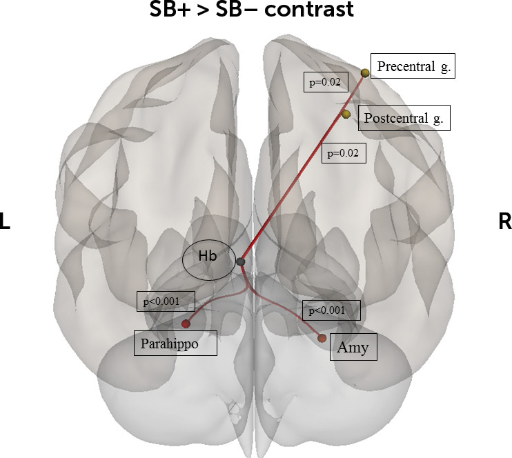
The present study investigated habenular rsFC differences between patients with a diagnosis of mood disorder, with and without a history of suicide related-behaviors, and healthy controls. To the best of our knowledge, it represents the largest study investigating neuroimaging correlates of suicidal-related behaviors in a population of mood disorder patients. As hypothesized, we found that the SB+ group had higher connectivity between the left habenula and several regions: left habenula and left parahippocampal gyrus, left and right amygdala, and right precentral and postcentral gyri. Moreover, patients with a diagnosis of mood disorder, independently of suicide-related status, displayed higher connectivity between the left habenula and middle temporal gyrus, angular gyrus, and PCC as well as decreased connectivity between the right habenula and left thalamus. The present results suggest that altered connectivity of the habenula with regions of the limbic and motor systems might be involved in the neural basis of suicide-related behaviors.
Typically described as composed by medial and lateral subregions, the habenula controls behavior, cognition, and emotion through extensive inputs from the basal ganglia and the limbic regions.33 Based on these inputs, the fasciculus retroflexus carries outputs that target dopaminergic and serotonergic neurons.12 By inhibiting the firing activity of the dopamine and serotonin systems, the habenula facilitates the evolutionary mechanism of action avoidance when the outcome is less rewarding than predicted.11
Our data support the evidence that altered activity of the habenula contributes to the etiology of psychiatric disorder, including depression.34 Evidence that the habenula is hyperactive during depression comes from animal models of depression33 and pharmacological studies.35 Neuroimaging studies of habenula activity in patients with MDD showed contrasting results. Although increased activity of the habenula was detected in remitted patients with MDD,36 reduced activity was observed in depressed MDD during the prediction of negative outcome37 and primary aversive conditioning.38 The investigation of an association between habenula connectivity and suicide is new to the literature. However, the higher connectivity of the left habenula we found in the SB+ group is somewhat in line with the disrupted habenula function observed in depression. Thus, it is tempting to speculate that in the SB+ group the higher connectivity with regions of the limbic and motor systems might be related, at least partially, to a possible increased sensitivity of habenula cells induced by chronic stress.12,39,40 Although the exact mechanism that links suicide and chronic stress is not fully understood, stress-related alterations in cell structure have been documented in suicide attempters.41 Chronic dysregulation of the habenula circuit may lead to long-term alterations of dopamine, serotonin, and norepinephrine release that impact behavioral functioning, particularly the learning of maladaptive coping strategies related to suicide-related behaviors.42 Interestingly, the parahippocampal gyrus has been linked to suicidality: in an fMRI experiment using a delay discount task, suicidal patients showed longer reward delays were associated with diminished parahippocampal response.43 The parahippocampal cortex has been hypothesized to be a critical part of a network of brain regions that processes contextual associations.44 This makes functional connectivity with the habenula a possibly important feature of contextual associations in general, and especially those that include reward as an important feature.
Interestingly, the left habenula was strongly associated with suicidality, while the right habenula was much less involved. Habenular asymmetry has been shown to be critically important for behavior in fish,45 and more recently, rsFC of the habenula in healthy humans was shown to be asymmetric.46 Our data suggest that habenular asymmetry may also be important for psychiatric disorder etiology, which may have implications in terms of future therapeutic intervention development, especially for neuromodulatory approaches. For example, deep brain stimulation of the habenula has been shown to be possibly therapeutic in treatment-resistant MDD.47 This might imply that unilateral habenula stimulation, which arguably could have lesser side effects, should be explored.
Not surprisingly, significant results were also found when the medial thalamic seed was considered. While we cannot exclude this might be partially attributable to the spatial distortion of the MRI images, it could also be attributable to shared connectivity patterns between the habenula and the medial nuclei of the thalamus. Overlapping and mutually interacting circuits (e.g., similar inputs from the ventral/limbic striatum) between the two regions have already been described.17,48
It is important to address several limitations in the present study. First, the reliability of in vivo study of the habenula through fMRI has been debated,17,49 considering the small size of this structure. We tried to temper this caveat using individual-specific habenula regions of interest, without attempting to isolate the medial and lateral subdivisions. However, we cannot exclude that head movement as well as spatial resolution and smoothing might have contributed to a signal contamination from the surrounding regions. Second, the current study examined suicide behavior in a mixed sample of depressed patients diagnosed with MDD or bipolar disorder. Although this study includes patients with diagnostic heterogeneity, the two disorders tend to be more similar than not when it comes to suicide-related behaviors.50 All individuals in the SB+ and SB– groups were prescribed multiple medications at the time of MRI. Although medication status was secondarily included as covariates in the analyses performed here, the potential effects of medication status on rsFC cannot be disentangled fully from those effects attributable to suicidal behavior. It is noteworthy that in a recent review of the literature,51 the authors concluded that medication effects do not seem to provide an alternative explanation for observed differences in BOLD signal.
In summary, the present findings suggest that the habenula might be involved in the neural circuitry of suicide. As such, the higher habenular rsFC we found in the suicide-related behaviors group may mediate a dysfunction in the critical survival mechanism by which the habenula works as a suppressor of motor activity. Nonetheless, additional research focused on the identification of neuroimaging differences between patients with only suicidal ideation from those with suicide-related behaviors is needed in order to identify patients at higher risk to attempt suicide.

Acknowledgments

The authors thank the Core for Advanced MRI at Baylor College of Medicine and Dr. Charles Neblett.

Footnote

The study sponsors were not involved in any aspect of research activities and did not approve the specific protocol or manuscript.

References

1.
Oquendo MA, Galfalvy H, Russo S, et al: Prospective study of clinical predictors of suicidal acts after a major depressive episode in patients with major depressive disorder or bipolar disorder. Am J Psychiatry 2004; 161:1433–1441
2.
Hawton K, van Heeringen K: Suicide. Lancet 2009; 373:1372–1381
3.
Dombrovski AY, Szanto K, Clark L, et al: Reward signals, attempted suicide, and impulsivity in late-life depression. JAMA Psychiatry 2013; 70:1020–1030
4.
Auerbach RP, Millner AJ, Stewart JG, et al: Identifying differences between depressed adolescent suicide ideators and attempters. J Affect Disord 2015; 186:127–133
5.
Oquendo MA, Sullivan GM, Sudol K, et al: Toward a biosignature for suicide. Am J Psychiatry 2014; 171:1259–1277
6.
Cox Lippard ET, Johnston JA, Blumberg HP: Neurobiological risk factors for suicide: insights from brain imaging. Am J Prev Med 2014; 47:S152–S162
7.
Serafini G, Pardini M, Pompili M, et al: Understanding suicidal behavior: the contribution of recent resting-state fMRI techniques. Front Psychiatry 2016; 7:69
8.
Oquendo MA, Galfalvy H, Sullivan GM, et al: Positron emission tomographic imaging of the serotonergic system and prediction of risk and lethality of future suicidal behavior. JAMA Psychiatry 2016; 73:1048–1055
9.
Spoletini I, Piras F, Fagioli S, et al: Suicidal attempts and increased right amygdala volume in schizophrenia. Schizophr Res 2011; 125:30–40
10.
Klemm WR: Habenular and interpeduncularis nuclei: shared components in multiple-function networks. Med Sci Monit 2004; 10:RA261–RA273
11.
Hikosaka O, Sesack SR, Lecourtier L, et al: Habenula: crossroad between the basal ganglia and the limbic system. J Neurosci 2008; 28:11825–11829
12.
Hikosaka O: The habenula: from stress evasion to value-based decision-making. Nat Rev Neurosci 2010; 11:503–513
13.
Salas R, Baldwin P, de Biasi M, et al: BOLD responses to negative reward prediction errors in human habenula. Front Hum Neurosci 2010; 4:36
14.
Fakhoury M: The habenula in psychiatric disorders: more than three decades of translational investigation. Neurosci Biobehav Rev 2017; 83:721–735.
15.
Velasquez KM, Molfese DL, Salas R: The role of the habenula in drug addiction. Front Hum Neurosci 2014; 8:174
16.
Proulx CD, Hikosaka O, Malinow R: Reward processing by the lateral habenula in normal and depressive behaviors. Nat Neurosci 2014; 17:1146–1152
17.
Ely BA, Xu J, Goodman WK, et al: Resting-state functional connectivity of the human habenula in healthy individuals: associations with subclinical depression. Hum Brain Mapp 2016; 37:2369–2384
18.
Aizawa H, Cui W, Tanaka K, et al: Hyperactivation of the habenula as a link between depression and sleep disturbance. Front Hum Neurosci 2013; 7:826
19.
Conway KP, Compton W, Stinson FS, et al: Lifetime comorbidity of DSM-IV mood and anxiety disorders and specific drug use disorders: results from the National Epidemiologic Survey on Alcohol and Related Conditions. J Clin Psychiatry 2006; 67:247–257
20.
Greene DJ, Black KJ, Schlaggar BL: Considerations for MRI study design and implementation in pediatric and clinical populations. Dev Cogn Neurosci 2016; 18:101–112
21.
First M, Gibbon M, Spitzer R: Structured Clinical Interview for DSM-IV Axis II Personality Disorders (SCID-II). Washington, DC, American Psychiatric Publishing, 1997
22.
First MB, Spitzer RL, Gibbon M, et al: Structured Clinical Interview for DSM-IV-TR Axis I Disorders. New York, New York State Psychiatric Institute, 2002
23.
Posner K, Brown GK, Stanley B, et al: The Columbia-Suicide Severity Rating Scale: initial validity and internal consistency findings from three multisite studies with adolescents and adults. Am J Psychiatry 2011; 168:1266–1277
24.
Madan A, Frueh BC, Allen JG, et al: Psychometric reevaluation of the Columbia-Suicide Severity Rating Scale: findings from a prospective, inpatient cohort of severely mentally ill adults. J Clin Psychiatry 2016; 77:e867–e873
25.
Minzenberg MJ, Lesh T, Niendam T, et al: Conflict-related anterior cingulate functional connectivity is associated with past suicidal ideation and behavior in recent-onset schizophrenia. J Psychiatr Res 2015; 65:95–101
26.
Minzenberg MJ, Lesh TA, Niendam TA, et al: Conflict-related anterior cingulate functional connectivity is associated with past suicidal ideation and behavior in recent-onset psychotic major mood disorders. J Neuropsychiatry Clin Neurosci 2016; 28:299–305
27.
Löwe B, Kroenke K, Herzog W, et al: Measuring depression outcome with a brief self-report instrument: sensitivity to change of the Patient Health Questionnaire (PHQ-9). J Affect Disord 2004; 81:61–66
28.
Kroenke K, Spitzer RL, Williams JBW: The PHQ-9: validity of a brief depression severity measure. J Gen Intern Med 2001; 16:606–613
29.
Whitfield-Gabrieli S, Nieto-Castanon A: Conn: a functional connectivity toolbox for correlated and anticorrelated brain networks. Brain Connect 2012; 2:125–141
30.
Behzadi Y, Restom K, Liau J, et al: A component based noise correction method (CompCor) for BOLD and perfusion based fMRI. Neuroimage 2007; 37:90–101
31.
Chai XJ, Castañón AN, Ongür D, et al: Anticorrelations in resting state networks without global signal regression. Neuroimage 2012; 59:1420–1428
32.
Tzourio-Mazoyer N, Landeau B, Papathanassiou D, et al: Automated anatomical labeling of activations in SPM using a macroscopic anatomical parcellation of the MNI MRI single-subject brain. Neuroimage 2002; 15:273–289
33.
Benarroch EE: Habenula: recently recognized functions and potential clinical relevance. Neurology 2015; 85:992–1000
34.
Loonen AJM, Ivanova SA: Circuits regulating pleasure and happiness: mechanisms of depression. Front Hum Neurosci 2016; 10:571
35.
Freo U, Ori C, Dam M, et al: Effects of acute and chronic treatment with fluoxetine on regional glucose cerebral metabolism in rats: implications for clinical therapies. Brain Res 2000; 854:35–41
36.
Roiser JP, Levy J, Fromm SJ, et al: The effects of tryptophan depletion on neural responses to emotional words in remitted depression. Biol Psychiatry 2009; 66:441–450
37.
Furman DJ, Gotlib IH: Habenula responses to potential and actual loss in major depression: preliminary evidence for lateralized dysfunction. Soc Cogn Affect Neurosci 2016; 11:843–851
38.
Lawson RP, Drevets WC, Roiser JP: Defining the habenula in human neuroimaging studies. Neuroimage 2013; 64:722–727
39.
Kaye A, Ross DA: The habenula: darkness, disappointment, and depression. Biol Psychiatry 2017; 81:e27–e28
40.
Li B, Piriz J, Mirrione M, et al: Synaptic potentiation onto habenula neurons in the learned helplessness model of depression. Nature 2011; 470:535–539
41.
Lindqvist D, Fernström J, Grudet C, et al: Increased plasma levels of circulating cell-free mitochondrial DNA in suicide attempters: associations with HPA-axis hyperactivity. Transl Psychiatry 2016; 6:e971
42.
Brüdern J, Berger T, Michel K, et al: Are suicide attempters wired differently? A comparison with nonsuicidal depressed individuals using plan analysis. J Nerv Ment Dis 2015; 203:514–521
43.
Vanyukov PM, Szanto K, Hallquist MN, et al: Paralimbic and lateral prefrontal encoding of reward value during intertemporal choice in attempted suicide. Psychol Med 2016; 46:381–391
44.
Aminoff EM, Kveraga K, Bar M: The role of the parahippocampal cortex in cognition. Trends Cogn Sci 2013; 17:379–390
45.
Aizawa H: Habenula and the asymmetric development of the vertebrate brain. Anat Sci Int 2013; 88:1–9
46.
Hétu S, Luo Y, Saez I, et al: Asymmetry in functional connectivity of the human habenula revealed by high-resolution cardiac-gated resting state imaging. Hum Brain Mapp 2016; 37:2602–2615
47.
Sartorius A, Kiening KL, Kirsch P, et al: Remission of major depression under deep brain stimulation of the lateral habenula in a therapy-refractory patient. Biol Psychiatry 2010; 67:e9–e11
48.
Ikemoto S, Yang C, Tan A: Basal ganglia circuit loops, dopamine and motivation: A review and enquiry. Behav Brain Res 2015; 290:17–31
49.
Loonen AJM, Ivanova SA: Commentary on “a non-reward attractor theory of depression”: a proposal to include the habenula connection. Neurosci Biobehav Rev 2017; 83:736–741
50.
Nock MK, Hwang I, Sampson NA, et al: Mental disorders, comorbidity and suicidal behavior: results from the National Comorbidity Survey Replication. Mol Psychiatry 2010; 15:868–876
51.
Hafeman DM, Chang KD, Garrett AS, et al: Effects of medication on neuroimaging findings in bipolar disorder: an updated review. Bipolar Disord 2012; 14:375–410

Information & Authors

Information

Published In

Go to The Journal of Neuropsychiatry and Clinical Neurosciences
The Journal of Neuropsychiatry and Clinical Neurosciences
Pages: 49 - 56
PubMed: 30282513

History

Received: 12 December 2017
Revision received: 19 March 2018
Accepted: 16 May 2018
Published online: 4 October 2018
Published in print: Winter 2019

Keywords

  1. Habenula
  2. Bipolar Disorder
  3. Major Depressive Disorder
  4. Suicide
  5. Resting-State fMRI

Authors

Affiliations

Elisa Ambrosi, M.D.
From the Menninger Department of Psychiatry and Behavioral Sciences, Baylor College of Medicine, Houston (EA, DAB, KNC, MAP, GiSp, JCF, AM, RS); Department of Neurosciences, Baylor College of Medicine, Houston; the Beth K. and Stuart C. Yudofsky Division of Neuropsychiatry, Baylor College of Medicine, Houston (EA, DAB, GiSp);the Menninger Clinic, Houston (MAP); the Laboratory of Neuropsychiatry, IRCCS Santa Lucia Foundation, Rome Italy (GiSp); Centro Lucio Bini, Roma, Italy (GaSa); the Michael E. DeBakey VA Medical Center, Houston (KNC, MAP, RS); the Department of Neurosciences, Mental Health, and Sensory Organs, Sapienza University, Rome (GaSa); the School of Medicine and Psychology, Sant’ Andrea Hospital, Rome (GaSa); Tufts Medical Center, Tufts University School of Medicine, Boston (GaSa); and the Department of Psychology, University of Hawaii, Hilo, HI (BCF), Trauma and Resilience Center, Department of Psychiatry and Behavioral Sciences, University of Texas Health Sciences Center, Houston (BCF).
David B. Arciniegas, M.D.
From the Menninger Department of Psychiatry and Behavioral Sciences, Baylor College of Medicine, Houston (EA, DAB, KNC, MAP, GiSp, JCF, AM, RS); Department of Neurosciences, Baylor College of Medicine, Houston; the Beth K. and Stuart C. Yudofsky Division of Neuropsychiatry, Baylor College of Medicine, Houston (EA, DAB, GiSp);the Menninger Clinic, Houston (MAP); the Laboratory of Neuropsychiatry, IRCCS Santa Lucia Foundation, Rome Italy (GiSp); Centro Lucio Bini, Roma, Italy (GaSa); the Michael E. DeBakey VA Medical Center, Houston (KNC, MAP, RS); the Department of Neurosciences, Mental Health, and Sensory Organs, Sapienza University, Rome (GaSa); the School of Medicine and Psychology, Sant’ Andrea Hospital, Rome (GaSa); Tufts Medical Center, Tufts University School of Medicine, Boston (GaSa); and the Department of Psychology, University of Hawaii, Hilo, HI (BCF), Trauma and Resilience Center, Department of Psychiatry and Behavioral Sciences, University of Texas Health Sciences Center, Houston (BCF).
Kaylah N. Curtis, M.P.H.
From the Menninger Department of Psychiatry and Behavioral Sciences, Baylor College of Medicine, Houston (EA, DAB, KNC, MAP, GiSp, JCF, AM, RS); Department of Neurosciences, Baylor College of Medicine, Houston; the Beth K. and Stuart C. Yudofsky Division of Neuropsychiatry, Baylor College of Medicine, Houston (EA, DAB, GiSp);the Menninger Clinic, Houston (MAP); the Laboratory of Neuropsychiatry, IRCCS Santa Lucia Foundation, Rome Italy (GiSp); Centro Lucio Bini, Roma, Italy (GaSa); the Michael E. DeBakey VA Medical Center, Houston (KNC, MAP, RS); the Department of Neurosciences, Mental Health, and Sensory Organs, Sapienza University, Rome (GaSa); the School of Medicine and Psychology, Sant’ Andrea Hospital, Rome (GaSa); Tufts Medical Center, Tufts University School of Medicine, Boston (GaSa); and the Department of Psychology, University of Hawaii, Hilo, HI (BCF), Trauma and Resilience Center, Department of Psychiatry and Behavioral Sciences, University of Texas Health Sciences Center, Houston (BCF).
Michelle A. Patriquin, Ph.D.
From the Menninger Department of Psychiatry and Behavioral Sciences, Baylor College of Medicine, Houston (EA, DAB, KNC, MAP, GiSp, JCF, AM, RS); Department of Neurosciences, Baylor College of Medicine, Houston; the Beth K. and Stuart C. Yudofsky Division of Neuropsychiatry, Baylor College of Medicine, Houston (EA, DAB, GiSp);the Menninger Clinic, Houston (MAP); the Laboratory of Neuropsychiatry, IRCCS Santa Lucia Foundation, Rome Italy (GiSp); Centro Lucio Bini, Roma, Italy (GaSa); the Michael E. DeBakey VA Medical Center, Houston (KNC, MAP, RS); the Department of Neurosciences, Mental Health, and Sensory Organs, Sapienza University, Rome (GaSa); the School of Medicine and Psychology, Sant’ Andrea Hospital, Rome (GaSa); Tufts Medical Center, Tufts University School of Medicine, Boston (GaSa); and the Department of Psychology, University of Hawaii, Hilo, HI (BCF), Trauma and Resilience Center, Department of Psychiatry and Behavioral Sciences, University of Texas Health Sciences Center, Houston (BCF).
Gianfranco Spalletta, M.D., Ph.D.
From the Menninger Department of Psychiatry and Behavioral Sciences, Baylor College of Medicine, Houston (EA, DAB, KNC, MAP, GiSp, JCF, AM, RS); Department of Neurosciences, Baylor College of Medicine, Houston; the Beth K. and Stuart C. Yudofsky Division of Neuropsychiatry, Baylor College of Medicine, Houston (EA, DAB, GiSp);the Menninger Clinic, Houston (MAP); the Laboratory of Neuropsychiatry, IRCCS Santa Lucia Foundation, Rome Italy (GiSp); Centro Lucio Bini, Roma, Italy (GaSa); the Michael E. DeBakey VA Medical Center, Houston (KNC, MAP, RS); the Department of Neurosciences, Mental Health, and Sensory Organs, Sapienza University, Rome (GaSa); the School of Medicine and Psychology, Sant’ Andrea Hospital, Rome (GaSa); Tufts Medical Center, Tufts University School of Medicine, Boston (GaSa); and the Department of Psychology, University of Hawaii, Hilo, HI (BCF), Trauma and Resilience Center, Department of Psychiatry and Behavioral Sciences, University of Texas Health Sciences Center, Houston (BCF).
Gabriele Sani, M.D.
From the Menninger Department of Psychiatry and Behavioral Sciences, Baylor College of Medicine, Houston (EA, DAB, KNC, MAP, GiSp, JCF, AM, RS); Department of Neurosciences, Baylor College of Medicine, Houston; the Beth K. and Stuart C. Yudofsky Division of Neuropsychiatry, Baylor College of Medicine, Houston (EA, DAB, GiSp);the Menninger Clinic, Houston (MAP); the Laboratory of Neuropsychiatry, IRCCS Santa Lucia Foundation, Rome Italy (GiSp); Centro Lucio Bini, Roma, Italy (GaSa); the Michael E. DeBakey VA Medical Center, Houston (KNC, MAP, RS); the Department of Neurosciences, Mental Health, and Sensory Organs, Sapienza University, Rome (GaSa); the School of Medicine and Psychology, Sant’ Andrea Hospital, Rome (GaSa); Tufts Medical Center, Tufts University School of Medicine, Boston (GaSa); and the Department of Psychology, University of Hawaii, Hilo, HI (BCF), Trauma and Resilience Center, Department of Psychiatry and Behavioral Sciences, University of Texas Health Sciences Center, Houston (BCF).
B. Christopher Frueh, Ph.D.
From the Menninger Department of Psychiatry and Behavioral Sciences, Baylor College of Medicine, Houston (EA, DAB, KNC, MAP, GiSp, JCF, AM, RS); Department of Neurosciences, Baylor College of Medicine, Houston; the Beth K. and Stuart C. Yudofsky Division of Neuropsychiatry, Baylor College of Medicine, Houston (EA, DAB, GiSp);the Menninger Clinic, Houston (MAP); the Laboratory of Neuropsychiatry, IRCCS Santa Lucia Foundation, Rome Italy (GiSp); Centro Lucio Bini, Roma, Italy (GaSa); the Michael E. DeBakey VA Medical Center, Houston (KNC, MAP, RS); the Department of Neurosciences, Mental Health, and Sensory Organs, Sapienza University, Rome (GaSa); the School of Medicine and Psychology, Sant’ Andrea Hospital, Rome (GaSa); Tufts Medical Center, Tufts University School of Medicine, Boston (GaSa); and the Department of Psychology, University of Hawaii, Hilo, HI (BCF), Trauma and Resilience Center, Department of Psychiatry and Behavioral Sciences, University of Texas Health Sciences Center, Houston (BCF).
J. Christopher Fowler, Ph.D.
From the Menninger Department of Psychiatry and Behavioral Sciences, Baylor College of Medicine, Houston (EA, DAB, KNC, MAP, GiSp, JCF, AM, RS); Department of Neurosciences, Baylor College of Medicine, Houston; the Beth K. and Stuart C. Yudofsky Division of Neuropsychiatry, Baylor College of Medicine, Houston (EA, DAB, GiSp);the Menninger Clinic, Houston (MAP); the Laboratory of Neuropsychiatry, IRCCS Santa Lucia Foundation, Rome Italy (GiSp); Centro Lucio Bini, Roma, Italy (GaSa); the Michael E. DeBakey VA Medical Center, Houston (KNC, MAP, RS); the Department of Neurosciences, Mental Health, and Sensory Organs, Sapienza University, Rome (GaSa); the School of Medicine and Psychology, Sant’ Andrea Hospital, Rome (GaSa); Tufts Medical Center, Tufts University School of Medicine, Boston (GaSa); and the Department of Psychology, University of Hawaii, Hilo, HI (BCF), Trauma and Resilience Center, Department of Psychiatry and Behavioral Sciences, University of Texas Health Sciences Center, Houston (BCF).
Alok Madan, Ph.D., M.P.H.
From the Menninger Department of Psychiatry and Behavioral Sciences, Baylor College of Medicine, Houston (EA, DAB, KNC, MAP, GiSp, JCF, AM, RS); Department of Neurosciences, Baylor College of Medicine, Houston; the Beth K. and Stuart C. Yudofsky Division of Neuropsychiatry, Baylor College of Medicine, Houston (EA, DAB, GiSp);the Menninger Clinic, Houston (MAP); the Laboratory of Neuropsychiatry, IRCCS Santa Lucia Foundation, Rome Italy (GiSp); Centro Lucio Bini, Roma, Italy (GaSa); the Michael E. DeBakey VA Medical Center, Houston (KNC, MAP, RS); the Department of Neurosciences, Mental Health, and Sensory Organs, Sapienza University, Rome (GaSa); the School of Medicine and Psychology, Sant’ Andrea Hospital, Rome (GaSa); Tufts Medical Center, Tufts University School of Medicine, Boston (GaSa); and the Department of Psychology, University of Hawaii, Hilo, HI (BCF), Trauma and Resilience Center, Department of Psychiatry and Behavioral Sciences, University of Texas Health Sciences Center, Houston (BCF).
Ramiro Salas, Ph.D. [email protected]
From the Menninger Department of Psychiatry and Behavioral Sciences, Baylor College of Medicine, Houston (EA, DAB, KNC, MAP, GiSp, JCF, AM, RS); Department of Neurosciences, Baylor College of Medicine, Houston; the Beth K. and Stuart C. Yudofsky Division of Neuropsychiatry, Baylor College of Medicine, Houston (EA, DAB, GiSp);the Menninger Clinic, Houston (MAP); the Laboratory of Neuropsychiatry, IRCCS Santa Lucia Foundation, Rome Italy (GiSp); Centro Lucio Bini, Roma, Italy (GaSa); the Michael E. DeBakey VA Medical Center, Houston (KNC, MAP, RS); the Department of Neurosciences, Mental Health, and Sensory Organs, Sapienza University, Rome (GaSa); the School of Medicine and Psychology, Sant’ Andrea Hospital, Rome (GaSa); Tufts Medical Center, Tufts University School of Medicine, Boston (GaSa); and the Department of Psychology, University of Hawaii, Hilo, HI (BCF), Trauma and Resilience Center, Department of Psychiatry and Behavioral Sciences, University of Texas Health Sciences Center, Houston (BCF).

Notes

Send correspondence to Dr. Salas; e-mail: [email protected]

Competing Interests

The authors report no financial relationships with commercial interests.

Funding Information

Office of Research and Development10.13039/100006379: VHA5I01CX000994
National Institute on Drug Abuse10.13039/100000026: DA026539, DA09167
Brain and Behavior Research Foundation10.13039/100000874: 19295
Robert and Janice McNair Foundation10.13039/100001234: MIND-MB
American Foundation for Suicide Prevention10.13039/100001455: SRG-2-125-14
Dr. Arciniegas is Editor of the Journal of Neuropsychiatry and Clinical Neurosciences.Deputy Editor C. Alan Anderson, M.D., oversaw peer review and served as decision editor.Dr. Madan is a McNair Scholar.Supported by the American Foundation for Suicide Prevention (grant SRG-2-125-14); the McNair Medical Institute; NARSAD (grant 19295); NIH (grants NIDA DA026539 and DA09167); and the Veterans Health Administration (grant VHA5I01CX000994). Supported in part by the Brown Foundation, the George and Mary Josephine Hamman Foundation, the Menninger Clinic Foundation, and the Toomim Family Fund. This article is partly the result of work supported with resources and use of facilities at the Michael E. DeBakey VA Medical Center, Houston.

Metrics & Citations

Metrics

Citations

Export Citations

If you have the appropriate software installed, you can download article citation data to the citation manager of your choice. Simply select your manager software from the list below and click Download.

For more information or tips please see 'Downloading to a citation manager' in the Help menu.

Format
Citation style
Style
Copy to clipboard

There are no citations for this item

View Options

View options

PDF/ePub

View PDF/ePub

Get Access

Login options

Already a subscriber? Access your subscription through your login credentials or your institution for full access to this article.

Personal login Institutional Login Open Athens login
Purchase Options

Purchase this article to access the full text.

PPV Articles - Journal of Neuropsychiatry and Clinical Neurosciences

PPV Articles - Journal of Neuropsychiatry and Clinical Neurosciences

Not a subscriber?

Subscribe Now / Learn More

PsychiatryOnline subscription options offer access to the DSM-5-TR® library, books, journals, CME, and patient resources. This all-in-one virtual library provides psychiatrists and mental health professionals with key resources for diagnosis, treatment, research, and professional development.

Need more help? PsychiatryOnline Customer Service may be reached by emailing [email protected] or by calling 800-368-5777 (in the U.S.) or 703-907-7322 (outside the U.S.).

Media

Figures

Other

Tables

Share

Share

Share article link

Share