
Abstract

Objective:

The selection of a bitemporal (BT) or right unilateral (RUL) electrode placement affects the efficacy and side effects of ECT. Previous studies have not entirely described the neurobiological underpinnings of such differential effects. Recent neuroimaging research on gray matter volumes is contributing to our understanding of the mechanism of action of ECT and could clarify the differential mechanisms of BT and RUL ECT.

Methods:

To assess the whole-brain gray matter volumetric changes observed after treating patients with treatment-resistant depression with BT or RUL ECT, the authors used MRI to assess 24 study subjects with treatment-resistant depression (bifrontotemporal ECT, N=12; RUL ECT, N=12) at two time points (before the first ECT session and after ECT completion).

Results:

Study subjects receiving BT ECT showed gray matter volume increases in the bilateral limbic system, but subjects treated with RUL ECT showed gray matter volume increases limited to the right hemisphere. The authors observed significant differences between the two groups in midtemporal and subcortical limbic structures in the left hemisphere.

Conclusions:

These findings highlight that ECT-induced gray matter volume increases may be specifically observed in the stimulated hemispheres. The authors suggest that electrode placement may relevantly contribute to the development of personalized ECT protocols.
ECT is an effective antidepressant treatment for patients with treatment-resistant depression.1 Therapeutic efficacy and cognitive side effects depend on a number of stimulation parameters (such as electrode placement, pulse width, etc.), but the specific effects of these variables on brain anatomy and physiology are poorly understood. Understanding the effects of treatment parameters on brain biology is a missing critical step toward the rational development of therapeutic innovations, particularly when considering individualization strategies focused on target engagement.
Electrode placement is a critical ECT parameter, with both therapeutic and side-effect implications. The two most common electrode placements are bitemporal (BT) and right unilateral (RUL [or d’Elia electrode placement]2) ECT. In 2011, the Food and Drug Administration (FDA)3 concluded that BT ECT is the most effective electrode placement. Nevertheless, RUL ECT has been increasingly prevalent in the past few decades given its more tolerable profile.4 Challenging common clinical assumptions, some studies5,6 have suggested that RUL ECT is not inferior in efficacy compared with BT ECT. Altogether, it seems that our knowledge of the comparative efficacy and side-effect profiles of RUL and BT treatments could be partially revised with additional nuances, and these clinical considerations would benefit from a mechanistic understanding of the distinct biological effects of these treatment parameters.
Gray matter volume increases, mainly in the limbic temporal lobe, have been observed after RUL and BT ECT.719 Although most studies have not found an association between volumetric and clinical changes, some have, although not always in the same directions.10,16,19,20 The nature of these changes may shed light on the mechanism of action of ECT, but recent studies do not allow a clear disambiguation between the mechanisms of BT versus RUL ECT. Indeed, only one recent structural neuroimaging study assessed the effect of RUL versus bilateral (that is, bitemporal and bifrontal) ECT. However, as opposed to our whole-brain approach, the authors limited their focus to volumetric changes in the hippocampus.20 Results from this study indicated that electrode placement determines the extent of volume change in the right and left hemispheres, with bilateral electrode placement being associated with bilateral hippocampal changes, whereas RUL electrode placement was preferentially associated with right hippocampal changes. In addition to these volumetric differences, previous neurophysiological studies have shown that slow-wave activity after RUL ECT was observed in the right hemisphere, whereas slow-wave activity was more prominent over the left hemisphere with BT ECT.21
In the present study, we aimed to assess the effects of ECT electrode placement, and specifically the differential impact of RUL versus bifrontotemporal ECT, on whole-brain volumetric changes in study subjects with treatment-resistant depression. We hypothesized that despite the common causal association with the ECT-induced generalized seizure, RUL and BT electrode placements would lead to different topographic distributions of the volumetric changes observed after ECT: specifically, study subjects treated with BT ECT, in comparison with subjects receiving RUL treatment, would show greater gray matter volume increases in the left hemisphere.

Methods

Participants

We recruited 24 study subjects with treatment-resistant depression from two research centers: the Mood Disorders Inpatient Unit of Bellvitge University Hospital, Barcelona, Spain (N=12), and the Department of Psychiatry at Massachusetts General Hospital (MGH), Boston (N=12). The mean age of study subjects from Bellvitge University Hospital was 59.17 years (SD=8.02), and the mean age of the study subjects from MGH was 42.25 years (SD=15.78).
Fifty percent of subjects from each cohort were men (Bellvitge, N=6; MGH, N=6).
Age and gender were used as covariates for statistical analyses. All study subjects met criteria for a major depressive episode according to DSM-IV-TR22 criteria as well as the following structural clinical interviews: the Structured Clinical Interview for DSM-IV Axis I Disorders–Clinician Version (SCID)23, for the Bellvitge University Hospital cohort, and the Mini-International Neuropsychiatric Interview 6.0 (MINI)24, for the MGH cohort. Exclusion criteria included 1) the presence or past history of a severe medical illness or neurological disorder, 2) contraindication to MRI scanning or abnormal MRI on visual inspection, and 3) a history of ECT during the past 12 months. Pharmacotherapy remained unchanged throughout the ECT protocol, with close monitoring for unwanted adverse effects (Table 1; see also Table S1 in the online supplement). Moreover, subjects in the Bellvitge University Hospital cohort underwent longitudinal clinical assessment using the 21-item Hamilton Depression Rating Scale (HAM-D),25 and subjects in the MGH cohort were assessed with the Quick Inventory of Depressive Symptomatology (QIDS).26 The study was approved by the local ethical review board of each center and was performed in accordance with the Declaration of Helsinki. All participants provided written, informed consent after a detailed description of the study was given.
TABLE 1. Concurrent Pharmacological Regimen for the Two Study Samplesa
DrugsBitemporal ECT (N=12)Right Unilateral ECT (N=11)b
N%N%
Antidepressants12100.01090.9
Antipsychotics975.0545.4
Lithium216.700
Anxiolytics650.0436.4
a
All study subjects had treatment-resistant depression.
b
Data were missing for one participant.

ECT

The 12 subjects in the Bellvitge University Hospital cohort were treated with bifrontotemporal brief-pulse (0.5–1 ms) ECT using a Thymatron System IV device (Somatics, Lake Bluff, Ill.). Anesthesia was induced with intravenous thiopental (2–2.5 mg kg−1), and succinylcholine (0.5 mg kg−1) was used for muscle paralysis. Initial stimulus dose was determined by using the half-age method,27 and subsequent dosing was determined according to seizure morphology adequacy. The 12 subjects in the MGH cohort were treated with ultra-brief pulse and brief pulse (0.3–0.5 ms) RUL ECT (d’Elia electrode placement2) using a MECTA spECTrum 5000Q machine (Portland, Ore.). Anesthesia for the procedure was provided using methohexital (0.8–1.2 mg kg−1) for induction and succinylcholine for muscle relaxation (0.5–1 mg kg−1). Initial stimulus dose was determined by titrating the seizure threshold, starting with 19 millicoulombs for women and 38 millicoulombs for men. Subsequent treatments were performed at six times the estimated seizure threshold. During an acute course of ECT, treatments were administered three times per week every other day at both centers.

Image Acquisition and Preprocessing

All study subjects were scanned two times: before the first ECT session (MRI 1) and after completion of the ECT course (MRI 2). A 3.0-T structural T1-weighted MRI high-resolution structural scan was locally acquired for each participant. Study subjects in the Bellvitge University Hospital cohort were scanned with a Philips Achieva 3.0-Tesla magnet scanner (Amsterdam) equipped with an eight-channel phased-array head coil (160 slices, repetition time=8.1 ms, echo time=3.7 ms, flip angle=8°, field of view=240×240 mm, matrix size=256×256 pixels, in-plane resolution=0.94×0.94 mm2, slice thickness=1 mm). Study subjects in the MGH cohort were scanned at the Martinos Center for Biomedical Imaging with a Siemens Skyra 3.0-Tesla magnet scanner (Munich, Germany) equipped with a 32-channel head coil (156 slices, repetition time=2,530 ms, multi-echo time=1.69, 3.55, 5.41, and 7.27 ms, flip angle=7°, field of view=256×256 mm, matrix size=256×256 pixels, in-plane resolution=1×1 mm2, slice thickness=1 mm).
All structural MRI data were processed on a Microsoft Windows platform using technical computing software (MATLAB, 7.14, MathWorks, Natick, Mass.) and Statistical Parametric Mapping (SPM12, Welcome Department of Imaging Neuroscience, London). The preprocessing consisted of an initial rigid-body within-subject coregistration to the first scan to ensure good starting estimates. This was followed by a pairwise longitudinal registration between the scans of each participant to obtain an average image and a Jacobian difference map. The average image was segmented, and the gray matter voxels were multiplied by the Jacobian difference map to obtain a gray matter volume change map for each participant. Next, we generated one specific template of both study samples (in Montreal Neurological Institute space) using Diffeomorphic Anatomical Registration Through Exponentiated Lie Algebra algorithm,28,29 which was used to spatially normalize the gray matter volume change maps. Finally, images were smoothed with a 6-mm full-width at half maximum isotropic Gaussian kernel.

Statistical Analyses

Sociodemographic and clinical data were analyzed with SPSS version 21 (SPSS, Chicago) using nonparametric tests.
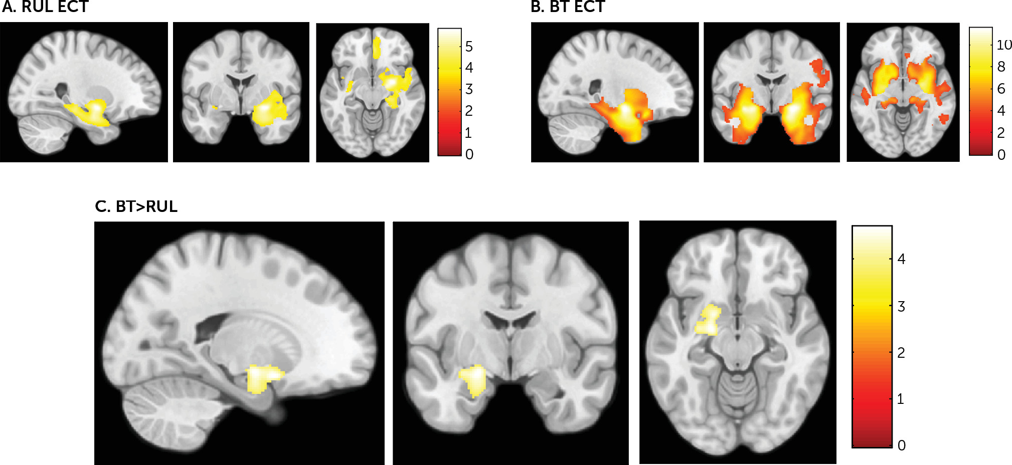
We used an independent two-sample model to derive a t statistic map comparing the gray matter volume change maps between participants with treatment-resistant depression treated with bifrontotemporal ECT (BT group) and participants with treatment-resistant depression treated with right unilateral ECT (RUL group). We initially estimated within-group volumetric changes (using two one-sample t tests at a p value <0.05, two-tailed, family-wise-error corrected for multiple comparisons across the whole brain) and created a combined mask (adding significant changes from both groups) in which we investigated between-group differences. This approach has been used as a strategy to increase the sensitivity directed toward our main objective (i.e., BT versus RUL ECT comparison). Age and gender were included as confounding covariates. Statistical significance was set at a p value <0.05 (two-tailed), family-wise-error corrected for multiple comparisons across all in-mask voxels (i.e., using small-volume correction procedures across all voxels showing volumetric changes in the RUL and BT groups). This mask contained 20,592 voxels.

Results

Sociodemographic and Clinical Characteristics

Twelve study subjects with treatment-resistant depression from the Bellvitge University Hospital cohort received an acute BT ECT course, averaging 11 ECT sessions per patient (mean ECT sessions, 11.08 [SD=1.50]). The mean HAM-D score before ECT initiation was 31.25 (SD=9.21), and the mean HAM-D score after completion of ECT was 2.92 (SD=2.54). At the end of treatment, all study subjects fulfilled clinical response criteria (>50% reduction in the HAM-D score), and all but one were in clinical remission (HAM-D score <8). Moreover, the reduction in depression severity (HAM-D score) between the MRI 1 and MRI 2 assessments was significant according to a Wilcoxon signed-rank test (z=–3.062, p=0.002, two-tailed).
On the other hand, 12 subjects with treatment-resistant depression from the MGH cohort received an acute RUL ECT course, averaging 10 ECT sessions per patient (mean ECT sessions, 10.33 [SD=2.42]). The mean QIDS score before ECT initiation was 17.42 (SD=3.34), and the mean QIDS score after completion of ECT was 11 (SD=4.90). At the end of treatment, four study subjects fulfilled clinical response criteria (>50% reduction in the QIDS score), and two were in clinical remission (QIDS score <6). The reduction in depression severity (QIDS score) between the MRI 1 and MRI 2 assessments was significant according to a Wilcoxon signed-rank test (z=–2.671, p=0.008, two-tailed).
Importantly, we observed a significantly (Mann-Whitney U test, z=–3.984, p<0.001, two-tailed) greater reduction in clinical severity among study subjects treated with BT ECT (89.26%, as measured by the percentage of change in HAM-D scores) compared with study subjects treated with RUL ECT (33.41%, as measured by the percentage of change in QIDS scores). Conversely, the number of ECT sessions did not significantly differ between groups (Mann-Whitney U test, z=–1.030, p<0.303, two-tailed).

Neuroimaging Analyses

The pattern of volumetric change between the MRI 1 and MRI 2 assessments for each group is shown in Figure 1A and 1B (p<0.001, uncorrected). Briefly, in the BT group, gray matter volume increases between both time points (before the first ECT session and after ECT completion) were symmetrically located in the bilateral limbic temporal lobes, bilateral insula, and bilateral striatum, whereas in the RUL group, gray matter volume changes were mainly located in the right limbic temporal lobe. Moreover, both groups showed gray matter volume increases in the right pregenual anterior cingulate cortex.
FIGURE 1. Brain Areas Showing Gray Matter Volume Increases in Subjects With Treatment-Resistant Depression Treated With Bitemporal (BT) ECT or Right Unilateral (RUL) ECTa
a Panel A shows the volume increases in the right limbic temporal lobe, the right pregenual anterior cingulate cortex, the left putamen, and the left insula in study subjects with treatment-resistant depression treated with RUL ECT. Panel B shows volume increases in the bilateral limbic temporal lobes, the bilateral insula, the bilateral striatum, and the right pregenual anterior cingulate cortex in study subjects with treatment-resistant depression treated with BT ECT. Panel C shows volume increases in the left limbic system (encompassing the ventral striatum and the amygdala and extending to the ventral hippocampus) in study subjects with treatment-resistant depression treated with BT ECT compared with study subjects treated with RUL ECT. Images are in anatomical norm. In panels A and B, the sagittal cut is of the right hemisphere; in panel C it is of the left hemisphere. The color bar represents the t value.
Importantly, the direct comparison between study subjects with treatment-resistant depression receiving BT and RUL ECT showed that subjects treated with bifrontotemporal ECT, compared with subjects who received right unilateral ECT, displayed a significantly greater gray matter volume change in the left medial-temporal region and other subcortical limbic structures, encompassing the ventral striatum (specifically, the ventral putamen portion) and the amygdala, extending to the ventral hippocampus (Table 2, Figure 1C). These results were significant after adjusting for the number of ECT sessions. Interestingly, the gray matter volume increase in the left medial-temporal structures was associated with clinical response in a previous study by our group (Cano et al.19).
TABLE 2. Brain Areas Showing Gray Matter Volume Increases in Subjects With Treatment-Resistant Depression Treated With Bitemporal ECT Compared With Subjects Treated With Right Unilateral ECT
ClusterMontreal Neurological Institute CoordinatesStatisticAnatomical Location
xyztdfpa
Left limbic system       
 –206–144.69200.027Left ventral striatum
 –21–6–124.65200.029Left amygdala
a
Family-wise-error corrected for multiple comparisons.

Discussion

This is the first study, to our knowledge, comparing the whole-brain volumetric correlates of BT and RUL ECT in study subjects with treatment-resistant depression. Consistent with previous research,719 we observed that ECT is associated with gray matter volume increases in limbic structures, including the ventral striatum, the amygdala, and the hippocampus. Indeed, neuroimaging, neuropathological, and lesion analysis studies have highlighted the importance of cortico-limbic circuit disruption in depression.30 However, our study provides novel insight regarding the gray matter volume increases associated with ECT electrode placement, because we described that limbic gray matter volume increases are specifically observed ipsilateral to the hemispheres stimulated: the pattern of gray matter change after BT treatment includes both hemispheres, compared with the unilateral right hemisphere changes observed after RUL ECT. In this sense, our results concur with the recent study by Oltedal et al.,20 who observed that while bilateral ECT accounted for similar volume changes in right and left hippocampi, RUL ECT led to more focal effects in the right hippocampus. It is noteworthy that although this study also demonstrated a dose-dependent effect of the number of ECT sessions on hippocampal volume, such ECT parameter did not significantly differ between our study groups, and regressing it out did not change our results. Therefore, our whole-brain analyses showed that these changes are not limited to the hippocampus but extend to other limbic structures critical for negative and positive affective processing and the pathophysiology of depression, such as the amygdala and the ventral striatum.31
The literature on BT ECT versus RUL ECT has shown that the seizure evoked by BT ECT is better generalized throughout the brain.32 Indeed, current density in unilateral ECT placements is substantially larger in the ipsilateral hemisphere than in the contralateral hemisphere.33,34 Accordingly, our brain volumetric findings appear to be related to the specific placement of the electrodes and, therefore, likely to be associated with the underlying current density distribution. Additionally, although some neuroimaging studies that mixed RUL ECT and BT ECT have reported that gray matter volume increases were not clearly lateralized to the stimulation side,7,11,14,16,35 when the brain volumetric effects of RUL ECT10,36 or BT ECT were specifically assessed,8,13,17,19 gray matter volume increases were consistently observed underneath the stimulation side (except in the study by Depping et al.,36 which limited the analysis to cerebellar volume changes). Overall, our findings, along with results from previous studies, clearly support the notion of a direct relationship between electrode placement and lateralization of ECT-induced volumetric increases. However, contrary to this line of argument, it is noteworthy that we also observed volumetric increases in the pregenual anterior cingulate cortex that were limited to the right hemisphere both in the BT group and the RUL group. Further research is warranted to ascertain the possible mechanisms underlying this finding, which may putatively involve interactions between current density and the specific structural plasticity features of the different brain regions.
Regarding clinical effects, we observed an 89.26% clinical response (as measured by the percentage of change in HAM-D score) among study subjects with treatment-resistant depression who received BT ECT, whereas study subjects who received RUL ECT showed a 33.41% clinical response (as measured by the percentage of change in QIDS score). These results are consistent with the 2011 conclusions presented by the FDA,3 indicating that RUL ECT is less effective than BT ECT. Although the greater generalization of the evoked seizure in BT ECT32 has been thought to predict greater clinical response,37,38 biophysical differences across the two electrode placements, such as induced electric field distribution or intensity in the brain, may be causally linked to the clinical differences between electrode placements. In this sense, the greater capability of BT ECT to induce comparable electric field strength in both hemispheres may explain the clinical superiority of this electrode placement.34 However, despite the characteristically low clinical response in the RUL ECT group, we also observed neuroanatomical changes associated with RUL ECT. In this sense, it is important that recent research suggests that volumetric changes should not be necessarily linked to treatment response.20
On the other hand, Abrams et al. suggested that slow-wave activity after BT ECT is accentuated in the left hemisphere21 and that left unilateral (LUL) electrode placement may be as effective as BT ECT,39 findings that dovetail with the positive association between volume increases in the left hemisphere and clinical improvement observed in our previous study.19 Therefore, another plausible interpretation of the clinical superiority of BT ECT may be, specifically, the modulation of the left hemisphere. Indeed, other effective brain stimulation treatments for treatment-resistant depression, such as transcranial magnetic stimulation, primarily target the left hemisphere.40 Moreover, despite the traditional association between left hemisphere stimulation and verbal memory disruptions, some reports suggest that patients who receive LUL may avoid specific side effects, such as disruption in nonverbal functions.41 Therefore, LUL may be a reasonable treatment alternative for specific subgroups of patients.
The present study has several limitations. Most importantly, there was a perfect collinear association between ECT electrode placement and scanner type, because these analyses were conducted independently (i.e., without possibility for a priori randomization) and our results stem from a post hoc analysis of the merged data sets. Although these confounders might have introduced nuisance between-group variance, the longitudinal processing applied to our data allowed us to minimize such putative interscan confounding effects: study subjects from the different scanners were never directly compared, and, instead, we exclusively compared the gray matter volume change maps that were obtained from each individual patient. Moreover, the anatomical correspondence between the stimulation side and structural changes would be difficult to explain by differences in scanner-specific artifacts and suggests that our findings are the consequence of ECT treatment. Nevertheless, a prospective study with a priori randomization would be needed to fully confirm our results. Second, we cannot determine what effect, if any, concurrent pharmacological treatment had on our results, although in an attempt to minimize this confounding effect, pharmacological treatment was not modified throughout the entire ECT course. Third, the putative differences between the standardized scales used at each study site (i.e., HAM-D and QIDS) in ascertaining remission from depression could have compromised our clinical findings. Finally, other ECT parameters in addition to electrode placement differed between the study groups (i.e., dosing method and pulse width). Therefore, our results should plausibly be better understood as depicting the neurobiological correlates of two different treatment approaches differing in various associated factors, of which electrode placement was the primary driver of other differences (e.g., BT ECT would be associated with lower stimulation intensity and greater therapeutic response). Nevertheless, despite being unable to specifically control for these other factors, due to their collinear association with electrode placement, our findings provide a framework to interpret previous results and stimulate further research on the relationships between ECT parameters with changes in brain biology and clinical efficacy in a prospective controlled manner.
In conclusion, our findings indicate that brain volumetric changes associated with ECT may be ipsilateral to the stimulation side (i.e., RUL ECT triggered gray matter volume increases limited to the right hemisphere, whereas BT ECT unleashed bilateral gray matter volume increases). Despite its diffuse biophysical properties, the biological effects of ECT are more specific than previously considered, and this specificity is parameter-dependent. Because the lateralization of gray matter volume changes may be associated with the clinical effects of ECT (i.e., efficacy and side effects), our results highlight the importance of carefully considering electrode placement in clinical settings in order to develop tailored treatment protocols, which may include RUL, BT, or even LUL approaches. Moreover, clinical research should carefully consider electrode placement in order to properly interpret the results from studies aimed toward unraveling the mechanisms of action of ECT.

Supplementary Material

File (appi.neuropsych.18080177.ds001.pdf)

References

1.
Lisanby SH: Electroconvulsive therapy for depression. N Engl J Med 2007; 357:1939–1945
2.
d’Elia G: Unilateral electroconvulsive therapy. Acta Psychiatr Scand Suppl 1970; 215:1–98
3.
Food and Drug Administration: Meeting to discuss the classification of electroconvulsive therapy devices (ECT), 2011. Silver Spring, Md, Food and Drug Administration. http://emord.com/blawg/wp-content/uploads/2016/08/ECT-Petition-Exhibits-Part-1.pdf
4.
Semkovska M, Keane D, Babalola O, et al: Unilateral brief-pulse electroconvulsive therapy and cognition: effects of electrode placement, stimulus dosage and time. J Psychiatr Res 2011; 45:770–780
5.
Sackeim HA, Prudic J, Devanand DP, et al: A prospective, randomized, double-blind comparison of bilateral and right unilateral electroconvulsive therapy at different stimulus intensities. Arch Gen Psychiatry 2000; 57:425–434
6.
Semkovska M, Landau S, Dunne R, et al: Bitemporal versus high-dose unilateral twice-weekly electroconvulsive therapy for depression (EFFECT-Dep): a pragmatic, randomized, non-inferiority trial. Am J Psychiatry 2016; 173:408–417
7.
Nordanskog P, Dahlstrand U, Larsson MR, et al: Increase in hippocampal volume after electroconvulsive therapy in patients with depression: a volumetric magnetic resonance imaging study. J ECT 2010; 26:62–67
8.
Tendolkar I, van Beek M, van Oostrom I, et al: Electroconvulsive therapy increases hippocampal and amygdala volume in therapy refractory depression: a longitudinal pilot study. Psychiatry Res 2013; 214:197–203
9.
Abbott CC, Jones T, Lemke NT, et al: Hippocampal structural and functional changes associated with electroconvulsive therapy response. Transl Psychiatry 2014; 4:e483
10.
Dukart J, Regen F, Kherif F, et al: Electroconvulsive therapy-induced brain plasticity determines therapeutic outcome in mood disorders. Proc Natl Acad Sci USA 2014; 111:1156–1161
11.
Nordanskog P, Larsson MR, Larsson EM, et al: Hippocampal volume in relation to clinical and cognitive outcome after electroconvulsive therapy in depression. Acta Psychiatr Scand 2014; 129:303–311
12.
Bouckaert F, De Winter FL, Emsell L, et al: Grey matter volume increase following electroconvulsive therapy in patients with late life depression: a longitudinal MRI study. J Psychiatry Neurosci 2016; 41:105–114
13.
Ota M, Noda T, Sato N, et al: Effect of electroconvulsive therapy on gray matter volume in major depressive disorder. J Affect Disord 2015; 186:186–191
14.
Bouckaert F, Dols A, Emsell L, et al: Relationship between hippocampal volume, serum BDNF, and depression severity following electroconvulsive therapy in late-life depression. Neuropsychopharmacology 2016; 41:2741–2748
15.
Jorgensen A, Magnusson P, Hanson LG, et al: Regional brain volumes, diffusivity, and metabolite changes after electroconvulsive therapy for severe depression. Acta Psychiatr Scand 2016; 133:154–164
16.
Joshi SH, Espinoza RT, Pirnia T, et al: Structural plasticity of the hippocampus and amygdala induced by electroconvulsive therapy in major depression. Biol Psychiatry 2016; 79:282–292
17.
Qiu H, Li X, Zhao W, et al: Electroconvulsive therapy-induced brain structural and functional changes in major depressive disorders: a longitudinal study. Med Sci Monit 2016; 22:4577–4586
18.
Sartorius A, Demirakca T, Böhringer A, et al: Electroconvulsive therapy increases temporal gray matter volume and cortical thickness. Eur Neuropsychopharmacol 2016; 26:506–517
19.
Cano M, Martínez-Zalacaín I, Bernabéu-Sanz Á, et al: Brain volumetric and metabolic correlates of electroconvulsive therapy for treatment-resistant depression: a longitudinal neuroimaging study. Transl Psychiatry 2017; 7:e1023
20.
Oltedal L, Narr KL, Abbott C, et al: Volume of the human hippocampus and clinical response following electroconvulsive therapy. Biol Psychiatry 2018; 84:574–581
21.
Abrams R, Dornbush RL, Feldstein S, et al: Unilateral and bilateral electroconvulsive therapy: effects on depression, memory, and the electroencephalogram. Arch Gen Psychiatry 1972; 27:88–91
22.
American Psychiatric Association: Diagnostic and Statistical Manual of Mental Disorders (DSM-IV-TR), 4th ed. Washington, DC, American Psychiatric Publishing, 2000
23.
First MB, Spitzer RL, Gibbon M, et al: Structured Clinical Interview for DSM-IV Axis I Disorders–Clinician Version (SCID-CV). Washington, DC, American Psychiatric Publishing, 1996
24.
Sheehan DV, Lecrubier Y, Sheehan KH, et al: The Mini-International Neuropsychiatric Interview (MINI): the development and validation of a structured diagnostic psychiatric interview for DSM-IV and ICD-10. J Clin Psychiatry 1998; 59(Suppl 20):22–33, quiz 34–57
25.
Hamilton M: A rating scale for depression. J Neurol Neurosurg Psychiatry 1960; 23:56–62
26.
Rush AJ, Trivedi MH, Ibrahim HM, et al: The 16-item Quick Inventory of Depressive Symptomatology (QIDS), clinician rating (QIDS-C), and self-report (QIDS-SR): a psychometric evaluation in patients with chronic major depression. Biol Psychiatry 2003; 54:573–583
27.
Petrides G, Fink M: The “half-age” stimulation strategy for ECT dosing. Convuls Ther 1996; 12:138–146
28.
Ashburner J: A fast diffeomorphic image registration algorithm. Neuroimage 2007; 38:95–113
29.
Ashburner J, Friston KJ: Computing average shaped tissue probability templates. Neuroimage 2009; 45:333–341
30.
Drevets WC, Price JL, Furey ML: Brain structural and functional abnormalities in mood disorders: implications for neurocircuitry models of depression. Brain Struct Funct 2008; 213:93–118
31.
Price JL, Drevets WC: Neurocircuitry of mood disorders. Neuropsychopharmacology 2010; 35:192–216
32.
Swartz CM, Larson G: Generalization of the effects of unilateral and bilateral ECT. Am J Psychiatry 1986; 143:1040–1041
33.
Sackeim HA: The efficacy of electroconvulsive therapy in the treatment of major depressive disorder, in The Limits of Biological Treatments for Psychological Distress: Comparisons with Psychotherapy and Placebo. Edited by Fisher S, Greenberg RP. New York, Lawrence Erlbaum Associates, 1989, pp 275–308
34.
Lee WH, Deng ZD, Kim TS, et al: Regional electric field induced by electroconvulsive therapy in a realistic finite element head model: influence of white matter anisotropic conductivity. Neuroimage 2012; 59:2110–2123
35.
Wade BS, Joshi SH, Njau S, et al: Effect of electroconvulsive therapy on striatal morphometry in major depressive disorder. Neuropsychopharmacology 2016; 41:2481–2491
36.
Depping MS, Nolte HM, Hirjak D, et al: Cerebellar volume change in response to electroconvulsive therapy in patients with major depression. Prog Neuropsychopharmacol Biol Psychiatry 2017; 73:31–35
37.
Ottosson JO: Experimental studies of the mode of action of electroconvulsive therapy: Introduction. Acta Psychiatr Scand Suppl 1960; 35:5–6
38.
Swartz CM, Nelson AI: Rational electroconvulsive therapy electrode placement. Psychiatry (Edgmont Pa) 2005; 2:37–43
39.
Abrams R (ed): Electroconvulsive therapy, 4th ed. New York, Oxford University Press, 2002
40.
O’Reardon JP, Solvason HB, Janicak PG, et al: Efficacy and safety of transcranial magnetic stimulation in the acute treatment of major depression: a multisite randomized controlled trial. Biol Psychiatry 2007; 62:1208–1216
41.
Kellner CH, Farber KG, Chen XR, et al: A systematic review of left unilateral electroconvulsive therapy. Acta Psychiatr Scand 2017; 136:166–176

Information & Authors

Information

Published In

Go to The Journal of Neuropsychiatry and Clinical Neurosciences
The Journal of Neuropsychiatry and Clinical Neurosciences
Pages: 152 - 158
PubMed: 30458664

History

Received: 10 August 2018
Revision received: 8 October 2018
Accepted: 12 October 2018
Published online: 21 November 2018
Published in print: Spring 2019

Keywords

  1. Treatment-Resistant Depression
  2. Bitemporal Electroconvulsive Therapy
  3. Right Unilateral Electroconvulsive Therapy
  4. Brain Morphometry

Authors

Affiliations

Marta Cano, M.Sc.
From the Department of Psychiatry, Bellvitge University Hospital-IDIBELL, L’Hospitalet de Llobregat, Barcelona (MC, IM-Z, RH-R, OC-R, JMM, MU, CS-M); the Department of Clinical Sciences, School of Medicine, University of Barcelona (MC, IM-Z, RH-R, JMM, MU); CIBERSAM, Carlos III Health Institute, Madrid (MC, NC, JP, EV, RH-R, OC-R, JMM, MU, CS-M); the Department of Psychiatry, Massachusetts General Hospital, Harvard Medical School, Boston (MC, EL, NM, MH, JAC); the Mental Health Department, Parc Taulí Sabadell, Universitat Autònoma de Barcelona (NC); the Radiology Department, MRI Research Unit, Hospital del Mar, Barcelona (JP); Sant Joan de Déu Barcelona-Children’s Hospital, Barcelona (EV); and the Department of Psychobiology and Methodology in Health Sciences, Universitat Autònoma de Barcelona (CS-M).
Erik Lee, B.S.
From the Department of Psychiatry, Bellvitge University Hospital-IDIBELL, L’Hospitalet de Llobregat, Barcelona (MC, IM-Z, RH-R, OC-R, JMM, MU, CS-M); the Department of Clinical Sciences, School of Medicine, University of Barcelona (MC, IM-Z, RH-R, JMM, MU); CIBERSAM, Carlos III Health Institute, Madrid (MC, NC, JP, EV, RH-R, OC-R, JMM, MU, CS-M); the Department of Psychiatry, Massachusetts General Hospital, Harvard Medical School, Boston (MC, EL, NM, MH, JAC); the Mental Health Department, Parc Taulí Sabadell, Universitat Autònoma de Barcelona (NC); the Radiology Department, MRI Research Unit, Hospital del Mar, Barcelona (JP); Sant Joan de Déu Barcelona-Children’s Hospital, Barcelona (EV); and the Department of Psychobiology and Methodology in Health Sciences, Universitat Autònoma de Barcelona (CS-M).
Narcís Cardoner, M.D., Ph.D.
From the Department of Psychiatry, Bellvitge University Hospital-IDIBELL, L’Hospitalet de Llobregat, Barcelona (MC, IM-Z, RH-R, OC-R, JMM, MU, CS-M); the Department of Clinical Sciences, School of Medicine, University of Barcelona (MC, IM-Z, RH-R, JMM, MU); CIBERSAM, Carlos III Health Institute, Madrid (MC, NC, JP, EV, RH-R, OC-R, JMM, MU, CS-M); the Department of Psychiatry, Massachusetts General Hospital, Harvard Medical School, Boston (MC, EL, NM, MH, JAC); the Mental Health Department, Parc Taulí Sabadell, Universitat Autònoma de Barcelona (NC); the Radiology Department, MRI Research Unit, Hospital del Mar, Barcelona (JP); Sant Joan de Déu Barcelona-Children’s Hospital, Barcelona (EV); and the Department of Psychobiology and Methodology in Health Sciences, Universitat Autònoma de Barcelona (CS-M).
Ignacio Martínez-Zalacaín, M.Sc.
From the Department of Psychiatry, Bellvitge University Hospital-IDIBELL, L’Hospitalet de Llobregat, Barcelona (MC, IM-Z, RH-R, OC-R, JMM, MU, CS-M); the Department of Clinical Sciences, School of Medicine, University of Barcelona (MC, IM-Z, RH-R, JMM, MU); CIBERSAM, Carlos III Health Institute, Madrid (MC, NC, JP, EV, RH-R, OC-R, JMM, MU, CS-M); the Department of Psychiatry, Massachusetts General Hospital, Harvard Medical School, Boston (MC, EL, NM, MH, JAC); the Mental Health Department, Parc Taulí Sabadell, Universitat Autònoma de Barcelona (NC); the Radiology Department, MRI Research Unit, Hospital del Mar, Barcelona (JP); Sant Joan de Déu Barcelona-Children’s Hospital, Barcelona (EV); and the Department of Psychobiology and Methodology in Health Sciences, Universitat Autònoma de Barcelona (CS-M).
Jesús Pujol, M.D., Ph.D.
From the Department of Psychiatry, Bellvitge University Hospital-IDIBELL, L’Hospitalet de Llobregat, Barcelona (MC, IM-Z, RH-R, OC-R, JMM, MU, CS-M); the Department of Clinical Sciences, School of Medicine, University of Barcelona (MC, IM-Z, RH-R, JMM, MU); CIBERSAM, Carlos III Health Institute, Madrid (MC, NC, JP, EV, RH-R, OC-R, JMM, MU, CS-M); the Department of Psychiatry, Massachusetts General Hospital, Harvard Medical School, Boston (MC, EL, NM, MH, JAC); the Mental Health Department, Parc Taulí Sabadell, Universitat Autònoma de Barcelona (NC); the Radiology Department, MRI Research Unit, Hospital del Mar, Barcelona (JP); Sant Joan de Déu Barcelona-Children’s Hospital, Barcelona (EV); and the Department of Psychobiology and Methodology in Health Sciences, Universitat Autònoma de Barcelona (CS-M).
Nikos Makris, M.D., Ph.D.
From the Department of Psychiatry, Bellvitge University Hospital-IDIBELL, L’Hospitalet de Llobregat, Barcelona (MC, IM-Z, RH-R, OC-R, JMM, MU, CS-M); the Department of Clinical Sciences, School of Medicine, University of Barcelona (MC, IM-Z, RH-R, JMM, MU); CIBERSAM, Carlos III Health Institute, Madrid (MC, NC, JP, EV, RH-R, OC-R, JMM, MU, CS-M); the Department of Psychiatry, Massachusetts General Hospital, Harvard Medical School, Boston (MC, EL, NM, MH, JAC); the Mental Health Department, Parc Taulí Sabadell, Universitat Autònoma de Barcelona (NC); the Radiology Department, MRI Research Unit, Hospital del Mar, Barcelona (JP); Sant Joan de Déu Barcelona-Children’s Hospital, Barcelona (EV); and the Department of Psychobiology and Methodology in Health Sciences, Universitat Autònoma de Barcelona (CS-M).
Michael Henry, M.D.
From the Department of Psychiatry, Bellvitge University Hospital-IDIBELL, L’Hospitalet de Llobregat, Barcelona (MC, IM-Z, RH-R, OC-R, JMM, MU, CS-M); the Department of Clinical Sciences, School of Medicine, University of Barcelona (MC, IM-Z, RH-R, JMM, MU); CIBERSAM, Carlos III Health Institute, Madrid (MC, NC, JP, EV, RH-R, OC-R, JMM, MU, CS-M); the Department of Psychiatry, Massachusetts General Hospital, Harvard Medical School, Boston (MC, EL, NM, MH, JAC); the Mental Health Department, Parc Taulí Sabadell, Universitat Autònoma de Barcelona (NC); the Radiology Department, MRI Research Unit, Hospital del Mar, Barcelona (JP); Sant Joan de Déu Barcelona-Children’s Hospital, Barcelona (EV); and the Department of Psychobiology and Methodology in Health Sciences, Universitat Autònoma de Barcelona (CS-M).
Esther Via, M.D., Ph.D.
From the Department of Psychiatry, Bellvitge University Hospital-IDIBELL, L’Hospitalet de Llobregat, Barcelona (MC, IM-Z, RH-R, OC-R, JMM, MU, CS-M); the Department of Clinical Sciences, School of Medicine, University of Barcelona (MC, IM-Z, RH-R, JMM, MU); CIBERSAM, Carlos III Health Institute, Madrid (MC, NC, JP, EV, RH-R, OC-R, JMM, MU, CS-M); the Department of Psychiatry, Massachusetts General Hospital, Harvard Medical School, Boston (MC, EL, NM, MH, JAC); the Mental Health Department, Parc Taulí Sabadell, Universitat Autònoma de Barcelona (NC); the Radiology Department, MRI Research Unit, Hospital del Mar, Barcelona (JP); Sant Joan de Déu Barcelona-Children’s Hospital, Barcelona (EV); and the Department of Psychobiology and Methodology in Health Sciences, Universitat Autònoma de Barcelona (CS-M).
Rosa Hernández-Ribas, M.D., Ph.D.
From the Department of Psychiatry, Bellvitge University Hospital-IDIBELL, L’Hospitalet de Llobregat, Barcelona (MC, IM-Z, RH-R, OC-R, JMM, MU, CS-M); the Department of Clinical Sciences, School of Medicine, University of Barcelona (MC, IM-Z, RH-R, JMM, MU); CIBERSAM, Carlos III Health Institute, Madrid (MC, NC, JP, EV, RH-R, OC-R, JMM, MU, CS-M); the Department of Psychiatry, Massachusetts General Hospital, Harvard Medical School, Boston (MC, EL, NM, MH, JAC); the Mental Health Department, Parc Taulí Sabadell, Universitat Autònoma de Barcelona (NC); the Radiology Department, MRI Research Unit, Hospital del Mar, Barcelona (JP); Sant Joan de Déu Barcelona-Children’s Hospital, Barcelona (EV); and the Department of Psychobiology and Methodology in Health Sciences, Universitat Autònoma de Barcelona (CS-M).
Oren Contreras-Rodríguez, Ph.D.
From the Department of Psychiatry, Bellvitge University Hospital-IDIBELL, L’Hospitalet de Llobregat, Barcelona (MC, IM-Z, RH-R, OC-R, JMM, MU, CS-M); the Department of Clinical Sciences, School of Medicine, University of Barcelona (MC, IM-Z, RH-R, JMM, MU); CIBERSAM, Carlos III Health Institute, Madrid (MC, NC, JP, EV, RH-R, OC-R, JMM, MU, CS-M); the Department of Psychiatry, Massachusetts General Hospital, Harvard Medical School, Boston (MC, EL, NM, MH, JAC); the Mental Health Department, Parc Taulí Sabadell, Universitat Autònoma de Barcelona (NC); the Radiology Department, MRI Research Unit, Hospital del Mar, Barcelona (JP); Sant Joan de Déu Barcelona-Children’s Hospital, Barcelona (EV); and the Department of Psychobiology and Methodology in Health Sciences, Universitat Autònoma de Barcelona (CS-M).
José M. Menchón, M.D., Ph.D.
From the Department of Psychiatry, Bellvitge University Hospital-IDIBELL, L’Hospitalet de Llobregat, Barcelona (MC, IM-Z, RH-R, OC-R, JMM, MU, CS-M); the Department of Clinical Sciences, School of Medicine, University of Barcelona (MC, IM-Z, RH-R, JMM, MU); CIBERSAM, Carlos III Health Institute, Madrid (MC, NC, JP, EV, RH-R, OC-R, JMM, MU, CS-M); the Department of Psychiatry, Massachusetts General Hospital, Harvard Medical School, Boston (MC, EL, NM, MH, JAC); the Mental Health Department, Parc Taulí Sabadell, Universitat Autònoma de Barcelona (NC); the Radiology Department, MRI Research Unit, Hospital del Mar, Barcelona (JP); Sant Joan de Déu Barcelona-Children’s Hospital, Barcelona (EV); and the Department of Psychobiology and Methodology in Health Sciences, Universitat Autònoma de Barcelona (CS-M).
Mikel Urretavizcaya, M.D., Ph.D.
From the Department of Psychiatry, Bellvitge University Hospital-IDIBELL, L’Hospitalet de Llobregat, Barcelona (MC, IM-Z, RH-R, OC-R, JMM, MU, CS-M); the Department of Clinical Sciences, School of Medicine, University of Barcelona (MC, IM-Z, RH-R, JMM, MU); CIBERSAM, Carlos III Health Institute, Madrid (MC, NC, JP, EV, RH-R, OC-R, JMM, MU, CS-M); the Department of Psychiatry, Massachusetts General Hospital, Harvard Medical School, Boston (MC, EL, NM, MH, JAC); the Mental Health Department, Parc Taulí Sabadell, Universitat Autònoma de Barcelona (NC); the Radiology Department, MRI Research Unit, Hospital del Mar, Barcelona (JP); Sant Joan de Déu Barcelona-Children’s Hospital, Barcelona (EV); and the Department of Psychobiology and Methodology in Health Sciences, Universitat Autònoma de Barcelona (CS-M).
Carles Soriano-Mas, Ph.D [email protected]
From the Department of Psychiatry, Bellvitge University Hospital-IDIBELL, L’Hospitalet de Llobregat, Barcelona (MC, IM-Z, RH-R, OC-R, JMM, MU, CS-M); the Department of Clinical Sciences, School of Medicine, University of Barcelona (MC, IM-Z, RH-R, JMM, MU); CIBERSAM, Carlos III Health Institute, Madrid (MC, NC, JP, EV, RH-R, OC-R, JMM, MU, CS-M); the Department of Psychiatry, Massachusetts General Hospital, Harvard Medical School, Boston (MC, EL, NM, MH, JAC); the Mental Health Department, Parc Taulí Sabadell, Universitat Autònoma de Barcelona (NC); the Radiology Department, MRI Research Unit, Hospital del Mar, Barcelona (JP); Sant Joan de Déu Barcelona-Children’s Hospital, Barcelona (EV); and the Department of Psychobiology and Methodology in Health Sciences, Universitat Autònoma de Barcelona (CS-M).
Joan A. Camprodon, M.D., Ph.D. [email protected]
From the Department of Psychiatry, Bellvitge University Hospital-IDIBELL, L’Hospitalet de Llobregat, Barcelona (MC, IM-Z, RH-R, OC-R, JMM, MU, CS-M); the Department of Clinical Sciences, School of Medicine, University of Barcelona (MC, IM-Z, RH-R, JMM, MU); CIBERSAM, Carlos III Health Institute, Madrid (MC, NC, JP, EV, RH-R, OC-R, JMM, MU, CS-M); the Department of Psychiatry, Massachusetts General Hospital, Harvard Medical School, Boston (MC, EL, NM, MH, JAC); the Mental Health Department, Parc Taulí Sabadell, Universitat Autònoma de Barcelona (NC); the Radiology Department, MRI Research Unit, Hospital del Mar, Barcelona (JP); Sant Joan de Déu Barcelona-Children’s Hospital, Barcelona (EV); and the Department of Psychobiology and Methodology in Health Sciences, Universitat Autònoma de Barcelona (CS-M).

Notes

Send correspondence to Dr. Soriano-Mas; e-mail: [email protected] or Dr. Camprodon; e-mail: [email protected]

Competing Interests

Ms. Cano receives research grant support from the Spanish Ministry for Education, Culture and Sport (grant FPU13/02141). Mr. Martínez-Zalacaín receives research grant support from the Carlos III Health Institute (P-FIS grant FI17/00294). Dr. Contreras-Rodríguez has received research grant support from the Catalan Government for a postdoctoral “PERIS” contract (grant SLT006/17/00236). Dr. Soriano-Mas has received research grant support from the Carlos III Health Institute for a Miguel Servet contract (grant CPII16/00048). Dr. Camprodon serves on the scientific advisory board for Apex Neuroscience. The other authors report no financial relationships with commercial interests.

Funding Information

Ministerio de Educación, Cultura y Deporte10.13039/501100003176: FPU13/02141
National Institute of Mental Health10.13039/100000025: R01 MH112737-01
Agència de Gestió d'Ajuts Universitaris i de Recerca: 2014SGR1672, 2017SGR1292
Instituto de Salud Carlos III10.13039/501100004587: CIBER-CB06/03/0034, CPII16/00048, EC08/00134, FI17/00294, PS09/01961
Generalitat de Catalunya10.13039/501100002809: SLT006/17/00236
Supported in part by NIMH (grant R01 MH112737-01) and the Harvard Medical School Dupont-Warren Award (to Dr. Camprodon), the Carlos III Health Institute (grants PS09/01961, EC08/0013, and CIBER-CB06/03/0034), FEDER funds, “A way to build Europe,” and the Agency of University and Research Funding Management of the Catalan Government (grants AGAUR, 2014SGR1672, and 2017SGR1292).

Metrics & Citations

Metrics

Citations

Export Citations

If you have the appropriate software installed, you can download article citation data to the citation manager of your choice. Simply select your manager software from the list below and click Download.

For more information or tips please see 'Downloading to a citation manager' in the Help menu.

Format
Citation style
Style
Copy to clipboard

There are no citations for this item

View Options

View options

PDF/ePub

View PDF/ePub

Get Access

Login options

Already a subscriber? Access your subscription through your login credentials or your institution for full access to this article.

Personal login Institutional Login Open Athens login
Purchase Options

Purchase this article to access the full text.

PPV Articles - Journal of Neuropsychiatry and Clinical Neurosciences

PPV Articles - Journal of Neuropsychiatry and Clinical Neurosciences

Not a subscriber?

Subscribe Now / Learn More

PsychiatryOnline subscription options offer access to the DSM-5-TR® library, books, journals, CME, and patient resources. This all-in-one virtual library provides psychiatrists and mental health professionals with key resources for diagnosis, treatment, research, and professional development.

Need more help? PsychiatryOnline Customer Service may be reached by emailing [email protected] or by calling 800-368-5777 (in the U.S.) or 703-907-7322 (outside the U.S.).

Media

Figures

Other

Tables

Share

Share

Share article link

Share