Full access
Windows to the Brain
Published Online: 15 October 2024

The Vagus Nerve and the Brain-Gut Axis: Implications for Neuropsychiatric Disorders

Publication: The Journal of Neuropsychiatry and Clinical Neurosciences
The primary role of cranial nerves is to supply sensory and motor innervation to the structures of the head and neck. Cranial nerves comprise nerve fibers associated with different cortical and other brain connections (e.g., brainstem nuclei and thalamic relays) (4). Compared with spinal nerves, the roots of which are neural fibers from the spinal gray matter, most cranial nerves stem from nuclei functionally organized throughout varying brainstem regions (4).
The vagus nerve is the 10th cranial nerve (cranial nerve X, or CN X) and the longest of all cranial nerves, originating in the medulla oblongata of the brainstem and extending into the gut region (1). It contains two main ganglia (superior and inferior), also known as the jugular and nodose ganglia, respectively (Figure 1). It exits the intracranial cavity via the jugular foramen, traveling inferiorly and embedded inside the carotid sheath within the anterior triangle of the neck (1).
FIGURE 1. MRI of the brain (sagittal) depicting the vagus nerve (cranial nerve X; CN X), associated ganglia, and brain nuclei. CN X is a mixed nerve structure, comprising both sensory (afferent) and motor (efferent) axons. The sensory axons constitute approximately 80% of the entire nerve bundle (efferent axons constitute approximately 20%). These afferent fibers stem from sensory ganglionic cells located within two ganglia (i.e., nodose and jugular) (1, 2). The afferent fibers originating from the nodose ganglion (red) carry two functional modalities, general visceral afferent (GVA) and special visceral afferent (SVA) axons, both terminating in the solitary nucleus (orange). The fibers originating from the jugular ganglion (blue) carry general somatic afferent (GSA) axons that terminate in the spinal trigeminal nucleus (magenta). The neurons giving rise to the efferent axons are in the brainstem, distributed among the dorsal motor nucleus of the vagus (cyan) and the ambiguous nucleus (green). These fibers carry general visceral efferent (GVE) and special visceral efferent (SVE) functions, respectively (13). Thus, CN X is associated with four brain nuclei and two peripheral ganglia, forming important neural circuits (1).
CN X consists of both sensory (afferent) and motor (efferent) axons (1). The sensory axons constitute approximately 80% of the entire nerve bundle. These afferent nerve fibers stem from sensory ganglionic cells located within its two ganglia (i.e., nodose and jugular). The afferent fibers originating from the nodose ganglion terminate in the solitary nucleus, whereas those that originate from the jugular ganglion terminate in the spinal trigeminal nucleus. The neurons giving rise to the efferent axons are located in the brainstem, distributed among the dorsal motor nucleus of the vagus and the ambiguous nucleus (1, 2). Thus, the CN X is associated with four brain nuclei and two peripheral ganglia, forming important neural circuits (Figure 1) (1).
CN X provides the main autonomic (parasympathetic) supply to most abdominal viscera and other organs (1). It extends the peripheral and autonomic domains of the nervous system throughout the tissues of the gut (via the enteric subdivision of the autonomic nervous system and complex ganglionated, submucosal, and myenteric plexuses), forming a bidirectional neural network that communicates through positive and negative feedback loops (2, 7).

Visceral Functions of CN X

CN X plays a key role in carrying general visceral afferents (except pain) and special visceral afferents from multiple organs and structures, including sensory signaling and reflexes from chemoreceptors, osmoreceptors, and mechanoreceptors. These signals travel along the vagal sensory branches (central and peripheral), carrying multiple stimuli from digestive system organs, as well as from the heart, lungs, and respiratory tract (8). The ascending nerve fibers communicate with the vagal sensory neurons in the nodose ganglia, transmitting signals from mechanical stimuli (e.g., distension and rubbing of the gut lumen). They also are influenced by hormones, neurotransmitters, neurotrophic factors, osmolytes, bacterial products, fatty acids, and other nutrients (2, 9, 10). Neurons in the nodose ganglia project to the caudal aspects of the nucleus of the solitary tract in the dorsal medulla oblongata of the brainstem (8). The nucleus of the solitary tract is involved in many visceral reflexes, such as the gag, cough, and emesis reflexes (11). In addition, CN X afferent signaling conveys appetite state, inflammation stages, and other physiologic processes (Figure 2) (10, 12).
FIGURE 2. Digital brain dissection illustrating the vagus nerve (CN X) innervation distribution. CN X provides the main autonomic (parasympathetic) supply to most abdominal viscera and other organs (1). CN X is a comprehensive peripheral nerve carrying five different functional modalities (i.e., GSA, GVA, SVA, GVE, and SVE), facilitating multiple signaling stimuli (to and from) the gut viscera, as well as the cardiovascular and pulmonary organs, pharynx, larynx, and other structures (3). The GVA and GVE modalities are the most relevant for the gastrointestinal tract and digestive associated organs (e.g., liver and gallbladder) because they carry all the afferent and efferent visceral functions of CN X within the gut tissues and organs (3, 4). GSA, general somatic afferent; GVA, general visceral afferent; GVE, general visceral efferent; SVA, special visceral afferent; SVE, special visceral efferent.
CN X also carries general visceral efferent fibers innervating the smooth muscle and glandular tissues (exocrine and endocrine) in the upper third of the esophagus, stomach walls, and small intestine as well as a portion of the colon (9). General visceral efferent fibers also provide parasympathetic innervation to the cardiac and pulmonary tissues. In addition, special visceral efferent fibers supply multiple muscles of the pharynx (Figure 2) (9).

Components of the Brain-Gut Axis

The brain-gut axis (BGA) is a two-way neurovisceral circuit, encompassing the central nervous system (CNS), the peripheral nervous system (PNS), the autonomic nervous system (ANS), the enteric nervous system (ENS) (the so-called second brain), and the hypothalamic-pituitary-adrenal (HPA) axis (5). Human microbiotas, a population of around 1013 to 1014 bacteria distributed among approximately 1,000 species, comprise another component of the BGA (Figure 3) (6).
FIGURE 3. Components of the brain-gut axis (BGA). The BGA is a two-way neurovisceral circuit, encompassing the central nervous system, the peripheral nervous system, the autonomic nervous system, the enteric nervous system (also known as the “second brain”), and the hypothalamic-pituitary-adrenal axis (5). Additionally, the human microbiotas containing around 1013 to 1014 bacteria population distributed among approximately 1,000 species of the gut’s microorganisms increase the axis (6).
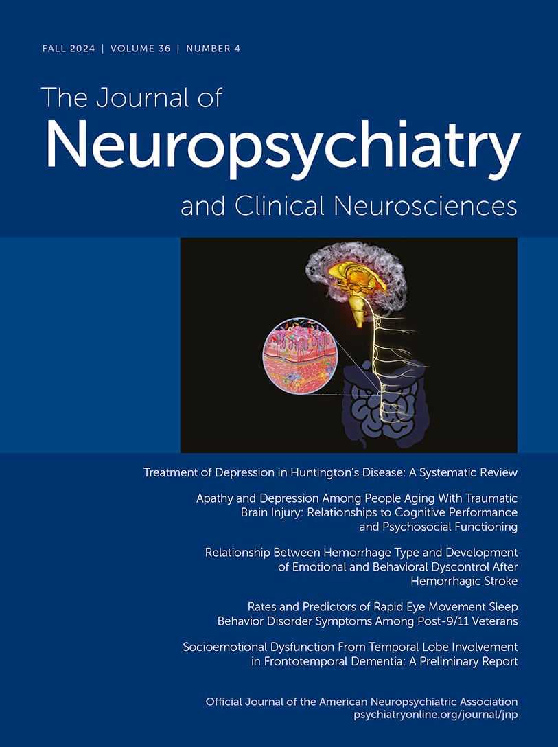
COVER. Artistic depiction of the main components of the brain-gut axis (simplified).
All images and figures were created with Canva, BioRender.com, and VH Dissector, for which the Mid-Atlantic (VISN 6) Mental Illness Research, Education and Clinical Center holds renewable registrations.
Regarding the BGA, the CN X seems to be the most relevant component of the PNS. It establishes the anatomic connection between the CNS and ENS, potentiating important neural processes, brain functions, and certain behaviors (e.g., feeding and mood) (1, 4, 13). This neuroanatomic communication system can process, regulate, and condition gut reflexes (e.g., in muscular tissues) as well as adjust to mood variations. Behaviors involved include promoting intuitive decision making, non–associative implicit learning (i.e., habituation and sensitization), redirecting neural signaling processes, and modulating the physiology of the gut, along with other homeostatic and systemic functions (7, 10, 14, 15).
Despite its length and complexity, CN X cannot reach and supply all the components of the intestinal mucosa directly. However, it can reach them indirectly via its conjoined functions with the ENS plexuses and enteroendocrine cells (EECs) (2, 7). EECs are specialized epithelial cells that chemically assess the intestinal lumen (neighboring cells and tissues) and its contents (nutritional macromolecules, microbiome, and others), signaling the brain via CN X and secreting important bioactive substances (e.g., cholecystokinin, ghrelin, peptide YY, neuropeptide Y, and glucagon-like peptide-1 and serotonin) (16). The EEC-vagal circuit is linked to the neuroendocrine pathway of the HPA axis and activated in response to environmental factors, emotions, and stress (5, 6, 16).
Serotonin is one of the most relevant neurotransmitters in mental health (e.g., depression) and is mostly synthesized outside the CNS by the enterochromaffin cells, a subset of specialized EECs in the gastrointestinal mucosa (1719). These cells regulate the mechanosensory and chemosensory functions that lead to digestion, secretion and absorption, and other metabolic functions (19).
In addition, EECs functionally interact with enteric glial cells, which further regulate hormone secretions and ENS signaling via the neuroimmune pathway of the BGA (6). Presently, the study of the BGA and its components has gained significant traction in clinical neuroscience. As a result, a growing body of evidence supports a putative role of the BGA in the pathophysiologic basis of selected mental disorders (6, 2025).

Unique Functions of CN X in Neuropsychiatric Disorders

CN X afferent circuits project to important neuroanatomic structures, including the prefrontal and motor cortices as well as the hippocampus, thalamus, hypothalamus, and insula (26, 27). CN X modulates the BGA, relevant to a number of neuropsychiatric conditions (e.g., anxiety, depression, posttraumatic stress disorder, Parkinson’s disease, and amyotrophic lateral sclerosis [ALS]), as well as other disorders (8, 14, 21, 22).
A plethora of scientific evidence indicates that CN X regulates the gut-microbiome-brain axis, playing an important role in the pathophysiology of depression, possibly mediating inflammatory processes between the visceral organs, the gut microbiome, and the brain (15, 25, 2830). A recent study investigated the role of the gut-liver-brain axis in depression-like phenotypes by using mice with liver cirrhosis induced by common bile duct ligations. The intervention caused multiple pathologic manifestations consistent with disrupted gut microbiome balance, depression-like behaviors, and decreased synaptic activation in the prefrontal cortex, among other changes. All these pathologic changes were reversed following a subdiaphragmatic vagotomy (31). The results indicated that in this model, CN X appears to be involved in regulating the neural mechanism that leads to dysbiosis (changes of the composition or functional alterations of the normal gut microbiome linked to a variety of diseases) among gut microbial species. In addition, depression-like phenotypes improved after a single injection of a new investigational compound, arketamine (31, 32).
Similar results have been reported with ablated visceral vagal communication in other experiments, resulting in anxiety- and fear-related behaviors, stress reactivity, and impaired cognition, which are linked to brain alterations observed in psychiatric disease (33, 34). Additional animal studies have shown that CN X–mediated processes are also influenced by the gut microbiome. Some bacterial strains (e.g., Lactobacillus rhamnosus JB-1 and Lactobacillus reuteri) interact with CN X, signaling the brain and altering behavior by reducing stress and anxiety– and depression-related conduct, as well as improving social interaction (35, 36).
In a preclinical study in mice that underwent fecal microbiota transplantation, inflammatory factors (e.g., interleukin 6, tumor necrosis factor alpha, and C-reactive protein) and intestinal mucosal permeability were increased, as were depressive and anxiety-like behaviors. The administration of probiotics (i.e., Clostridium butyricum) ameliorated the depressive and anxiety-like behaviors (37).
Human studies of treatment-resistant depression show results consistent with the preclinical data (38, 39). A recent study confirmed that dysbiosis is an important culprit in major depressive disorder, suggesting that the microbiota among patients with treatment-resistant depression significantly differs from that among patients who respond to routine antidepressant treatments (39).
In addition, there is evidence that proinflammatory species are overexpressed in the gut microbiome of people with Parkinson’s disease (32, 40, 41). Dysbiosis may result from spreading of systemic inflammatory agents (e.g., cytokines) or altered permeability of the intestinal barrier via a leaky blood-brain barrier and CN X. These problems can generate CNS synucleinopathy that itself potentiates further neuroinflammatory processes in vagal neurons and other brain areas (i.e., in the brainstem and substantia nigra) (24, 4042). The evidence indicates that in Parkinson’s disease, certain dysregulated intestinal pathogen–associated molecules homeostatically expressed in epithelial cells, such as Toll-like receptors 2 and 4, facilitate detection of gut microbial flora and contribute to gut dysbiosis and neuroinflammation (40, 43).
Gut microbiome imbalances consistent with dysbiosis have been observed in experimental models of ALS and among patients with ALS (4446). This condition is characterized by the progressive loss of motor neurons in the CNS, voluntary muscle wasting, and paralysis (47). Motor neurons are very susceptible to homeostatic changes and other factors (e.g., stress) (48). Why gut dysbiosis contributes to ALS remains unknown. However, the metabolic dysfunction, immune dysregulation, and altered gut barrier integrity linked to dysbiosis are potential pathologic mechanisms (49).

Vagal Nerve Stimulation

Vagal nerve stimulation (VNS) is a neuromodulatory technique approved by the U.S. Food and Drug Administration for the treatment of epilepsy and depression among patients ≥12 years (5053). Clinical evidence indicates that VNS, with implantable and noninvasive devices, can modulate essential functions within the body and treat various systemic and brain conditions, such as treatment-resistant depression and epilepsy (23, 5052). VNS appears beneficial for many patients, including expectant mothers manifesting pharmaco-resistant seizures and children with medically intractable epilepsy (53). In addition, VNS has shown positive results as an intervention for treatment-resistant depression. A pilot study (N=6) indicated prolonged antidepressive effects after VNS that were linked to the modulation of inflammation responses, relevant neuropsychiatric functions, and blood-brain barrier activity (51).
VNS appears to induce neural activity (plasticity, modulation, etc.) with positive therapeutic effects. The exact physiologic mechanisms by which VNS exerts its neuromodulatory effects via CN X remain unclear (23, 27, 54). However, it is believed that VNS potentiates regulation of specific neurotransmitters (e.g., acetylcholine, gamma-aminobutyric acid, dopamine, norepinephrine, and serotonin) and other neuroactive substances (e.g., brain-derived neurotrophic factor and cytokines). These substances are pivotal in multiple brain structures (limbic system, locus coeruleus, cortical and subcortical regions, etc.). They can foster cognition, protect against gut hyperpermeability (preventing gut microbiome translocation), decrease neuroinflammation, promote healthy aging, and facilitate neuronal plasticity (7, 27, 54, 55).

Conclusions

In the context of the BGA, CN X has a broad role in mental health beyond its wide anatomic distribution and well-known somatic and visceral functions. CN X comprises a direct anatomic connection by which the PNS, ANS, and ENS can communicate with the CNS, integrating all functional circuits of the BGA, including the neural pathway (e.g., CN X, ENS, and neurotransmitters), the immune pathway (e.g., cytokines), and the neuroendocrine pathway (e.g., HPA axis, EECs, and gut microbiome).
CN X–mediated processes are also influenced by bacterial subpopulations in the gut microbiome, positioning CN X as a major conduit for brain health as well as disease. In this role, CN X can alter behaviors (e.g., feeding, satiety, and mood), employing adaptive mechanisms that potentiate homeostasis. However, proinflammatory gut bacteria can transmit inflammatory responses to the brain via the CN X, contributing to serious mental health and neurodegenerative disorders, such as depression, Parkinson’s disease, and ALS.
CN X is also a potential target for innovative interventions for brain disorders. VNS (invasive and noninvasive) is a therapeutic approach that capitalizes on CN X’s neuromodulatory role (via the BGA), inducing neuroplasticity and neuromodulation with beneficial therapeutic effects.
Taken altogether, significant evidence supports CN X’s neuromodulatory role in the BGA relevant to mental disorders. However, many important questions related to the pathogenesis, pathophysiology, and further potential treatments (nerve stimulation modalities, probiotics, symbiotics, and others) for neuropsychiatric and neurodegenerative diseases remain unanswered.

References

1.
Kenny BJ, Bordoni B: Neuroanatomy, cranial nerve 10 (vagus nerve); in StatPearls. Treasure Island, Fla., StatPearls Publishing, 2024. http://www.ncbi.nlm.nih.gov/books/NBK537171
2.
Cao Y, Li R, Bai L: Vagal sensory pathway for the gut-brain communication. Semin Cell Dev Biol 2024; 156:228–243
3.
Hansen JT: Head and neck; in Netter’s Clinical Anatomy. Amsterdam, Elsevier, 2023, pp 435–554. https://www.clinicalkey.com/#!/content/book/3-s2.0-B9780323826624000084?scrollTo=%23hl0002383
4.
Sonne J, Lopez-Ojeda W: Neuroanatomy, cranial nerve; in StatPearls. Treasure Island, Fla., StatPearls Publishing, 2024. http://www.ncbi.nlm.nih.gov/books/NBK470353
5.
Carabotti M, Scirocco A, Maselli MA, et al: The gut-brain axis: interactions between enteric microbiota, central and enteric nervous systems. Ann Gastroenterol 2015; 28:203–209
6.
Peterson CT: Dysfunction of the microbiota-gut-brain axis in neurodegenerative disease: the promise of therapeutic modulation with prebiotics, medicinal herbs, probiotics, and synbiotics. J Evid Based Integr Med 2020; 25:2515690X20957225
7.
Tan C, Yan Q, Ma Y, et al: Recognizing the role of the vagus nerve in depression from microbiota-gut brain axis. Front Neurol 2022; 13:1015175
8.
Prescott SL, Liberles SD: Internal senses of the vagus nerve. Neuron 2022; 110:579–599
9.
Mtui E, Gruener G, Dockery P: Autonomic nervous system; in Fitzgerald’s Clinical Neuroanatomy and Neuroscience. Amsterdam, Elsevier, 2021, pp 148–163. https://www.clinicalkey.com/#!/content/book/3-s2.0-B9780702079092000139?scrollTo=%23hl0000668
10.
Moțățăianu A, Şerban G, Andone S: The role of short-chain fatty acids in microbiota-gut-brain cross-talk with a focus on amyotrophic lateral sclerosis: a systematic review. Int J Mol Sci 2023; 24:15094
11.
AbuAlrob MA, Tadi P: Neuroanatomy, nucleus solitarius; in StatPearls. Treasure Island, Fla., StatPearls Publishing, 2024. http://www.ncbi.nlm.nih.gov/books/NBK549831
12.
Zheng Y, Bonfili L, Wei T, et al: Understanding the gut-brain axis and its therapeutic implications for neurodegenerative disorders. Nutrients 2023; 15:4631
13.
Schemann M, Frieling T, Enck P: To learn, to remember, to forget—how smart is the gut? Acta Physiol 2020; 228:e13296
14.
Suganya K, Koo BS: Gut-brain axis: role of gut microbiota on neurological disorders and how probiotics/prebiotics beneficially modulate microbial and immune pathways to improve brain functions. Int J Mol Sci 2020; 21:7551
15.
Mayer EA: Gut feelings: the emerging biology of gut-brain communication. Nat Rev Neurosci 2011; 12:453–466
16.
Dockray GJ: Enteroendocrine cell signalling via the vagus nerve. Curr Opin Pharmacol 2013; 13:954–958
17.
Lin SH, Lee LT, Yang YK: Serotonin and mental disorders: a concise review on molecular neuroimaging evidence. Clin Psychopharmacol Neurosci 2014; 12:196–202
18.
Moncrieff J, Cooper RE, Stockmann T, et al: The serotonin theory of depression: a systematic umbrella review of the evidence. Mol Psychiatry 2023; 28:3243–3256
19.
Alcaino C, Knutson KR, Treichel AJ, et al: A population of gut epithelial enterochromaffin cells is mechanosensitive and requires Piezo2 to convert force into serotonin release. Proc Natl Acad Sci U S A 2018; 115:E7632–E7641
20.
Morais LH, Schreiber HL, IV, Mazmanian SK: The gut microbiota-brain axis in behaviour and brain disorders. Nat Rev Microbiol 2021; 19:241–255
21.
Bonaz B: The gut-brain axis in Parkinson’s disease. Rev Neurol 2024; 180:65–78
22.
Breit S, Kupferberg A, Rogler G, et al: Vagus nerve as modulator of the brain-gut axis in psychiatric and inflammatory disorders. Front Psychiatry 2018; 9:44
23.
Cimpianu CL, Strube W, Falkai P, et al: Vagus nerve stimulation in psychiatry: a systematic review of the available evidence. J Neural Transm 2017; 124:145–158
24.
Gorecki AM, Anyaegbu CC, Anderton RS: TLR2 and TLR4 in Parkinson’s disease pathogenesis: the environment takes a toll on the gut. Transl Neurodegener 2021; 10:47
25.
Johnson KV, Steenbergen L: Gut feelings: vagal stimulation reduces emotional biases. Neuroscience 2022; 494:119–131
26.
Berthoud HR, Neuhuber WL: Functional and chemical anatomy of the afferent vagal system. Auton Neurosci 2000; 85:1–17
27.
Trifilio E, Shortell D, Olshan S, et al: Impact of transcutaneous vagus nerve stimulation on healthy cognitive and brain aging. Front Neurosci 2023; 17:1184051
28.
Chang L, Wei Y, Hashimoto K: Brain-gut-microbiota axis in depression: a historical overview and future directions. Brain Res Bull 2022; 182:44–56
29.
Hashimoto K: Chapter 3: neuroinflammation through the vagus nerve–dependent gut-microbiota-brain axis in treatment-resistant depression; in Progress in Brain Research. Edited by Li C-T, Cheng C-M. Amsterdam, Elsevier, 2023, pp 61–77. https://www.sciencedirect.com/science/article/pii/S0079612323000031
30.
Kamel LY, Xiong W, Gott BM, et al: Vagus nerve stimulation: an update on a novel treatment for treatment-resistant depression. J Neurol Sci 2022; 434:120171
31.
Yang Y, Eguchi A, Mori C, et al: Depression-like phenotypes in mice following common bile duct ligation: insights into the gut-liver-brain axis via the vagus nerve. Neurobiol Dis 2024; 192:106433
32.
Wang Q, Luo Y, Ray Chaudhuri K, et al: The role of gut dysbiosis in Parkinson’s disease: mechanistic insights and therapeutic options. Brain 2021; 144:2571–2593
33.
Fülling C, Dinan TG, Cryan JF: Gut microbe to brain signaling: what happens in vagus. Neuron 2019; 101:998–1002
34.
Décarie-Spain L, Hayes AMR, Lauer LT, et al: The gut-brain axis and cognitive control: a role for the vagus nerve. Semin Cell Dev Biol 2024; 156:201–209
35.
Bravo JA, Forsythe P, Chew MV, et al: Ingestion of Lactobacillus strain regulates emotional behavior and central GABA receptor expression in a mouse via the vagus nerve. Proc Natl Acad Sci U S A 2011; 108:16050–16055
36.
Sgritta M, Dooling SW, Buffington SA, et al: Mechanisms underlying microbial-mediated changes in social behavior in mouse models of autism spectrum disorder. Neuron 2019; 101:246–259.e6
37.
Liu P, Liu Z, Wang J, et al: Immunoregulatory role of the gut microbiota in inflammatory depression. Nat Commun 2024; 15:3003
38.
Gao M, Tu H, Liu P, et al: Association analysis of gut microbiota and efficacy of SSRIs antidepressants in patients with major depressive disorder. J Affect Disord 2023; 330:40–47
39.
Fontana A, Manchia M, Panebianco C, et al: Exploring the role of gut microbiota in major depressive disorder and in treatment resistance to antidepressants. Biomedicines 2020; 8:311
40.
Perez-Pardo P, Dodiya HB, Engen PA, et al: Role of TLR4 in the gut-brain axis in Parkinson’s disease: a translational study from men to mice. Gut 2019; 68:829–843
41.
Shannon KM: Gut-derived sterile inflammation and Parkinson’s disease. Front Neurol 2022; 13:831090
42.
Braak H, Del Tredici K: Invited article: nervous system pathology in sporadic Parkinson disease. Neurology 2008; 70:1916–1925
43.
Burgueño JF, Abreu MT: Epithelial Toll-like receptors and their role in gut homeostasis and disease. Nat Rev Gastroenterol Hepatol 2020; 17:263–278
44.
Boddy SL, Giovannelli I, Sassani M, et al: The gut microbiome: a key player in the complexity of amyotrophic lateral sclerosis (ALS). BMC Med 2021; 19:13
45.
Wright ML, Fournier C, Houser MC, et al: Potential role of the gut microbiome in ALS: a systematic review. Biol Res Nurs 2018; 20:513–521
46.
Guo K, Figueroa-Romero C, Noureldein MH, et al: Gut microbiome correlates with plasma lipids in amyotrophic lateral sclerosis. Brain 2024; 147:665–679
47.
Taylor JP, Brown RH, Jr., Cleveland DW: Decoding ALS: from genes to mechanism. Nature 2016; 539:197–206
48.
Vandoorne T, De Bock K, Van Den Bosch L: Energy metabolism in ALS: an underappreciated opportunity? Acta Neuropathol 2018; 135:489–509
49.
Noor Eddin A, Alfuwais M, Noor Eddin R, et al: Gut-modulating agents and amyotrophic lateral sclerosis: current evidence and future perspectives. Nutrients 2024; 16:590
50.
Guiraud D, Andreu D, Bonnet S, et al: Vagus nerve stimulation: state of the art of stimulation and recording strategies to address autonomic function neuromodulation. J Neural Eng 2016; 13:041002
51.
Lespérance P, Desbeaumes Jodoin V, Drouin D, et al: Vagus nerve stimulation modulates inflammation in treatment-resistant depression patients: a pilot study. Int J Mol Sci 2024; 25:2679
52.
Carreno FR, Frazer A: Vagal nerve stimulation for treatment-resistant depression. Neurotherapeutics 2017; 14:716–727
53.
Johnson RL, Wilson CG: A review of vagus nerve stimulation as a therapeutic intervention. J Inflamm Res 2018; 11:203–213
54.
Aniwattanapong D, List JJ, Ramakrishnan N, et al: Effect of vagus nerve stimulation on attention and working memory in neuropsychiatric disorders: a systematic review. Neuromodulation 2022; 25:343–355
55.
Driskill CM, Childs JE, Phensy AJ, et al: Vagus nerve stimulation (VNS) modulates synaptic plasticity in the rat infralimbic cortex via Trk-B receptor activation to reduce drug-seeking. bioRxiv [Preprint] 2024; 2024.01.25.577293

Information & Authors

Information

Published In

Go to The Journal of Neuropsychiatry and Clinical Neurosciences
The Journal of Neuropsychiatry and Clinical Neurosciences
Pages: 278 - 282

History

Received: 12 June 2024
Accepted: 5 August 2024
Published in print: Fall 2024
Published online: 15 October 2024

Keywords

  1. Brain-Gut Axis
  2. Gut Microbiota
  3. Neuropsychiatric Disorders
  4. Neuroanatomy
  5. Neuroinflammation
  6. Vagus Nerve

Authors

Details

Wilfredo López-Ojeda, Ph.D., M.S.
Veterans Affairs Mid-Atlantic Mental Illness Research, Education and Clinical Center and the Research and Academic Affairs Service Line, W. G. Hefner Veterans Affairs Medical Center, Salisbury, N.C. (López-Ojeda, Hurley); Departments of Psychiatry and Behavioral Medicine (López-Ojeda, Hurley) and Radiology (Hurley), Wake Forest University School of Medicine, Winston-Salem, N.C.
Robin A. Hurley, M.D., F.A.N.P.A. [email protected]
Veterans Affairs Mid-Atlantic Mental Illness Research, Education and Clinical Center and the Research and Academic Affairs Service Line, W. G. Hefner Veterans Affairs Medical Center, Salisbury, N.C. (López-Ojeda, Hurley); Departments of Psychiatry and Behavioral Medicine (López-Ojeda, Hurley) and Radiology (Hurley), Wake Forest University School of Medicine, Winston-Salem, N.C.

Notes

Send correspondence to Dr. Hurley ([email protected]).

Competing Interests

The authors report no financial relationships with commercial interests.

Funding Information

This study was supported by the U.S. Department of Veterans Affairs Veterans Integrated Services Network 6, Mental Illness Research, Education and Clinical Center.

Metrics & Citations

Metrics

Citations

Export Citations

If you have the appropriate software installed, you can download article citation data to the citation manager of your choice. Simply select your manager software from the list below and click Download.

For more information or tips please see 'Downloading to a citation manager' in the Help menu.

Format
Citation style
Style
Copy to clipboard

View Options

View options

PDF/EPUB

View PDF/EPUB

Get Access

Login options

Already a subscriber? Access your subscription through your login credentials or your institution for full access to this article.

Personal login Institutional Login Open Athens login

Not a subscriber?

Subscribe Now / Learn More

PsychiatryOnline subscription options offer access to the DSM-5-TR® library, books, journals, CME, and patient resources. This all-in-one virtual library provides psychiatrists and mental health professionals with key resources for diagnosis, treatment, research, and professional development.

Need more help? PsychiatryOnline Customer Service may be reached by emailing [email protected] or by calling 800-368-5777 (in the U.S.) or 703-907-7322 (outside the U.S.).
Media

Figures

Other

Tables

Share

Share

Share article link

Share