Full access
Special Articles
Published Online: 21 March 2022

Behavioral Variant Frontotemporal Dementia and Social and Criminal Transgressions

Publication: The Journal of Neuropsychiatry and Clinical Neurosciences

Abstract

Objective:

Behavioral variant frontotemporal dementia (bvFTD) is associated with social and criminal transgressions; studies from countries around the world have documented such behavior in persons with this condition. An overview and analysis of social and criminal transgressions in bvFTD and their potential neurobiological mechanisms can provide a window for understanding the relationship of antisocial behavior and the brain.

Methods:

This review evaluated the literature on the frequency of social and criminal transgressions in bvFTD and the neurobiological disturbances that underlie them.

Results:

There is a high frequency of transgressions among patients with bvFTD due to impairments in neurocognition, such as social perception, behavioral regulation, and theory of mind, and impairments in social emotions, such as self-conscious emotions and empathy. Additionally, there is significant evidence for a specific impairment in an innate sense of morality. Alterations in these neurobiological processes result from predominantly right-hemisphere pathology in frontal (ventromedial, orbitofrontal, inferolateral frontal), anterior temporal (amygdala, temporal pole), limbic (anterior cingulate, amygdala), and insular regions.

Conclusions:

Overlapping disturbances in neurocognition, social emotions, and moral reasoning result from disease in the mostly mesial and right-sided frontotemporal network necessary for responding emotionally to others and for behavioral control. With increased sophistication in neurobiological interventions, future goals may be the routine evaluation of these processes among individuals with bvFTD who engage in social and criminal transgressions and the targeting of these neurobiological mechanisms with behavioral, pharmacological, and other interventions.
Behavioral variant frontotemporal dementia (bvFTD) is a disorder associated with social and criminal transgressions. BvFTD is a progressive neurodegeneration of frontal and anterior temporal regions that usually presents with personality changes in midlife (1). In addition to apathy, disinhibition, loss of empathy, stereotypies, and altered dietary behavior, the personality changes of bvFTD include a tendency for violation of social norms and even antisocial behavior. These transgressions, which range from shoplifting to the inappropriate touching of children, can result in criminal prosecution and incarceration (2).
Although clinicians and investigators have long known that persons with bvFTD may engage in behaviors that constitute criminal transgressions, there is no critical review of this association and all the potential reasons for it. Understanding the processes that underlie antisocial acts in bvFTD could clarify the processes that underlie antisocial acts in general. The neuropathological localization of bvFTD is centered in mesial frontolimbic, frontoinsular, and anterior cingulate cortices, which contain Von Economo neurons evident in humans and other socially complex mammals (3), and anterior temporal lobe structures involved in socioemotional processing (4, 5). This neuropathological localization indicates a number of disturbances that can potentially result in violations of social norms and rules in bvFTD. This literature review gives a broad overview of criminal transgressions in bvFTD and the neurobiological processes that can contribute to their sociopathic acts.

CRIMINAL TRANSGRESSIONS IN bvFTD

Reports from around the world have established the association of increased criminal transgressions among persons with bvFTD when compared with healthy persons or those with Alzheimer’s disease (AD) or other dementias (613) (Table 1). In an early report from the United States (6), 16 of 28 (57%) persons with bvFTD had sociopathic behavior such as unsolicited sexual acts and traffic violations, compared with only 7% of similarly impaired individuals with AD (6). A subsequent, much larger U.S. study of the medical records of 2,397 persons with neurodegenerative diseases found prior criminal behavior among 37% (N=64/171) of those with bvFTD, compared with only 8% (N=42/545) of those with AD (7). Those with semantic variant primary progressive aphasia (svPPA) or semantic dementia (SD), a related frontotemporal dementia syndrome confined to the anterior temporal lobes, were also prone to criminal violations (27%; N=24/89). Persons with bvFTD and svPPA were about a decade younger than those with AD when their criminal transgressions were first noted. Moreover, 14% of the bvFTD group were first referred for diagnosis because of criminal behavior, compared with only 2% of those with AD.
TABLE 1. Studies of criminal behavior among persons with frontotemporal dementia (FTD)a
    With criminal behavior
StudyCountryMethodologyTotal numberN%
Mendez et al. (6)U.S.Clinical comparison56  
 bvFTDb   16/2857.0
 AD   2/287.0
Diehl-Schmid et al. (9)GermanyClinical comparison83  
 bvFTD   12/3254.0
 SD   10/1856.0
 AD   4/3312.0
Liljegren et al. (7)U.S.Retrospective; medical record review2,397 (830 with main neurodegenerative diagnoses)  
 bvFTD   64/17137.4
 svPPA   24/8927.0
 HD   6/3020.0
 AD   42/5457.7
Shinagawa et al. (10)JapanRetrospective; clinical comparison412  
 bvFTD   24/7333.0
 svPPA   18/8421.0
 AD   14/2556.0
Liljegren et al. (12)cSwedenNeuropathological dementia with police interactions281  
 FTD syndromes   25/9726.0
 AD   9/1019.0
 VaD plus mixedd   16/8319.0
Liljegren et al. (11)cSwedenNeuropathological dementia with medical records220  
 FTD syndromes   50/11942.0
 AD   15/10114.9
Talaslahti et al., 2021 (13)FinlandData from hospital discharge register versus data from police register92,191  
 FTD syndromes     
  Men   107/45623.5
  Women   32/6045.3
 AD     
  Men   3,531/27,51212.8
  Women   835/53,0281.6
 DLB     
  Men   628/5,32611.8
  Women   156/5,2653.0
a
AD=Alzheimer’s disease; bvFTD=behavioral variant frontotemporal dementia; DLB=dementia with Lewy bodies; HD=Huntington's disease; SD=semantic dementia; svPPA=semantic variant primary progressive aphasia; VaD=vascular dementia.
b
Met subsequent criteria for bvFTD.
c
Overlap of patients included in study.
d
Various neuropathologies included.
Other investigators have reported similar findings from Europe and Japan. A German study of 83 patients found a history of criminal behavior in more than half of those with bvFTD (53%; N=17/32) or SD (56%; N=10/18), but only 12% (N=4/33) of those with AD (9). In a Swedish cohort of 220 patients with pathologically verified dementia, higher criminal behavior and social inappropriateness occurred in 42% (N=50/119) of their patients with FTD syndrome, especially those with non-tau pathology, compared with only 15% (N=15/101) of their patients with AD (11). A further evaluation of police interactions among 281 patients with pathologically verified dementia found that 26% (N=25/97) of the patients with FTD syndrome had police reports of criminal behavior versus only 9% (N=9/101) of the patients with AD (12). Although some of the patients with bvFTD had early criminal behavior even before being examined by a physician (12), criminal behavior occurred throughout the course of bvFTD, with over 80% with at least one or more offenses (11). In one study, investigators were able to cross-check data for all Finnish patients from hospital discharges between 1998 and 2015 with all data from their police registry on criminal offenses from that period (13). The crime rate was higher among 92,191 patients with dementia, compared with the general population, with at least one crime committed by 23.5% of men with FTD syndrome and 5.3% of women with FTD syndrome versus 12.8% of men with AD and 1.6% of women with AD, and 11.8% of men with dementia with Lewy bodies (DLB) and 3.0% of women with dementia with Lewy bodies. During the 4 years preceding their FTD syndrome diagnosis, the incidence of crimes showed a substantially increasing trend among the men in this sample, with the first crime occurring on average 2.7 years (SD=1.1) before their diagnosis. Finally, Japanese investigators have reported high rates of law violations before initial consultation among 33% (N=24/73) of persons with bvFTD and 21% (N=18/84) among persons with svPPA, compared with only 6% (N=14/255) of those with AD (10). In this study, 10 of the individuals with bvFTD had four or five violations before consultation, consistent with a high rate of recidivism (10, 11).
The crimes committed by persons with bvFTD have included theft, sexual behavior, and traffic violations, as well as a range of other transgressions (14). In a large U.S. study, crimes included trespassing, public urination, sexual advances or exposure, violence toward others, and driving infractions such as speeding, hit-and-run accidents, and driving under the influence (7). Perhaps the most common crime in bvFTD is pathological stealing, such as taking something from someone or from a store without paying (6, 7, 10, 15, 16). These acts are distinct from kleptomania in that the individuals with bvFTD lack anxiety beforehand or relief afterward, lack obsessional thinking, do not usually seek out opportunities to steal, and commit thefts only when items of interest are present.
Sexual transgressions are most commonly associated with either general disinhibition and poor impulse control or with compulsive tendencies, such as a need for repetitive kissing or an addiction to pornography (1719). One report found a significant increase in sexual frequency among 13% (N=6/47) of those with bvFTD versus none of 58 with AD (20). Paraphilic advances, including pedophilia, may emerge from disinhibition of lifelong and previously repressed predispositions (19, 2124). Primary hypersexuality seems otherwise rare; however, some persons with bvFTD with right temporal involvement have manifested a dramatic increase in sexual frequency, actively seeking sexual stimulation, widened sexual interests, and sexual arousal from previously unexciting stimuli (19).
In the Finnish study (13), traffic offenses and crimes against property constituted 94% of all transgressions; violence, mainly assaults and illegal threats, was unusual. Several other reports indicate more violent or aggressive acts among persons with bvFTD compared with those with other dementias (7, 25), but these acts appear reactive, not instrumental or planned. The Finnish researchers described one murder and one attempted murder in their FTD group (13); in the Swedish series (12), there was an attempt to use a car to injure others and the presence of homicidal and suicidal threats (12). Nevertheless, despite historical interpretations of bvFTD as the source of prior notorious murders (26), carefully thought-out or premeditated murder is unlikely in bvFTD.

Neurobiological Processes

These reports indicate that bvFTD is associated with social and criminal transgressions, but they do not elucidate the underlying neurobiological processes that result in this behavior. Understanding these processes can clarify the relationship of antisocial behavior and the brain in healthy people as well as in those with brain disorders. Persons with bvFTD appear to violate social norms and rules while retaining knowledge of their behavior and of moral and conventional rules. Their offenses often occur at a time when they lack major nonsocial cognitive deficits. They can describe the nature and potential consequences of their transgressions, but they may not feel sufficiently concerned to be deterred. The developing literature on socioemotional behavioral changes in bvFTD suggests that a combination of overlapping neurobiological changes contributes to their social and criminal transgressions. These include impairments in social cognition (perception of emotions, animacy, and humanness; behavioral regulation and control, theory of mind [ToM]), social emotions (self-conscious emotions, empathy), and an additional sense of morality.

Social Cognition

Social Perception

Persons with bvFTD have deficits in the perception of facial, body, and other social and emotional cues, especially subtle social signals. Many studies of bvFTD have documented impaired recognition of emotions displayed in both static and video images of faces (2735). Compared with individuals with AD, persons with bvFTD have trouble labeling facial emotional expressions, particularly subtle expressions of low emotional intensity (36, 37), and they have trouble tracking emotions portrayed in film characters (37). Beyond faces, persons with bvFTD have difficulty deciphering ambiguous emotions when watching videotaped interviews (38), and they even have difficulty recognizing emotions conveyed in art (39). In bvFTD, the impaired monitoring of emotion recognition, especially facial emotions, is associated with reduced volume of anterior temporal, frontal, and insular regions (37, 40). Among those with bvFTD, poor emotional recognition and attribution performance are particularly strongly associated with atrophy and decreased synaptic activity in the right anterior temporal regions, specifically the amygdala and temporal pole (28, 41), and impaired recognition of negative facial emotional expressions is associated with damage to the right lateral inferior and middle temporal gyri (Brodmann’s areas 20 and 21) (42). Among those with SD, emotional processing deficits are associated with decreased gray matter in the right lateral and medial temporal regions, possibly impairing the derivation of semantic meaning from emotional information (43, 44). In addition to the right anterior temporal lobe, decreased gray matter in the right lateral orbitofrontal cortex (OFC) is associated with impaired tracking of changing emotions (45), and the anterior insula (AI) participates in emotional awareness (46).
A second and more complex aspect of social perception is the detection of animacy, or the characteristic of being living or sentient, and even whether a living being is human. The impaired attribution of animacy may contribute to the social behavior disturbances in bvFTD (47). Several reports describe those with FTD syndromes as having difficulty recognizing something as living (i.e., animate versus nonanimate) (48), unrelated to the detection of agency (capability of intrinsically driven action) from more posterior cortical regions (47). For example, when viewing moving geometric shapes (47), persons with bvFTD, but not those with AD, are significantly impaired in animacy attribution and have decreased sympathetic (skin conductance) responses, a physiological indicator of attribution of a social “story” to the moving shapes. On analysis of cortical thicknesses, animacy scores correlate with the right pars orbitalis and opercularis, suggesting that impaired animacy perception in bvFTD is associated with atrophy in the right inferior frontal gyrus (IFG). In this group, the cerebellum and its frontal connections may also be involved in viewing abstract animation with geometric shapes (49, 50).
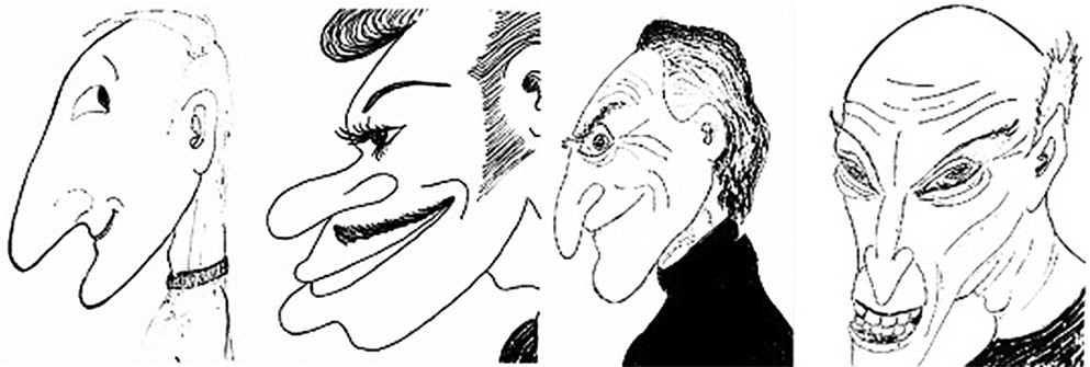
Humanness appears to be a specific animacy property, part of a dedicated person-recognition network involving the right temporal lobe, which can be impaired in bvFTD (51, 52). The detection of humanness involves not only the processing of human faces but also semantic information organized into a person-specific animate subcategory in the right temporal region (51, 53, 54). In one study, bvFTD with predominant right frontotemporal involvement was associated with overinclusion in describing morphed or distorted faces as human (51), independent of the ability to recognize faces, interpret facial emotions, or appreciate the distinction between animate and inanimate things. Further evidence for impaired perception of humanness is that persons with FTD who are artists may, as their disease progresses, draw others in a progressively distorted, nonhuman, or alien fashion (Figure 1) (52).
FIGURE 1. Disrupted facial “humanness” in caricatures of artists as they progress from a normal, premorbid state (first drawing) to gradually worsening stages of behavioral variant frontotemporal dementia

Behavioral Regulation and Control

Another important cognitive defect that contributes to social and criminal transgressions in bvFTD involves deficits in regulation or control. Most individuals with bvFTD are predisposed to choose the immediate option for an immediate reward without considering the future consequences of their choice. They are unable to stop themselves, suppress prepotent responses, or resist impulses; this results in behavioral disinhibition (55). Compared with those with AD or healthy control subjects, persons with bvFTD usually perform worse on cognitive inhibition tests such as the Stroop Color-Word Interference Test, the Hayling Sentence Completion Test, and the Trail Making B Test, with more perseveration errors on the Wisconsin Card Sorting Test and the Graphic Alternating Patterns (55). A sensitive index for bvFTD is decreased error sensitivity or the correction of errors on cognitive inhibition tasks (56). Behavioral scales, such as the Barratt Impulsiveness Scale, also show significantly worse behavioral disinhibition in bvFTD versus AD (57). Investigators have shown disinhibition in bvFTD to be mostly socially inappropriate person-based transgressions, such as making personal referent comments, rather than generalized impulsivity (58).
In bvFTD, the difficulty controlling behavior results from pathology involving the OFC and IFG, which anatomically overlap at the pars orbitalis portion of the IFG. The OFC is important for regulating behavior by operant conditioning to rewards versus punishments. The medial OFC may be more involved in processing reward, while the lateral OFC may be more involved in processing punishment and aversion to harm (59, 60). In bvFTD, OFC atrophy (especially of the medial part) is associated with increased caregiver ratings of inappropriate behavior, such as acting impulsively, speaking openly of private matters, and physical touching beyond appropriate boundaries (6163). In one study that included bvFTD, generalized disinhibition was correlated with a smaller OFC volume specifically on the right (58). Temporal lobe involvement in bvFTD may contribute through the right uncinate fasciculus and other connections with the OFC (55). The IFG also plays a fundamental role in response inhibition and behavioral control (6466). The IFG, especially on the right (6467), provides a stop signal for internally suppressing automatic prepotent or preplanned responses (67).

Theory of Mind

Mentalizing or ToM is the ability to represent the thoughts, beliefs, attitudes, and feelings of others. ToM is possibly the most critical aspect of social cognition, and ToM deficits can be among the first manifestations of bvFTD (68, 69). Studies implicate deficits in ToM as contributing to neuropsychiatric symptoms in bvFTD, such as social inappropriateness, and thereby increasing caregiver burden (70, 71).
ToM has both a cognitive component for the identification of others’ beliefs and intentions and an affective component for the identification of others’ emotions. It is the affective component that is impaired early in bvFTD (68, 7279), particularly when compared with AD (72, 73, 75, 76, 78, 80, 81). Affective ToM difficulty is generally dissociated from classic executive functions or tests (69, 80, 8288), and affective ToM tasks are significantly better than executive tasks at distinguishing those with bvFTD from healthy persons and from those with AD (82). Affective ToM is also arguably associated with difficulties in emotional recognition (75, 76, 80, 81, 89). In contrast, impairments in cognitive ToM, especially higher levels or orders of ToM (80, 81), develop later in the course of bvFTD and are associated with executive impairment (90), increasing cognitive impairment, and dementia severity (76). Good tests for ToM in bvFTD include the affective ToM aspects of the faux pas (“wrong behavior”) test (78, 81, 90, 91), The Awareness of Social Inference Test (TASIT) with its measures of comprehension of lies and recognition of sarcasm (78, 9294), the Reading the Mind in the Eyes Test (68, 82), the Story-Based Empathy Test, which is a nonverbal task for inferring others’ emotions (72, 95), and a number of other measures (72, 9597).
Impaired ToM in bvFTD results from atrophy affecting an extensive network for mentalizing (98). The entire ToM network extends from posterior areas (posterior superior temporal sulcus, precuneus/posterior cingulate cortex, right temporoparietal junction) to anterior areas (medial prefrontal cortex, amygdala, anterior temporal pole) (99, 100). ToM abnormalities in bvFTD result from early degeneration in the anterior areas of this network where affective states are determined (79, 101). Damage to ventromedial prefrontal cortex (VMPFC), especially on the right and extending to medial Brodmann’s area 10 (97, 102), is associated with decreased performance on tasks that assess affective ToM, such as identifying faux pas and irony (103106) and recognizing sarcasm on the TASIT (81, 107), but not on tasks which are more strictly cognitive ToM (second-order false belief) (81, 108). Nevertheless, as the disease progresses, cognitive ToM is also impaired in bvFTD from involvement of dorsomedial prefrontal regions and a right frontal-temporal neural network for resolving social dilemmas (109112). Right frontal pathology in bvFTD may impair cognitive ToM from the inability to suppress self-perspective and self-reflection sufficiently to take another’s perspective (113, 114). Finally, bvFTD alters resting state networks involved in ToM, such as the salience network, and results in compensatory changes in other related resting state networks (41, 92).

Social Emotions

Most persons with bvFTD develop hypoemotionality—evidenced as decreased emotional displays and emotional reactivity—that significantly affects their caregivers (115). Diagnostic criteria for bvFTD include many behaviors suggesting hypoemotionality, such as decreased engagement in previously rewarding activities, inability to initiate or sustain projects, diminished responses to interesting or novel events, and blunted emotional reactions in general (1, 116). Persons with bvFTD have impairments in facial expressivity, especially in self-conscious reactions to negative facial emotions or to acoustic startle (44, 117, 118). Others have described anosodiaphoria, or lack of emotional concern, in bvFTD, especially for how they are perceived or evaluated by others (119, 120). Hypoemotionality in bvFTD is further evidenced as decreased psychophysiological arousal to affectively charged stimuli (30, 121132), and there is decreased sympathetic nervous reactivity, particularly to complex or self-conscious emotions, even in the context of intact emotional and error awareness (44, 121, 128, 131, 133136). In contrast, individuals with bvFTD may not reliably report their hypoemotionality, showing either alexithymia or altered emotional awareness despite intact or even exaggerated self-reports of their emotions based on interpretations of contextual cues or external rules (128, 137, 138).

Self-Conscious Emotions

Not all emotions are affected equally in bvFTD. The most salient hypoemotionality in this condition involves self-conscious emotions, which require an evaluation of the self in social situations (44, 134, 139). The experience of negative emotions, such as anger, disgust, fear, or sadness, are also affected but somewhat less so than self-conscious emotions (27, 117, 134, 135, 140144), whereas positive emotions may be relatively preserved in bvFTD (31, 44, 145, 146). Self-conscious emotions arise from the need to belong and conform, and they mediate obedience to social norms and rules (147). Individuals with bvFTD have an overall diminished need or concern for social conformity and decreased self-conscious emotions, including the absence of guilt when engaging in transgressions (2, 148). Other prototypical self-conscious emotions impaired in bvFTD include embarrassment, shame, pride, gratitude, compassion, fear of negative evaluation by others, and outrage at unfair treatment. They further include “fortune-of-other” emotions evoked via social comparison between one’s current status and that of another’s (147), such as envy, jealousy, and schadenfreude, or pleasure at another’s misfortune.
Self-conscious emotional processing deficits in bvFTD are related to hypoactivity of a frontal-cingulate-insular circuit which includes the VMPFC, the pregenual and middle anterior cingulate cortex (ACC), the AI, and other right-hemisphere regions, particularly the temporal pole (41, 140, 149151). Individuals with lesions of the VMPFC, especially on the right with extension to the OFC, exhibit diminished emotional responsivity, intentional emotional imitation, and self-conscious emotions such as guilt, shame, and compassion (152155). Pathology affecting the left frontal emotional regulatory circuits may heighten the more positive emotions and decrease the more negative ones (145). Loss of gray matter in the right pregenual ACC in bvFTD is also associated with decreased embarrassment, guilt, and other self-conscious emotions (127), and damage to the right AI is associated with a loss of emotional expressions (44). Finally, extensive bilateral anterior temporal-amygdala involvement in bvFTD can produce the Klüver-Bucy syndrome with, among other symptoms, a pronounced unemotional passivity.

Empathy

A core diagnostic criterion for bvFTD, and a source of great distress for caregivers and others, is loss of empathy (1, 2, 116, 156, 157). Empathy is the ability to recognize and appreciate the subjective experience of others (to share an isomorphic emotion of another human being) and reference it to oneself (158). Caregivers and others describe persons with bvFTD, and especially those with prominent right anterior temporal involvement, as uncaring and lacking personal warmth (117, 159). Empathy has both emotional aspects (affect sharing, emotional contagion, empathic concern, personal distress) and cognitive aspects (perspective taking, ToM, mental representations), with the most central aspect of empathy being emotional affective sharing or feeling “as” another (160162). Although both emotional and cognitive empathy are impaired in bvFTD, this disorder is particularly associated with decreased affective sharing early in its course (159, 163167). Affective sharing requires corepresenting or coregistering of perceived third-party emotions onto oneself (168, 169). In other words, observing affective states in others activates corresponding first-person emotion areas in the observer, along with the associated physiological changes involved in the firsthand experience of those states (170). In contrast, deficits in cognitive empathy in bvFTD seem to depend on the development of deficits in executive functions and in ToM (171173).
Loss of empathy is a prominent feature of bvFTD not explained by loss of other social emotions, and loss of empathy goes beyond a general hypoemotionality. In bvFTD, loss of empathy persists despite covarying out for low emotionality to pictures (174). Studies using the Interpersonal Reactivity Index, which indicate reductions in both emotional and cognitive empathy in persons with bvFTD relative to those with AD and to neurologically healthy individuals (72, 159, 164, 165, 175, 176), reveal lower empathic concern ratings in bvFTD, which cannot be explained by deficits in executive function, social cognition, or emotional recognition (38, 159, 171, 175, 177). The absence of a clear relationship between the lack of empathic concern and facial affective expressions, skin conduction responses, or valence of stimuli in bvFTD further suggests a separation between empathy and basic emotional processing (129, 164). In a study that used the performance-based Multifaceted Empathy Test, empathy for negative stimuli appeared particularly impaired in bvFTD (160), and some investigators have proposed that in bvFTD the relatively preserved positive emotional reactivity undermines empathy by mitigating the negative emotional reactivity that more often motivates empathy (117, 160).
Loss of empathy in bvFTD is related to pathology in a distributed network of regions centered on the frontal-cingulate-insular and temporal structures. These include frontal lobe (fronto-operculum, OFC, IFG), ACC (dorsal middle region; right subgenual), AI and right posterior insula, and right temporal lobe regions (amygdala, temporal pole, fusiform gyrus) (133, 159, 164, 178181). Damage to both the AI and ACC is especially associated with impaired affective empathy (182), as affective sharing depends on the salience network, an AI-ACC predominant network (117, 131). Representations in AI underlie interoceptive awareness and representations of one’s own feeling states (46), which in turn form the basis for the simulation of the feelings of others (168). The ACC supports the integration of interoception and emotional salience for motivation and action choice (46, 183). Finally, empathic processing involves the temporal lobe, including a semantic appraisal network for “emotional tuning” of semantic information in the right, more than the left, medial and lateral temporal cortex and amygdala (149, 159, 181). Persons with bvFTD, particularly those with right temporal involvement, may incorrectly overestimate their capacity for empathy (128, 160, 164). They can even show exaggerated displays of empathy, again reflecting an overdependence of empathic responsiveness on external context or environmental cues (137, 138, 184).

Morality

Persons with bvFTD show defective moral reasoning and may engage in immoral acts despite their ability to describe the wrongness of their behavior and any potential consequences and punishments (6, 185, 186). Morality is a code of values and customs that guides social conduct. Beyond codes of conduct held by societies, religions, legal systems, or groups, there is neurobiological evidence for an underlying universal moral code held by all rational people and driven by innate emotional drives, such as doing no harm within one’s own group, a sense of fairness and reciprocity, and group or community loyalty (187). Moral emotional drives are rapid, involuntary, and intuitive reactions, which are distinct from deliberate moral reasoning (188). Neurobiological evidence points to this automatic, emotionally mediated morality as being centered in the VMPFC, particularly on the right (188), a focus of the neuropathology of bvFTD.
An early observation of persons with bvFTD was their tendency away from intuitive moral emotional reactions toward deliberate, utilitarian moral decisions based on “the greater good for the greater many” (2, 185). When presented with a moral behavior inventory, persons with bvFTD described having normal moral responses despite engaging in social transgressions (185). When presented with the most famous moral dilemma, the trolley problem, their “normal” moral responses were unemotional and utilitarian (185).
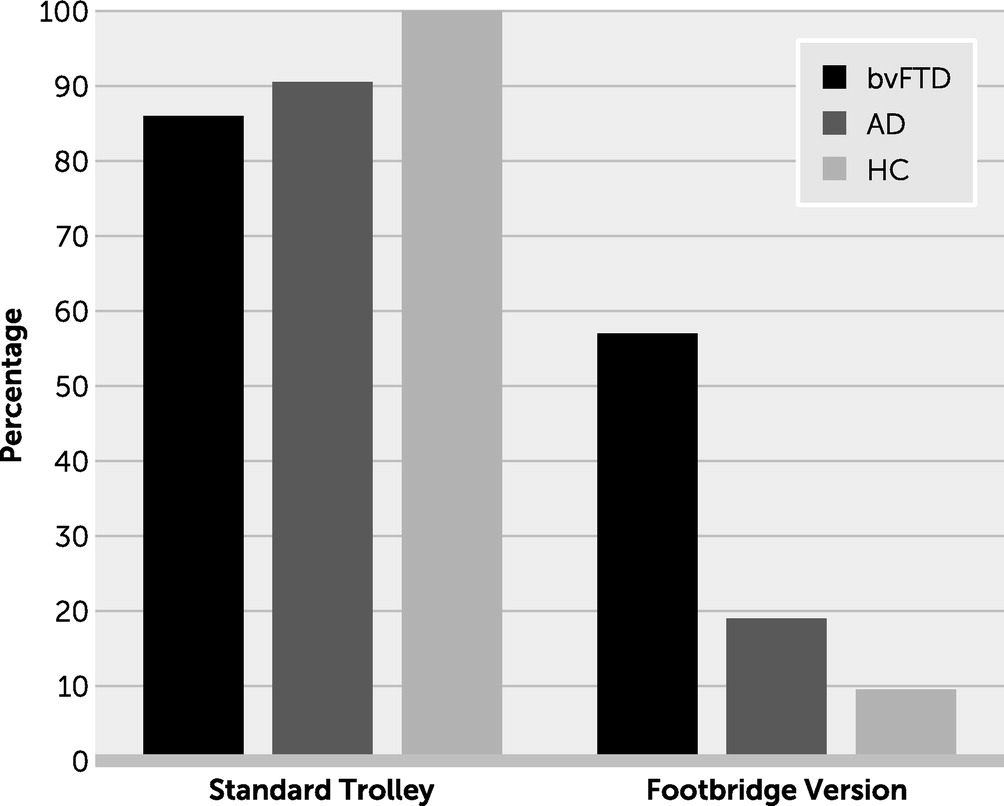
The two basic versions of the trolley problem presented participants with a runaway trolley that may kill five workers on the track. In the first version, the participant had to decide whether to flip a switch that would divert the trolley down a sidetrack, resulting in the death of only one large man lying on the sidetrack. This impersonal dilemma lacked the direct infliction of harm on another. In the second, “footbridge” version, the participant, who is now standing on a footbridge next to a large man, had to decide whether to push the large man onto the track, thereby killing him but stopping the trolley and saving the workers. This personal moral dilemma emphasized the participant’s own involvement and agency in inflicting harm. These trolley studies did not reveal significant differences between persons with bvFTD, those with AD, and healthy participants in preferring to pull the switch in the first version; however, in the second version only those with bvFTD were significantly more likely to be willing to push the large man in order to save the workers (Figure 2) (2, 185). Persons with bvFTD and those with traumatic brain injury made similar, significantly different utilitarian decisions during other personal scenarios involving the direct infliction of harm, whereas healthy individuals hesitated and reported discomfort (2, 47, 185, 189193). When further studied, affective ToM and emotional arousal (skin conduction responses) were significantly decreased among those with bvFTD facing moral dilemmas (156, 189). In these individuals, decreased conflict or discomfort with personal moral dilemmas was also correlated with a measure of social norms violations (14, 188).
FIGURE 2. Percentage positive responses on trolley moral dilemmas: behavioral variant frontotemporal dementia (bvFTD) group versus Alzheimer’s disease (AD) group versus healthy control (HC) groupa
a The bar graph shows the standard trolley problem: no significant differences. The graph also shows data for the footbridge version: three groups (χ2=13.07, df=2, p=0.001); bvFTD versus AD groups (χ2=4.95, df=1, p=0.026). The graph was developed with data described by Mendez and Shapira (185).
Morality involves a number of frontal lobe regions: the VMPFC, OFC, and dorsolateral prefrontal cortex (194, 195). On the moral dilemmas, increased endorsement of personal harm is particularly associated with VMPFC damage, especially on the right, in persons with bvFTD and traumatic brain injury (185, 196199). Early-life lesions in the VMPFC and adjacent areas can impair the development of moral knowledge or decisions that avoid harm to others (200). Individuals with damage to the VMPFC may not foresee negative emotional reactions when contemplating directly harming another person (201, 202); however, when faced with indirect or incidental harm to others, they are able to use reason for the greater good (185, 196198, 200, 203). It appears that the VMPFC assigns emotional valence to social scenarios; when damaged, it prevents the normal emotional aversion for social or criminal transgressions.
There are several conclusions from the studies of moral reasoning in bvFTD (Box 1). First, in the absence of a strong negative emotional reaction to the perception of harming another, persons with bvFTD and those with damage to the VMPFC do not experience an emotional aversion and instead rely on cognitive processes to access apparent moral decisions. They lack discomfort or distress or physiological arousal in making moral decisions. Second, these individuals perform comparably to control subjects on impersonal harm dilemmas, yet they display an abnormal pattern of utilitarian judgment during personal harm scenarios that normally evoke strong negative reactions. Third, persons with bvFTD compared with control subjects have impaired integration of others’ intentions, which are necessary to override responses to negative outcomes, and they judge attempted harm as more permissible and accidental harm as less permissible (204). Fourth, they are more rule-based and dependent on social rules or context rather than care-based and dependent on another’s perceived welfare (205). Finally, despite knowing the nature and potential negative results of their actions, their moral reasoning lacks responsiveness to the consequences of their decisions (206).

BOX 1. Characteristics of moral behavior among persons with behavioral variant frontotemporal dementia

1. Willing to inflict direct harm in personal moral dilemmas
2. Not guided by emotions in moral decision making
 a. Absence of emotional (discomfort, distress) in personal moral dilemmas
 b. Absence of sympathetic or physiological arousal in moral dilemmas
 c. Similar to ventromedial prefrontal cortex lesions, lack emotional aversion to perceived harm
3. Fall back on deliberate, cognitive utilitarian reasoning for correct moral decisions
 a. Moral decisions more rule-based than care-based
 b. Moral decisions more dependent on external context than internal concern
 c. Moral decisions emphasize outcomes (e.g., regardless if accidental) and deemphasize intentions (e.g., attempted harm)
4. Know nature, consequences, and right and wrong of their actions
5. Not responsive or deterred by nature, consequences, or right and wrong of actions
6. Not disturbed by the consequences of their moral transgressions

Conclusions

The literature indicates multiple (albeit overlapping) mechanisms for the predisposition to social and criminal transgressions among persons with bvFTD. These include disturbances in basic aspects of social cognition, such as abnormalities in social perception, behavioral regulation and control, and ToM or mentalizing. Disturbed social perception in bvFTD goes beyond just impaired recognition of facial or other emotional cues. It includes a disturbance in the appreciation of animacy or of being a living entity and the state of being a living human. Social emotions are disturbed, including not only self-conscious emotions such as guilt or embarrassment but also empathy and affect sharing. Furthermore, bvFTD impairs an innate universal morality despite retaining knowledge of right and wrong and of social rules and norms. Critically affected structures that predominantly involve the right hemisphere include the VMPFC for emotional labeling, the OFC and the IFG for inhibition and operant learning, the AI for emotional awareness, the ACC for limbic responsiveness, and the anterior temporal lobe, which can contribute through emotional recognition and other mechanisms.
This review suggests that impairment in brain mechanisms involved in the emotional processing of others, along with disinhibition, constitute the necessary elements for social and criminal transgressions in bvFTD. The contribution of individual neurobiological processes can vary across individuals and may indicate clinical subtypes of bvFTD. Investigators need to disentangle the contributions of each of these processes to antisocial behavior. Understanding these processes may lead to a battery for assessment that can be applied to individuals with antisocial behavior in general, but especially those with new-onset antisocial behavior in later life, who may have neuropathology. It is conceivable that this could lead to neurobiological profiles underlying antisocial acts, so that in the future there could be behavioral strategies, pharmacological treatment, preventive measures, better support for caregivers, and other methods specifically targeted to the neurobiological mechanisms that underly antisocial behavior not only in bvFTD but in all individuals who engage in antisocial acts.
References

1.
Rascovsky K, Hodges JR, Knopman D, et al: Sensitivity of revised diagnostic criteria for the behavioural variant of frontotemporal dementia. Brain 2011; 134:2456–2477
2.
Mendez MF, Anderson E, Shapira JS: An investigation of moral judgement in frontotemporal dementia. Cogn Behav Neurol 2005; 18:193–197
3.
Seeley WW, Carlin DA, Allman JM, et al: Early frontotemporal dementia targets neurons unique to apes and humans. Ann Neurol 2006; 60:660–667
4.
Seeley WW: Selective functional, regional, and neuronal vulnerability in frontotemporal dementia. Curr Opin Neurol 2008; 21:701–707
5.
Rosen HJ, Allison SC, Schauer GF, et al: Neuroanatomical correlates of behavioural disorders in dementia. Brain 2005; 128:2612–2625
6.
Mendez MF, Chen AK, Shapira JS, et al: Acquired sociopathy and frontotemporal dementia. Dement Geriatr Cogn Disord 2005; 20:99–104
7.
Liljegren M, Naasan G, Temlett J, et al: Criminal behavior in frontotemporal dementia and Alzheimer disease. JAMA Neurol 2015; 72:295–300
8.
Miller BL, Ikonte C, Ponton M, et al: A study of the Lund-Manchester research criteria for frontotemporal dementia: clinical and single-photon emission CT correlations. Neurology 1997; 48:937–942
9.
Diehl-Schmid J, Perneczky R, Koch J, et al: Guilty by suspicion? Criminal behavior in frontotemporal lobar degeneration. Cogn Behav Neurol 2013; 26:73–77
10.
Shinagawa S, Shigenobu K, Tagai K, et al: Violation of laws in frontotemporal dementia: a multicenter study in Japan. J Alzheimers Dis 2017; 57:1221–1227
11.
Liljegren M, Landqvist Waldö M, Frizell Santillo A, et al: Association of neuropathologically confirmed frontotemporal dementia and Alzheimer disease with criminal and socially inappropriate behavior in a Swedish cohort. JAMA Netw Open 2019; 2:e190261
12.
Liljegren M, Landqvist Waldo M, Rydbeck R, et al: Police interactions among neuropathologically confirmed dementia patients: prevalence and cause. Alzheimer Dis Assoc Disord 2018; 32(4):346–350
13.
Talaslahti T, Ginters M, Kautiainen H, et al: Criminal behavior in the four years preceding diagnosis of neurocognitive disorder: a nationwide register study in Finland. Am J Geriatr Psychiatry 2021; 29:657–665
14.
Mendez MF: The unique predisposition to criminal violations in frontotemporal dementia. J Am Acad Psychiatry Law 2010; 38:318–323
15.
Birkhoff JM, Garberi C, Re L: The behavioral variant of frontotemporal dementia: An analysis of the literature and a case report. Int J Law Psychiatry 2016; 47:157–163
16.
Mendez MF: Pathological stealing in dementia: poor response to SSRI medications. J Clin Psychiatry 2011; 72:418–419
17.
Mendez MF, Shapira JS: Kissing or “osculation” in frontotemporal dementia. J Neuropsychiatry Clin Neurosci 2014; 26:258–261
18.
Mendez MF, Shapira JS: Internet pornography and frontotemporal dementia. J Neuropsychiatry Clin Neurosci 2011; 23:E3
19.
Mendez MF, Chow T, Ringman J, et al: Pedophilia and temporal lobe disturbances. J Neuropsychiatry Clin Neurosci 2000; 12:71–76
20.
Mendez MF, Shapira JS: Hypersexual behavior in frontotemporal dementia: a comparison with early-onset Alzheimer’s disease. Arch Sex Behav 2013; 42:501–509
21.
Cipriani G, Lucetti C, Danti S, et al: Violent and criminal manifestations in dementia patients. Geriatr Gerontol Int 2016; 16:541–549
22.
Cipriani G, Ulivi M, Danti S, et al: Sexual disinhibition and dementia. Psychogeriatrics 2016; 16:145–153
23.
Booth BD: Elderly sexual offenders. Curr Psychiatry Rep 2016; 18:34
24.
Fazel S, Hope T, O’Donnell I, et al: Psychiatric, demographic and personality characteristics of elderly sex offenders. Psychol Med 2002; 32:219–226
25.
Liljegren M, Landqvist Waldö M, Englund E: Physical aggression among patients with dementia, neuropathologically confirmed post-mortem. Int J Geriatr Psychiatry 2018; 33:e242–e248
26.
Zago S, Scarpazza C, Difonzo T, et al: Behavioral variant of frontotemporal dementia and homicide in a historical case. J Am Acad Psychiatry Law 2021; 49:219–227
27.
Werner KH, Roberts NA, Rosen HJ, et al: Emotional reactivity and emotion recognition in frontotemporal lobar degeneration. Neurology 2007; 69:148–155
28.
Rosen HJ, Perry RJ, Murphy J, et al: Emotion comprehension in the temporal variant of frontotemporal dementia. Brain 2002; 125:2286–2295
29.
Lavenu I, Pasquier F, Lebert F, et al: Perception of emotion in frontotemporal dementia and Alzheimer disease. Alzheimer Dis Assoc Disord 1999; 13:96–101
30.
Marshall CR, Hardy CJD, Allen M, et al: Cardiac responses to viewing facial emotion differentiate frontotemporal dementias. Ann Clin Transl Neurol 2018; 5:687–696
31.
Diehl-Schmid J, Pohl C, Ruprecht C, et al: The Ekman 60 Faces Test as a diagnostic instrument in frontotemporal dementia. Arch Clin Neuropsychol 2007; 22:459–464
32.
Miller LA, Hsieh S, Lah S, et al: One size does not fit all: face emotion processing impairments in semantic dementia, behavioural-variant frontotemporal dementia and Alzheimer’s disease are mediated by distinct cognitive deficits. Behav Neurol 2012; 25:53–60
33.
Dara C, Kirsch-Darrow L, Ochfeld E, et al: Impaired emotion processing from vocal and facial cues in frontotemporal dementia compared to right hemisphere stroke. Neurocase 2013; 19:521–529
34.
Kumfor F, Irish M, Leyton C, et al: Tracking the progression of social cognition in neurodegenerative disorders. J Neurol Neurosurg Psychiatry 2014; 85:1076–1083
35.
Snowden JS, Austin NA, Sembi S, et al: Emotion recognition in Huntington’s disease and frontotemporal dementia. Neuropsychologia 2008; 46:2638–2649
36.
Jiskoot LC, Poos JM, Vollebergh ME, et al: Emotion recognition of morphed facial expressions in presymptomatic and symptomatic frontotemporal dementia, and Alzheimer’s dementia. J Neurol 2021; 268:102–113
37.
Brown CL, Hua AY, De Coster L, et al: Comparing two facets of emotion perception across multiple neurodegenerative diseases. Soc Cogn Affect Neurosci 2020; 15:511–522
38.
Fernandez-Duque D, Hodges SD, Baird JA, et al: Empathy in frontotemporal dementia and Alzheimer’s disease. J Clin Exp Neuropsychol 2010; 32:289–298
39.
Shiota MN, Simpson ML, Kirsch HE, et al: Emotion recognition in objects in patients with neurological disease. Neuropsychology 2019; 33:1163–1173
40.
Garcia-Cordero I, Migeot J, Fittipaldi S, et al: Metacognition of emotion recognition across neurodegenerative diseases. Cortex 2021; 137:93–107
41.
Caminiti SP, Canessa N, Cerami C, et al: Affective mentalizing and brain activity at rest in the behavioral variant of frontotemporal dementia. Neuroimage Clin 2015; 9:484–497
42.
Rosen HJ, Wilson MR, Schauer GF, et al: Neuroanatomical correlates of impaired recognition of emotion in dementia. Neuropsychologia 2006; 44:365–373
43.
Kamminga J, Kumfor F, Burrell JR, et al: Differentiating between right-lateralised semantic dementia and behavioural-variant frontotemporal dementia: an examination of clinical characteristics and emotion processing. J Neurol Neurosurg Psychiatry 2015; 86:1082–1088
44.
Kumfor F, Hazelton JL, Rushby JA, et al: Facial expressiveness and physiological arousal in frontotemporal dementia: phenotypic clinical profiles and neural correlates. Cogn Affect Behav Neurosci 2019; 19:197–210
45.
Goodkind MS, Sollberger M, Gyurak A, et al: Tracking emotional valence: the role of the orbitofrontal cortex. Hum Brain Mapp 2012; 33:753–762
46.
Craig AD: How do you feel--now? The anterior insula and human awareness. Nat Rev Neurosci 2009; 10:59–70
47.
Fong SS, Paholpak P, Daianu M, et al: The attribution of animacy and agency in frontotemporal dementia versus Alzheimer’s disease. Cortex 2017; 92:81–94
48.
Mendez MF, Kremen SA, Tsai PH, et al: Interhemispheric differences in knowledge of animals among patients with semantic dementia. Cogn Behav Neurol 2010; 23:240–246
49.
Synn A, Mothakunnel A, Kumfor F, et al: Mental states in moving shapes: distinct cortical and subcortical contributions to theory of mind impairments in dementia. J Alzheimers Dis 2018; 61:521–535
50.
Van den Stock J, De Winter FL, Stam D, et al: Reduced tendency to attribute mental states to abstract shapes in behavioral variant frontotemporal dementia links with cerebellar structural integrity. Neuroimage Clin 2019; 22:101770
51.
Mendez MF, Lim GT: Alterations of the sense of “humanness” in right hemisphere predominant frontotemporal dementia patients. Cogn Behav Neurol 2004; 17:133–138
52.
Mendez MF, Perryman KM: Disrupted facial empathy in drawings from artists with frontotemporal dementia. Neurocase 2003; 9:44–50
53.
Gainotti G, Barbier A, Marra C: Slowly progressive defect in recognition of familiar people in a patient with right anterior temporal atrophy. Brain 2003; 126:792–803
54.
Mendez MF, Ringman JM, Shapira JS: Impairments in the face-processing network in developmental prosopagnosia and semantic dementia. Cogn Behav Neurol 2015; 28:188–197
55.
Migliaccio R, Tanguy D, Bouzigues A, et al: Cognitive and behavioural inhibition deficits in neurodegenerative dementias. Cortex 2020; 131:265–283
56.
Ranasinghe KG, Rankin KP, Lobach IV, et al: Cognition and neuropsychiatry in behavioral variant frontotemporal dementia by disease stage. Neurology 2016; 86:600–610
57.
Mariano LI, O’Callaghan C, Guimarães HC, et al: Disinhibition in frontotemporal dementia and Alzheimer’s disease: a neuropsychological and behavioural investigation. J Int Neuropsychol Soc 2020; 26:163–171
58.
Paholpak P, Carr AR, Barsuglia JP, et al: Person-based versus generalized impulsivity disinhibition in frontotemporal dementia and Alzheimer disease. J Geriatr Psychiatry Neurol 2016; 29:344–351
59.
Berridge KC, Kringelbach ML: Neuroscience of affect: brain mechanisms of pleasure and displeasure. Curr Opin Neurobiol 2013; 23:294–303
60.
Roberts S, Henry JD, Molenberghs P: Immoral behaviour following brain damage: a review. J Neuropsychol 2019; 13:564–588
61.
Peters F, Perani D, Herholz K, et al: Orbitofrontal dysfunction related to both apathy and disinhibition in frontotemporal dementia. Dement Geriatr Cogn Disord 2006; 21:373–379
62.
Hornberger M, Geng J, Hodges JR: Convergent grey and white matter evidence of orbitofrontal cortex changes related to disinhibition in behavioural variant frontotemporal dementia. Brain 2011; 134:2502–2512
63.
Knutson KM, Dal Monte O, Schintu S, et al: Areas of brain damage underlying increased reports of behavioral disinhibition. J Neuropsychiatry Clin Neurosci 2015; 27:193–198
64.
Aron AR, Robbins TW, Poldrack RA: Right inferior frontal cortex: addressing the rebuttals. Front Hum Neurosci 2014; 8:905
65.
Aron AR, Robbins TW, Poldrack RA: Inhibition and the right inferior frontal cortex: one decade on. Trends Cogn Sci 2014; 18:177–185
66.
Swick D, Ashley V, Turken AU: Left inferior frontal gyrus is critical for response inhibition. BMC Neurosci 2008; 9:102
67.
Aron AR, Fletcher PC, Bullmore ET, et al: Stop-signal inhibition disrupted by damage to right inferior frontal gyrus in humans. Nat Neurosci 2003; 6:115–116
68.
Pardini M, Emberti Gialloreti L, Mascolo M, et al: Isolated theory of mind deficits and risk for frontotemporal dementia: a longitudinal pilot study. J Neurol Neurosurg Psychiatry 2013; 84:818–821
69.
Girardi A, MacPherson SE, Abrahams S: Deficits in emotional and social cognition in amyotrophic lateral sclerosis. Neuropsychology 2011; 25:53–65
70.
Brioschi Guevara A, Knutson KM, Wassermann EM, et al: Theory of mind impairment in patients with behavioural variant fronto-temporal dementia (bv-FTD) increases caregiver burden. Age Ageing 2015; 44:891–895
71.
Desmarais P, Lanctôt KL, Masellis M, et al: Social inappropriateness in neurodegenerative disorders. Int Psychogeriatr 2018; 30:197–207
72.
Dodich A, Cerami C, Crespi C, et al: Differential impairment of cognitive and affective mentalizing abilities in neurodegenerative dementias: evidence from behavioral variant of frontotemporal dementia, Alzheimer’s disease, and mild cognitive impairment. J Alzheimers Dis 2016; 50:1011–1022
73.
Poletti M, Enrici I, Adenzato M: Cognitive and affective theory of mind in neurodegenerative diseases: neuropsychological, neuroanatomical and neurochemical levels. Neurosci Biobehav Rev 2012; 36:2147–2164
74.
Poletti M, Lucetti C, Logi C, et al: Cognitive correlates of negative symptoms in behavioral variant frontotemporal dementia: implications for the frontal lobe syndrome. Neurol Sci 2013; 34:1893–1896
75.
Henry JD, Phillips LH, von Hippel C: A meta-analytic review of theory of mind difficulties in behavioural-variant frontotemporal dementia. Neuropsychologia 2014; 56:53–62
76.
Torralva T, Gleichgerrcht E, Torres Ardila MJ, et al: Differential cognitive and affective theory of mind abilities at mild and moderate stages of behavioral variant frontotemporal dementia. Cogn Behav Neurol 2015; 28:63–70
77.
Orso B, Mattei C, Arnaldi D, et al: Clinical and MRI predictors of conversion from mild behavioural impairment to dementia. Am J Geriatr Psychiatry 2020; 28:755–763
78.
Bora E, Walterfang M, Velakoulis D: Theory of mind in behavioural-variant frontotemporal dementia and Alzheimer’s disease: a meta-analysis. J Neurol Neurosurg Psychiatry 2015; 86:714–719
79.
Adenzato M, Cavallo M, Enrici I: Theory of mind ability in the behavioural variant of frontotemporal dementia: an analysis of the neural, cognitive, and social levels. Neuropsychologia 2010; 48:2–12
80.
Fernandez-Duque D, Baird JA, Black SE: False-belief understanding in frontotemporal dementia and Alzheimer’s disease. J Clin Exp Neuropsychol 2009; 31:489–497
81.
Gregory C, Lough S, Stone V, et al: Theory of mind in patients with frontal variant frontotemporal dementia and Alzheimer’s disease: theoretical and practical implications. Brain 2002; 125:752–764
82.
Schroeter ML, Pawelke S, Bisenius S, et al: A modified Reading the Mind in the Eyes Test predicts behavioral variant frontotemporal dementia better than executive function tests. Front Aging Neurosci 2018; 10:11
83.
Ramanan S, de Souza LC, Moreau N, et al: Determinants of theory of mind performance in Alzheimer’s disease: a data-mining study. Cortex 2017; 88:8–18
84.
Bertoux M, O’Callaghan C, Dubois B, et al: In two minds: executive functioning versus theory of mind in behavioural variant frontotemporal dementia. J Neurol Neurosurg Psychiatry 2016; 87:231–234
85.
Narvid J, Gorno-Tempini ML, Slavotinek A, et al: Of brain and bone: the unusual case of Dr. A. Neurocase 2009; 15:190–205
86.
Torralva T, Kipps CM, Hodges JR, et al: The relationship between affective decision-making and theory of mind in the frontal variant of fronto-temporal dementia. Neuropsychologia 2007; 45:342–349
87.
Lough S, Gregory C, Hodges JR: Dissociation of social cognition and executive function in frontal variant frontotemporal dementia. Neurocase 2001; 7:123–130
88.
Lough S, Hodges JR: Measuring and modifying abnormal social cognition in frontal variant frontotemporal dementia. J Psychosom Res 2002; 53:639–646
89.
Freedman M, Binns MA, Black SE, et al: Theory of mind and recognition of facial emotion in dementia: challenge to current concepts. Alzheimer Dis Assoc Disord 2013; 27:56–61
90.
Giovagnoli AR, Bell B, Erbetta A, et al: Analyzing theory of mind impairment in patients with behavioral variant frontotemporal dementia. Neurol Sci 2019; 40:1893–1900
91.
Delbeuck X, Pollet M, Pasquier F, et al: The clinical value of the Faux Pas Test for diagnosing behavioral-variant frontotemporal dementia. J Geriatr Psychiatry Neurol 2020; 35:62–65
92.
Rijpma MG, Shdo SM, Shany-Ur T, et al: Salience driven attention is pivotal to understanding others’ intentions. Cogn Neuropsychol 2021; 38:88–106
93.
Kumfor F, Honan C, McDonald S, et al: Assessing the “social brain” in dementia: applying TASIT-S. Cortex 2017; 93:166–177
94.
Shany-Ur T, Poorzand P, Grossman SN, et al: Comprehension of insincere communication in neurodegenerative disease: lies, sarcasm, and theory of mind. Cortex 2012; 48:1329–1341
95.
Dodich A, Cerami C, Canessa N, et al: A novel task assessing intention and emotion attribution: Italian standardization and normative data of the Story-Based Empathy task. Neurol Sci 2015; 36:1907–1912
96.
Bertoux M, Delavest M, de Souza LC, et al: Social Cognition and Emotional Assessment differentiates frontotemporal dementia from depression. J Neurol Neurosurg Psychiatry 2012; 83:411–416
97.
Bertoux M, Funkiewiez A, O’Callaghan C, et al: Sensitivity and specificity of ventromedial prefrontal cortex tests in behavioral variant frontotemporal dementia. Alzheimers Dement 2013; 9(Suppl):S84–S94
98.
Couto B, Manes F, Montañés P, et al: Structural neuroimaging of social cognition in progressive non-fluent aphasia and behavioral variant of frontotemporal dementia. Front Hum Neurosci 2013; 7:467
99.
Ruby P, Decety J: How would you feel versus how do you think she would feel? A neuroimaging study of perspective-taking with social emotions. J Cogn Neurosci 2004; 16:988–999
100.
Gallagher HL, Frith CD: Functional imaging of ‘theory of mind’. Trends Cogn Sci 2003; 7:77–83
101.
Downey LE, Blezat A, Nicholas J, et al: Mentalising music in frontotemporal dementia. Cortex 2013; 49:1844–1855
102.
Bertoux M, Volle E, Funkiewiez A, et al: Social Cognition and Emotional Assessment (SEA) is a marker of medial and orbital frontal functions: a voxel-based morphometry study in behavioral variant of frontotemporal degeneration. J Int Neuropsychol Soc 2012; 18:972–985
103.
Geraci A, Surian L, Ferraro M, et al: Theory of mind in patients with ventromedial or dorsolateral prefrontal lesions following traumatic brain injury. Brain Inj 2010; 24:978–987
104.
Leopold A, Krueger F, dal Monte O, et al: Damage to the left ventromedial prefrontal cortex impacts affective theory of mind. Soc Cogn Affect Neurosci 2012; 7:871–880
105.
Shamay-Tsoory SG, Aharon-Peretz J, Levkovitz Y: The neuroanatomical basis of affective mentalizing in schizophrenia: comparison of patients with schizophrenia and patients with localized prefrontal lesions. Schizophr Res 2007; 90:274–283
106.
Shamay-Tsoory SG, Tibi-Elhanany Y, Aharon-Peretz J: The ventromedial prefrontal cortex is involved in understanding affective but not cognitive theory of mind stories. Soc Neurosci 2006; 1:149–166
107.
Shamay-Tsoory SG, Tomer R, Aharon-Peretz J: The neuroanatomical basis of understanding sarcasm and its relationship to social cognition. Neuropsychology 2005; 19:288–300
108.
Shamay-Tsoory SG, Aharon-Peretz J: Dissociable prefrontal networks for cognitive and affective theory of mind: a lesion study. Neuropsychologia 2007; 45:3054–3067
109.
Bejanin A, Chételat G, Laisney M, et al: Distinct neural substrates of affective and cognitive theory of mind impairment in semantic dementia. Soc Neurosci 2017; 12:287–302
110.
Irish M, Hodges JR, Piguet O: Right anterior temporal lobe dysfunction underlies theory of mind impairments in semantic dementia. Brain 2014; 137:1241–1253
111.
Duval C, Bejanin A, Piolino P, et al: Theory of mind impairments in patients with semantic dementia. Brain 2012; 135:228–241
112.
Eslinger PJ, Moore P, Troiani V, et al: Oops! Resolving social dilemmas in frontotemporal dementia. J Neurol Neurosurg Psychiatry 2007; 78:457–460
113.
Le Bouc R, Lenfant P, Delbeuck X, et al: My belief or yours? Differential theory of mind deficits in frontotemporal dementia and Alzheimer’s disease. Brain 2012; 135:3026–3038
114.
Gregory S, ffytche D, Simmons A, et al: The antisocial brain: psychopathy matters. Arch Gen Psychiatry 2012; 69:962–972
115.
Brown CL, Wells JL, Hua AY, et al: Emotion recognition and reactivity in persons with neurodegenerative disease are differentially associated with caregiver health. Gerontologist 2020; 60:1233–1243
116.
Mendez MF, Fong SS, Shapira JS, et al: Observation of social behavior in frontotemporal dementia. Am J Alzheimers Dis Other Demen 2014; 29:215–221
117.
Hua AY, Sible IJ, Perry DC, et al: Enhanced positive emotional reactivity undermines empathy in behavioral variant frontotemporal dementia. Front Neurol 2018; 9:402
118.
Marshall CR, Hardy CJD, Russell LL, et al: Motor signatures of emotional reactivity in frontotemporal dementia. Sci Rep 2018; 8:1030
119.
Mendez MF, Shapira JS: Loss of emotional insight in behavioral variant frontotemporal dementia or “frontal anosodiaphoria”. Conscious Cogn 2011; 20:1690–1696
120.
Gainotti G: Anosognosia in degenerative brain diseases: the role of the right hemisphere and of its dominance for emotions. Brain Cogn 2018; 127:13–22
121.
Joshi A, Mendez MF, Kaiser N, et al: Skin conductance levels may reflect emotional blunting in behavioral variant frontotemporal dementia. J Neuropsychiatry Clin Neurosci 2014; 26:227–232
122.
Fletcher PD, Downey LE, Golden HL, et al: Pain and temperature processing in dementia: a clinical and neuroanatomical analysis. Brain 2015; 138:3360–3372
123.
Guo CC, Sturm VE, Zhou J, et al: Dominant hemisphere lateralization of cortical parasympathetic control as revealed by frontotemporal dementia. Proc Natl Acad Sci USA 2016; 113:E2430–E2439
124.
Marshall CR, Hardy CJD, Russell LL, et al: The functional neuroanatomy of emotion processing in frontotemporal dementias. Brain 2019; 142:2873–2887
125.
Marshall CR, Hardy CJD, Russell LL, et al: Impaired interoceptive accuracy in semantic variant primary progressive aphasia. Front Neurol 2017; 8:610
126.
Eckart JA, Sturm VE, Miller BL, et al: Diminished disgust reactivity in behavioral variant frontotemporal dementia. Neuropsychologia 2012; 50:786–790
127.
Sturm VE, Sollberger M, Seeley WW, et al: Role of right pregenual anterior cingulate cortex in self-conscious emotional reactivity. Soc Cogn Affect Neurosci 2013; 8:468–474
128.
Balconi M, Cotelli M, Brambilla M, et al: Understanding emotions in frontotemporal dementia: the explicit and implicit emotional cue mismatch. J Alzheimers Dis 2015; 46:211–225
129.
Joshi A, Jimenez E, Mendez MF: Pavlov’s orienting response in frontotemporal dementia. J Neuropsychiatry Clin Neurosci 2017; 29:351–356
130.
Clark CN, Nicholas JM, Henley SM, et al: Humour processing in frontotemporal lobar degeneration: a behavioural and neuroanatomical analysis. Cortex 2015; 69:47–59
131.
Sturm VE, Sible IJ, Datta S, et al: Resting parasympathetic dysfunction predicts prosocial helping deficits in behavioral variant frontotemporal dementia. Cortex 2018; 109:141–155
132.
Sturm VE, Brown JA, Hua AY, et al: Network architecture underlying basal autonomic outflow: evidence from frontotemporal dementia. J Neurosci 2018; 38:8943–8955
133.
Joshi A, Barsuglia JP, Mather MJ, et al: Evaluation of emotional blunting in behavioral variant frontotemporal dementia compared to Alzheimer’s disease. Dement Geriatr Cogn Disord 2014; 38:79–88
134.
Sturm VE, Rosen HJ, Allison S, et al: Self-conscious emotion deficits in frontotemporal lobar degeneration. Brain 2006; 129:2508–2516
135.
Sturm VE, Ascher EA, Miller BL, et al: Diminished self-conscious emotional responding in frontotemporal lobar degeneration patients. Emotion 2008; 8:861–869
136.
Scherling CS, Zakrzewski J, Datta S, et al: Mistakes, too few to mention? Impaired self-conscious emotional processing of errors in the behavioral variant of frontotemporal dementia. Front Behav Neurosci 2017; 11:189
137.
Kumfor F, Ibañez A, Hutchings R, et al: Beyond the face: how context modulates emotion processing in frontotemporal dementia subtypes. Brain 2018; 141:1172–1185
138.
Carr AR, Paholpak P, Daianu M, et al: An investigation of care-based vs. rule-based morality in frontotemporal dementia, Alzheimer’s disease, and healthy controls. Neuropsychologia 2015; 78:73–79
139.
Goodkind MS, Sturm VE, Ascher EA, et al: Emotion recognition in frontotemporal dementia and Alzheimer’s disease: a new film-based assessment. Emotion 2015; 15:416–427
140.
Kumfor F, Irish M, Hodges JR, et al: Discrete neural correlates for the recognition of negative emotions: insights from frontotemporal dementia. PLoS One 2013; 8:e67457
141.
Kumfor F, Miller L, Lah S, et al: Are you really angry? The effect of intensity on facial emotion recognition in frontotemporal dementia. Soc Neurosci 2011; 6:502–514
142.
Kumfor F, Piguet O: Disturbance of emotion processing in frontotemporal dementia: a synthesis of cognitive and neuroimaging findings. Neuropsychol Rev 2012; 22:280–297
143.
Fernandez-Duque D, Black SE: Impaired recognition of negative facial emotions in patients with frontotemporal dementia. Neuropsychologia 2005; 43:1673–1687
144.
Kessels RP, Gerritsen L, Montagne B, et al: Recognition of facial expressions of different emotional intensities in patients with frontotemporal lobar degeneration. Behav Neurol 2007; 18:31–36
145.
Sturm VE, Yokoyama JS, Eckart JA, et al: Damage to left frontal regulatory circuits produces greater positive emotional reactivity in frontotemporal dementia. Cortex 2015; 64:55–67
146.
Lough S, Kipps CM, Treise C, et al: Social reasoning, emotion and empathy in frontotemporal dementia. Neuropsychologia 2006; 44:950–958
147.
Jankowski KF, Takahashi H: Cognitive neuroscience of social emotions and implications for psychopathology: examining embarrassment, guilt, envy, and schadenfreude. Psychiatry Clin Neurosci 2014; 68:319–336
148.
Mendez MF, Shapira JS, Saul RE: The spectrum of sociopathy in dementia. J Neuropsychiatry Clin Neurosci 2011; 23:132–140
149.
Gainotti G: A historical review of investigations on laterality of emotions in the human brain. J Hist Neurosci 2019; 28:23–41
150.
Gainotti G: The role of the right hemisphere in emotional and behavioral disorders of patients with frontotemporal lobar degeneration: an updated review. Front Aging Neurosci 2019; 11:55
151.
Kipps CM, Nestor PJ, Acosta-Cabronero J, et al: Understanding social dysfunction in the behavioural variant of frontotemporal dementia: the role of emotion and sarcasm processing. Brain 2009; 132:592–603
152.
Gola KA, Shany-Ur T, Pressman P, et al: A neural network underlying intentional emotional facial expression in neurodegenerative disease. Neuroimage Clin 2017; 14:672–678
153.
Beer JS, Heerey EA, Keltner D, et al: The regulatory function of self-conscious emotion: insights from patients with orbitofrontal damage. J Pers Soc Psychol 2003; 85:594–604
154.
Beer JS, Lombardo MV, Bhanji JP: Roles of medial prefrontal cortex and orbitofrontal cortex in self-evaluation. J Cogn Neurosci 2010; 22:2108–2119
155.
Anderson SW, Bechara A, Damasio H, et al: Impairment of social and moral behavior related to early damage in human prefrontal cortex. Nat Neurosci 1999; 2:1032–1037
156.
Fong SS, Navarrete CD, Perfecto SE, et al: Behavioral and autonomic reactivity to moral dilemmas in frontotemporal dementia versus Alzheimer’s disease. Soc Neurosci 2017; 12:409–418
157.
Takeda A, Sturm VE, Rankin KP, et al: Relationship turmoil and emotional empathy in frontotemporal dementia. Alzheimer Dis Assoc Disord 2019; 33:260–265
158.
de Vignemont F, Singer T: The empathic brain: how, when and why? Trends Cogn Sci 2006; 10:435–441
159.
Rankin KP, Gorno-Tempini ML, Allison SC, et al: Structural anatomy of empathy in neurodegenerative disease. Brain 2006; 129:2945–2956
160.
Oliver LD, Mitchell DG, Dziobek I, et al: Parsing cognitive and emotional empathy deficits for negative and positive stimuli in frontotemporal dementia. Neuropsychologia 2015; 67:14–26
161.
Lamm C, Rütgen M, Wagner IC: Imaging empathy and prosocial emotions. Neurosci Lett 2019; 693:49–53
162.
Shamay-Tsoory SG, Tomer R, Berger BD, et al: Characterization of empathy deficits following prefrontal brain damage: the role of the right ventromedial prefrontal cortex. J Cogn Neurosci 2003; 15:324–337
163.
Carr AR, Mendez MF: Affective empathy in behavioral variant frontotemporal dementia: a meta-analysis. Front Neurol 2018; 9:417
164.
Dermody N, Wong S, Ahmed R, et al: Uncovering the neural bases of cognitive and affective empathy deficits in Alzheimer’s disease and the behavioral-variant of frontotemporal dementia. J Alzheimers Dis 2016; 53:801–816
165.
Rankin KP, Kramer JH, Miller BL: Patterns of cognitive and emotional empathy in frontotemporal lobar degeneration. Cogn Behav Neurol 2005; 18:28–36
166.
Sturm VE, Yokoyama JS, Seeley WW, et al: Heightened emotional contagion in mild cognitive impairment and Alzheimer’s disease is associated with temporal lobe degeneration. Proc Natl Acad Sci USA 2013; 110:9944–9949
167.
Bartochowski Z, Gatla S, Khoury R, et al: Empathy changes in neurocognitive disorders: a review. Ann Clin Psychiatry 2018; 30:220–232
168.
Bernhardt BC, Singer T: The neural basis of empathy. Annu Rev Neurosci 2012; 35:1–23
169.
Jackson PL, Rainville P, Decety J: To what extent do we share the pain of others? Insight from the neural bases of pain empathy. Pain 2006; 125:5–9
170.
Preston SD, de Waal FB: Empathy: its ultimate and proximate bases. Behav Brain Sci 2002; 25:1–20
171.
Baez S, Manes F, Huepe D, et al: Primary empathy deficits in frontotemporal dementia. Front Aging Neurosci 2014; 6:262
172.
Mackes NK, Golm D, O’Daly OG, et al: Tracking emotions in the brain: revisiting the Empathic Accuracy Task. Neuroimage 2018; 178:677–686
173.
Brown CL, Hua AY, De Coster L, et al: Comparing two facets of emotion perception across multiple neurodegenerative diseases. Soc Cogn Affect Neurosci 2020; 15(5):511–522
174.
Mendez MF, Carr AR, Jimenez EE, et al: Impaired empathy versus general hypoemotionality in frontotemporal dementia. J Neuropsychiatry Clin Neurosci 2019; 31:378–385
175.
Hsieh S, Irish M, Daveson N, et al: When one loses empathy: its effect on carers of patients with dementia. J Geriatr Psychiatry Neurol 2013; 26:174–184
176.
Narme P, Mouras H, Roussel M, et al: Assessment of socioemotional processes facilitates the distinction between frontotemporal lobar degeneration and Alzheimer’s disease. J Clin Exp Neuropsychol 2013; 35:728–744
177.
Eslinger PJ, Moore P, Anderson C, et al: Social cognition, executive functioning, and neuroimaging correlates of empathic deficits in frontotemporal dementia. J Neuropsychiatry Clin Neurosci 2011; 23:74–82
178.
Cerami C, Dodich A, Canessa N, et al: Neural correlates of empathic impairment in the behavioral variant of frontotemporal dementia. Alzheimers Dement 2014; 10:827–834
179.
Lamm C, Decety J, Singer T: Meta-analytic evidence for common and distinct neural networks associated with directly experienced pain and empathy for pain. Neuroimage 2011; 54:2492–2502
180.
Fan Y, Duncan NW, de Greck M, et al: Is there a core neural network in empathy? An fMRI based quantitative meta-analysis. Neurosci Biobehav Rev 2011; 35:903–911
181.
Shdo SM, Ranasinghe KG, Gola KA, et al: Deconstructing empathy: Neuroanatomical dissociations between affect sharing and prosocial motivation using a patient lesion model. Neuropsychologia 2018; 116(Pt A):126–135
182.
Leigh R, Oishi K, Hsu J, et al: Acute lesions that impair affective empathy. Brain 2013; 136:2539–2549
183.
Taylor KS, Seminowicz DA, Davis KD: Two systems of resting state connectivity between the insula and cingulate cortex. Hum Brain Mapp 2009; 30:2731–2745
184.
Clark CN, Warren JD: Emotional caricatures in frontotemporal dementia. Cortex 2016; 76:134–136
185.
Mendez MF, Shapira JS: Altered emotional morality in frontotemporal dementia. Cogn Neuropsychiatry 2009; 14:165–179
186.
Teichmann M, Daigmorte C, Funkiewiez A, et al: Moral emotions in frontotemporal dementia. J Alzheimers Dis 2019; 69:887–896
187.
Graham J, Nosek BA, Haidt J, et al: Mapping the moral domain. J Pers Soc Psychol 2011; 101:366–385
188.
Mendez MF: The neurobiology of moral behavior: review and neuropsychiatric implications. CNS Spectr 2009; 14:608–620
189.
Gleichgerrcht E, Torralva T, Roca M, et al: The role of social cognition in moral judgment in frontotemporal dementia. Soc Neurosci 2011; 6:113–122
190.
Chiong W, Wilson SM, D’Esposito M, et al: The salience network causally influences default mode network activity during moral reasoning. Brain 2013; 136:1929–1941
191.
Martins AT, Faísca L, Esteves F, et al: Changes in social emotion recognition following traumatic frontal lobe injury. Neural Regen Res 2012; 7:101–108
192.
Njomboro P, Humphreys GW, Deb S: Exploring social cognition in patients with apathy following acquired brain damage. BMC Neurol 2014; 14:18
193.
van Noordt S, Chiappetta K, Good D: Moral decision-making in university students with self-reported mild head injury. Soc Neurosci 2017; 12:541–550
194.
Darby RR, Horn A, Cushman F, et al: Lesion network localization of criminal behavior. Proc Natl Acad Sci USA 2018; 115:601–606
195.
Adolphs R, Gläscher J, Tranel D: Searching for the neural causes of criminal behavior. Proc Natl Acad Sci USA 2018; 115:451–452
196.
Koenigs M, Young L, Adolphs R, et al: Damage to the prefrontal cortex increases utilitarian moral judgements. Nature 2007; 446:908–911
197.
Ciaramelli E, Muccioli M, Làdavas E, et al: Selective deficit in personal moral judgment following damage to ventromedial prefrontal cortex. Soc Cogn Affect Neurosci 2007; 2:84–92
198.
Moretto G, Làdavas E, Mattioli F, et al: A psychophysiological investigation of moral judgment after ventromedial prefrontal damage. J Cogn Neurosci 2010; 22:1888–1899
199.
Thomas BC, Croft KE, Tranel D: Harming kin to save strangers: further evidence for abnormally utilitarian moral judgments after ventromedial prefrontal damage. J Cogn Neurosci 2011; 23:2186–2196
200.
Taber-Thomas BC, Asp EW, Koenigs M, et al: Arrested development: early prefrontal lesions impair the maturation of moral judgement. Brain 2014; 137:1254–1261
201.
Greene J, Haidt J: How (and where) does moral judgment work? Trends Cogn Sci 2002; 6:517–523
202.
Greene JD, Sommerville RB, Nystrom LE, et al: An fMRI investigation of emotional engagement in moral judgment. Science 2001; 293:2105–2108
203.
Rowley DA, Rogish M, Alexander T, et al: Counter-intuitive moral judgement following traumatic brain injury. J Neuropsychol 2018; 12:200–215
204.
Baez S, Kanske P, Matallana D, et al: Integration of intention and outcome for moral judgment in frontotemporal dementia: brain structural signatures. Neurodegener Dis 2016; 16:206–217
205.
Carr AR, Paholpak P, Daianu M, et al: An investigation of care-based vs. rule-based morality in frontotemporal dementia, Alzheimer’s disease, and healthy controls. Neuropsychologia 2015; 78:73–79
206.
Darby RR, Edersheim J, Price BH: What patients with behavioral-variant frontotemporal dementia can teach us about moral responsibility. AJOB Neurosci 2016; 7:193–201

Information & Authors

Information

Published In

Go to The Journal of Neuropsychiatry and Clinical Neurosciences
The Journal of Neuropsychiatry and Clinical Neurosciences
Pages: 328 - 340
PubMed: 35306828

History

Received: 31 August 2021
Revision received: 24 October 2021
Revision received: 13 November 2021
Accepted: 24 November 2021
Published online: 21 March 2022
Published in print: Fall 2022

Keywords

  1. Cognition
  2. Dementia
  3. Delirium
  4. Perception
  5. Social Brain

Authors

Details

Mario F. Mendez, M.D., Ph.D. [email protected]
Departments of Neurology and Psychiatry and Behavioral Sciences, David Geffen School of Medicine, University of California, Los Angeles; and Neurology Service, Neurobehavior Unit, VA Greater Los Angeles Healthcare System.

Notes

Send correspondence to Dr. Mendez ([email protected]).

Funding Information

This research was supported by the U.S. National Institute on Aging (grant 1RF1AG050967).Dr. Mendez reports no financial relationships with commercial interests.

Metrics & Citations

Metrics

Citations

Export Citations

If you have the appropriate software installed, you can download article citation data to the citation manager of your choice. Simply select your manager software from the list below and click Download.

For more information or tips please see 'Downloading to a citation manager' in the Help menu.

Format
Citation style
Style
Copy to clipboard

View Options

View options

PDF/EPUB

View PDF/EPUB

Login options

Already a subscriber? Access your subscription through your login credentials or your institution for full access to this article.

Personal login Institutional Login Open Athens login
Purchase Options

Purchase this article to access the full text.

PPV Articles - Journal of Neuropsychiatry and Clinical Neurosciences

PPV Articles - Journal of Neuropsychiatry and Clinical Neurosciences

Not a subscriber?

Subscribe Now / Learn More

PsychiatryOnline subscription options offer access to the DSM-5-TR® library, books, journals, CME, and patient resources. This all-in-one virtual library provides psychiatrists and mental health professionals with key resources for diagnosis, treatment, research, and professional development.

Need more help? PsychiatryOnline Customer Service may be reached by emailing [email protected] or by calling 800-368-5777 (in the U.S.) or 703-907-7322 (outside the U.S.).

Media

Figures

Other

Tables

Share

Share

Share article link

Share