Full access
REGULAR
Published Online: 1 January 2008

Nondysphoric Depression Following Stroke

Publication: The Journal of Neuropsychiatry and Clinical Neurosciences
A large body of scientific literature illustrates prevalence, clinical correlates, and outcome associated with clinical conditions characterized by depressive symptoms sufficient to produce social dysfunction but not to meet DSM-IV criteria for either major depression or dysthymia. 110 At least two subsyndromal conditions have been observed depending on patients’ endorsement of sadness or anhedonia (i.e., dysphoria). When dysphoric symptoms are endorsed, the condition overlaps with the construct of minor depression. 1112 A clinical condition presenting with depressive ideation (as opposed to depressive emotion) and vegetative symptoms in individuals not endorsing dysphoria has been long recognized by many astute investigators and given different names including masked, 13 atypical, 14 subclinical, 15 subthreshold, 16 subdysthymic, 17 and subsyndromal depression. 11 The denominations given by Broadhead et al. 1 (minor depression without mood disturbance) and Gallo et al. 3 (depression without sadness) underscore the apparent paradox of a depressive disorder without reported sadness.
We refer to this condition as nondysphoric subsyndromal depression because it highlights both that the threshold number of symptoms for a diagnosis of major depressive episode or dysthymia is not reached and the absence of sad, despondent mood or anhedonia. Several empirical studies have provided external validation of the construct of nondysphoric subsyndromal depression in community-dwelling individuals 3, 11 and in primary care. These studies have found that this condition is associated with poverty and physical disability and may be a long-term risk factor for cognitive decline and mortality. 3, 11 While these studies suggest some hypotheses on mechanisms, there has been no systematic attempt to examine the possible neural mechanisms of nondysphoric subsyndromal depression.
The poststroke depression model has postulated possible mechanisms of depressive disorders. 18 For instance, the association between major depression in the acute poststroke period and damage to the left frontal lobe has generated productive hypotheses for analysis of functional imaging of mood disorders 19 and has suggested the location for focal transcranial magnetic stimulation 20 that resulted in successful treatment studies.
In order to begin to develop a hypothesis for brain mechanisms in nondysphoric subsyndromal depression, we examined its association with the location of brain damage following stroke. We tested the general hypothesis that absence of endorsement of dysphoria is a “negative” symptom in Jacksonian terms 21 and as such stems from impairment in emotional processing. For many years, investigators have recognized that emotional indifference was frequently the consequence of right frontal brain lesions. 2224 We posited that a prerequisite of reporting sadness or anhedonia in persons who otherwise suffer cognitive (e.g., hopelessness, worthlessness, thoughts of death) and vegetative (e.g., appetite loss, sleep problems) symptoms of depression is the integrity of brain regions subserving emotion processing and self-awareness. These regions may be partly overlapping with sectors of the brain that have been associated with alexithymia. 25 While the exact nature of alexithymia has been debated, 26 its conceptualization as a disorder of emotional awareness has gained consensus. 27 Consistent with this view, evidence is accumulating that alexithymic features are associated with dysfunction of the right anterior hemisphere structures including the inferior, middle, and orbital frontal cortex, 28 and the anterior cingulate gyrus. 29 The anterior cingulate cortex appears to be particularly suited to be a candidate region associated with alexithymic features since it subserves the conscious experience of emotions. 30 We have recently shown that smaller right anterior cingulate gyrus gray matter volumes are associated with greater alexithymic features. 31 Based on these premises, we predicted that nondysphoric subsyndromal depression in the acute poststroke period will be associated with damage to anterior portions of the right hemisphere including frontal, cingulate, basal ganglia, and anterior temporal structures. Finding such associations will begin to validate the hypothesis that reduced emotional awareness is a possible mechanism for the presentation of nondysphoric subsyndromal depression.
METHOD

Study Population

Three-hundred and one consecutive admissions to a university hospital for the treatment of acute stroke over a period of ten years constituted the study sample. About one third of all admissions were not included in the group study due to subjects’ comprehension deficits or markedly decreased levels of consciousness. This population has previously been examined for anxiety disorders 32 and cognitive impairment related to poststroke depression. 33 Missing data were not replaced.

Inclusion Criteria

Based on a modification of criteria found to be effective in discriminating dysphoric from nondysphoric depression in community-dwelling individuals, 3 subjects were categorized into three groups. Using symptoms assessed with the Present State Examination (PSE), 34 a semistructured mental status interview, patients were initially categorized as depressed (dysphoric and nondysphoric) if they endorsed at least one psychological symptom (i.e., hopelessness, worthlessness, thoughts of death, or suicidal ideation), and at least two vegetative symptoms (i.e., loss or increase in appetite, gain or loss of weight, excess sleeping or insomnia, psychomotor retardation, loss of energy, difficulty concentrating or making decisions) during the past month. In this group of depressed persons, individuals endorsing sadness or anhedonia fell into the dysphoric category, while individuals who did not endorse sadness or anhedonia fell into the nondysphoric depression category. Subjects who did not meet any of the above criteria were included in a nondepressed group that served as a comparison group (noncases).

CT Scan Examination

A neurologist who was blind to all of the psychopathological and cognitive measures read the computerized tomography (CT) scans. All CT scans had consistent slice thickness and were performed without contrast at 0° angle from the canthomeatal line using GE 9900, AS&E 500, or EMI 1010 scanners. About two thirds of patients received a CT scan performed on the day of admission. If no lesion was visible, a repeat scan 2 days to 3 weeks later was obtained. On the repeat scan 21% of subjects did not show an observable lesion.
There were no significant differences in age, education, social-economic status, or measures of physical impairment using the Johns Hopkins Functional Inventory (JHFI), 3536 or of cognitive impairment using the Mini-Mental State Examination (MMSE) between subjects who did and did not receive a head CT. More men (60.8%) than women (47.3%) received a head CT (chi-square [1]= 5.25, p<0.03).
There were no significant age, gender, education, or social economic status differences between subjects with positive and negative head CT. Persons with a lesion on head CT were also more depressed—the Hamilton Depression Rating Scale (HAM-D) score for positive CT was 6.5 (SD=6.6) versus a negative CT score of 4.6 (SD=3.9), F (1, 190)=4.32, p<0.05; more physically impaired—Johns Hopkins Functional Inventory score for positive CT was 7.06 (SD=5.5) versus a negative CT score of 4.4 (SD=4.7), F (1, 190)=4.32, p<0.05; and more cognitively impaired—MMSE score for positive CT was 21.8 (SD=6.5) versus a negative CT score of 24.1 (SD=4.6), F (1, 180)=5.98, p<0.02.
The area of brain damage was localized following the procedure of Levine and Grek 37 described extensively elsewhere. 38 This method allows accurate localization of each lesion along the anteroposterior axis of the brain defined as the distance of the anterior border of the damaged area from the frontal pole averaged over all the slices where the lesion was visible. This distance was expressed as a percentage of the maximum distance between the frontal and occipital poles of the brain on the CT slice in which the body of the lateral ventricles was visible. The posterior border of the damaged area was calculated with the same methods. Lesion volume was determined by measuring the area of the largest cross section of the lesion divided by the area of the brain at the level of the body of the lateral ventricles. Reliability of this procedure and correlation with other methods of determining lesion volume have been previously published. 3839 The main hypothesis of an association between right hemisphere lesion and nondysphoric subsyndromal depression was tested using all subjects that showed a lesion on the initial or repeat CT examination while excluding subjects who did not show a lesion. Lesions were divided into left, right, and bilateral hemisphere damage and brain stem, cerebellar, or other lesions.

Psychiatric and Neurological Examinations

About 2 weeks after the acute episode, patients were administered a series of standardized quantitative measures of mood and measures of impairment that have previously been shown to be reliable and valid 4041 in private rooms between 11:00 a.m. and 2:00 p.m., to minimize the possible effect of diurnal variations of mood on interview response. A trained psychiatrist administered the modified Present State Examination (PSE), 34 a semistructured psychiatric interview with good interrater agreement and interscale correlation 42 modified to examine all DSM symptoms for major depression and to include primarily symptoms related to mood disorders or anxiety.
In addition to their use for determining group membership, some PSE items were used to test the hypothesis that emotional psychopathology features would distinguish dysphoric from nondysphoric depression. PSE item 964 - “Emptiness of all feelings and lack of ability to react emotionally” - was selected to examine subjects’ ability to report disturbed emotional experience, while PSE item 979 - “Sad affect” - was selected to examine subjects’ affective presentation based on the psychiatrist judgment of posture and facial expression.
The MMSE, the Johns Hopkins Functioning Inventory, and the HAM-D 43 were also administered to all patients. The MMSE has been shown to be reliable and valid in assessing a limited range of cognitive functions in patients with stroke. 39 Scores range from 0 to 30, and a score of 23 or below indicates significant cognitive impairment. The Johns Hopkins Functioning Inventory contains 10 items and evaluates the degree of independence in activities of daily living. 3536 Scores range from 0 to 27, and higher scores indicate a greater degree of impairment. In conjunction with the psychiatric evaluation, quantitative assessments of personal satisfaction with social functioning were made using the Social Functioning Examination. 41 Scores range from 0 to 1.0 with higher scores indicating greater impairment. All patients received a neurological exam and diagnosis based on criteria established by the Stroke Data Bank Project of the National Institute of Neurological and Communicative Disorders and Stroke in Bethesda, Maryland. The location of the acute stroke lesions on brain imaging was judged by the neurologist to be consistent with the neurological examination. Written informed consent was obtained, after complete description of the study.

Analyses

Subjects with dysphoric depression and nondysphoric depression, and nondepressed comparison subjects (noncases) were compared using means and standard deviations and one-way analysis of variance (ANOVA). When F values showed a significant overall effect, post-hoc tests (least-square differences) were used to determine the significance of the difference between any two groups. For nominal data Pearson chi-square (Fisher’s exact test for expected cell size less than 5) and frequency distributions were used. P values represent two-tailed tests.

RESULTS

Prevalence, Demographic, and Clinical Variables

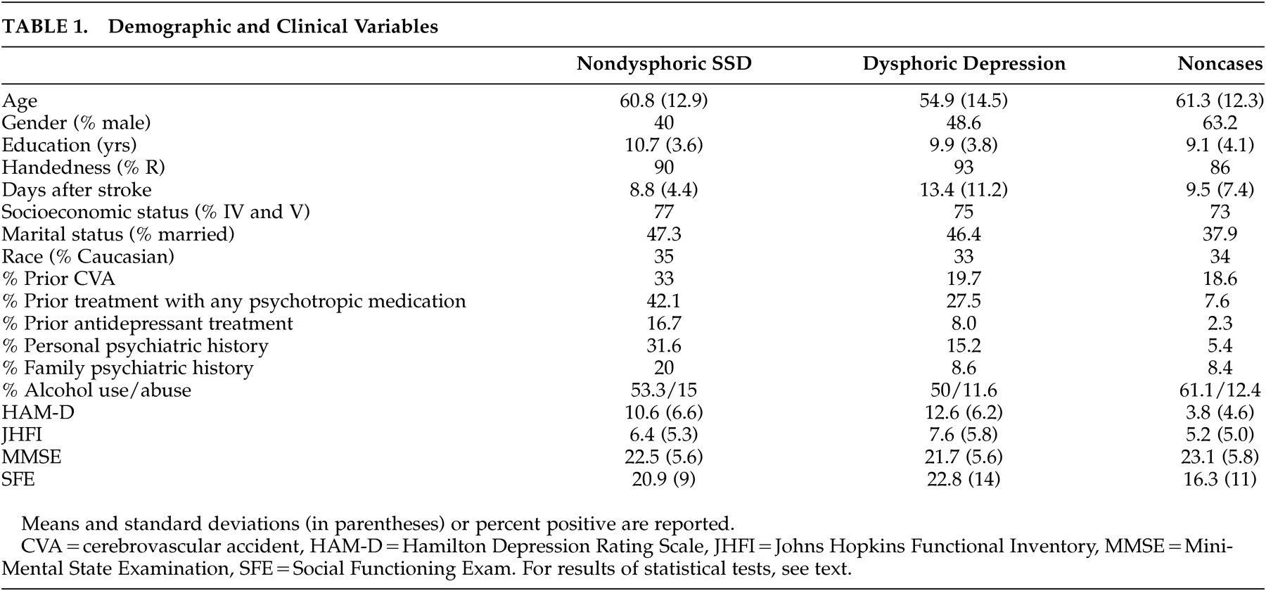
There were 20 (6.6%) subjects meeting criteria for nondysphoric subsyndromal depression, whereas 107 (35%) constituted the dysphoric group. Of these, 55 met criteria for minor depression and 52 met criteria for major depression. Demographic and clinical variables data are shown in Table 1 .
TABLE 1. Demographic and Clinical Variables
A significant group effect of age was found ( F [2, 290]=7.7, p<0.0001), with dysphoric patients being younger than noncases (p<0.05). No other post-hoc test reached statistical significance. Female sex was significantly more frequent in both depression groups compared to nondepressed comparison subjects (chi-square [2]=8.1, p<0.02), but dysphoric and nondysphoric depression did not differ (chi-square [1]=.5, p>0.4).
No group effect of race, handedness, education, socioeconomic and marital status, history of prior stroke, psychiatric family history, or alcohol consumption or abuse was found to be statistically significant. A group effect for personal psychiatric diagnosis was found be statistically significant (chi-square [2]=16, p<0.001), but the difference between dysphoric and nondysphoric did not reach statistical significance (chi-square [1]=3.2, p=0.074). Consistent with this finding, a significant group effect of prior treatment with any psychotropic medications was found (chi-square [2]=33, p<0.0001, but there was no difference between dysphoric and nondysphoric depression (chi-square [1]=1.1, p>0.2). Twice as many nondysphoric subsyndromal depression subjects were treated with antidepressants before the occurrence of the stroke compared with persons with dysphoric depression (16.7% versus 8%). Subjects with this condition were also treated more frequently (5% versus 0%) with antipsychotics and other psychotropic medications (11% versus 1%) (whole model statistics for all drug categories, chi-square [8]=26.8, p<0.001).

Psychopathology and Impairment Variables

As expected based on our inclusion criteria, a significant group effect for severity of depressive symptoms was found ( F [2, 294]=92, p<0.0001), with both dysphoric and nondysphoric disorders showing greater symptom severity compared to nondepressed controls (p<0.05) ( Table 1 ). The difference between dysphoric and nondysphoric depression groups was not statistically significant (p>0.05).
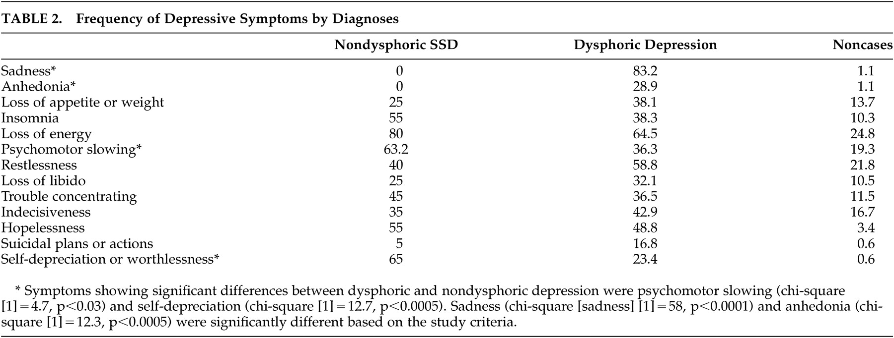
The frequency of affective, cognitive, and vegetative symptoms of depression is shown in Table 2 . Affective symptoms were significantly more frequent in the dysphoric than the nondysphoric subsyndromal depression group. All overall effects were highly significant, as expected (chi-square values between 16 and 87, p values <0.0003 or better in all cases). Statistically significant differences between nondysphoric subsyndromal depression and dysphoric depression included psychomotor slowing and self-depreciation ( Table 2 ).
TABLE 2. Frequency of Depressive Symptoms by Diagnoses
In addition, there were quantitative and qualitative group differences in the emotion-related psychopathology. While there was no subject in the nondysphoric subsyndromal depression and comparison groups acknowledging either moderate or severe “emptiness of all feelings and lack of ability to react emotionally,” 36% (24.8% moderate and 11.4% severe) of subjects in the dysphoric depression group did endorse this symptom (chi-square [4]=87.0, p<0.0001); the difference between dysphoric and nondysphoric depression was also statistically significant (two-tailed Fisher’s exact p<0.003). In addition to this symptom, there was a significant overall effect for the psychopathological sign “sad affect,” which was present in 52.6% of nondysphoric subsyndromal depression patients (11.5% moderate and 42.1% severe), in 65% of dysphoric depression subjects (49% moderate and 16.3% severe), and in 14.6% (12.7% moderate and 2.9% severe) of controls (two-tailed Fisher’s exact p<0.002). The difference between dysphoric and nondysphoric depression on moderate or severe sad affect was also statistically significant (two-tailed Fisher’s exact p<0.002), meaning that nondysphoric subsyndromal depression patients had greater likelihood to show more severe sad affect compared to dysphoric depressed patients. In aggregate these data signify that while patients with nondysphoric depression did not endorse emotional psychopathology, observation of their affect was consistent with a depressive disorder.
Severity of impairment in activities of daily living also showed a significant group effect ( F [2, 294]=6.6, p<0.002), with nondysphoric subsyndromal depression lying between dysphoric depression and noncases, but only the dysphoric group was significantly different compared with noncases (p<0.05). Satisfaction with social functioning showed a significant group effect ( F [2, 212]=6.51, p<0.002). While nondysphoric subsyndromal depression social functioning was between the values of the dysphoric group and noncases, only dysphoric depression was significantly different from noncases in post-hoc tests (p<0.05). In addition, differences in cognitive functioning were not found to be significantly different ( F [2, 281]=1.6, p>0.2).

Localization of Brain Damage

For subjects who received a head CT, a brain lesion was found in eight nondysphoric subjects (57.1%), 36 dysphoric depressed subjects (83.7%), and 87 controls (62.6%) (chi-square [2]=7.2, p<0.03). There were six subjects (42.8%) in the nondysphoric, seven in the dysphoric (16.8%), and 52 in the control group (37.4%) where a repeat head CT did not reveal a brain lesion.
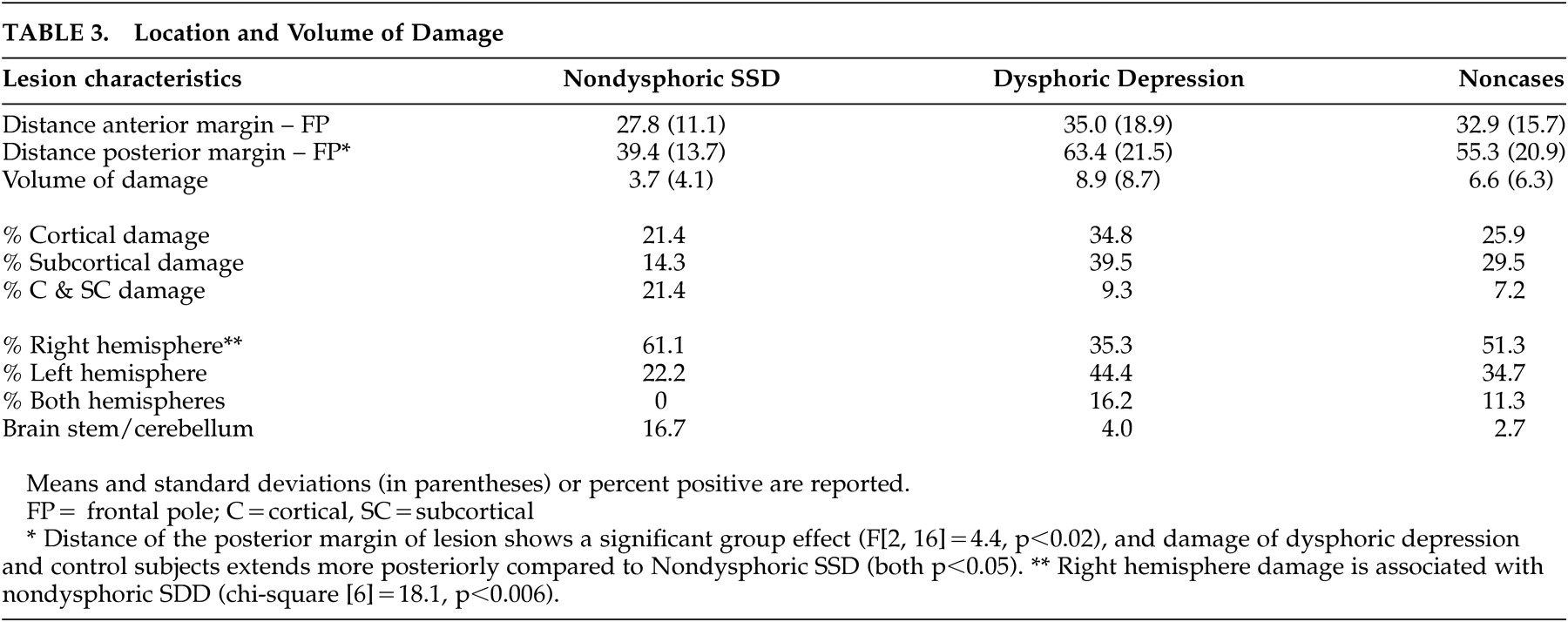
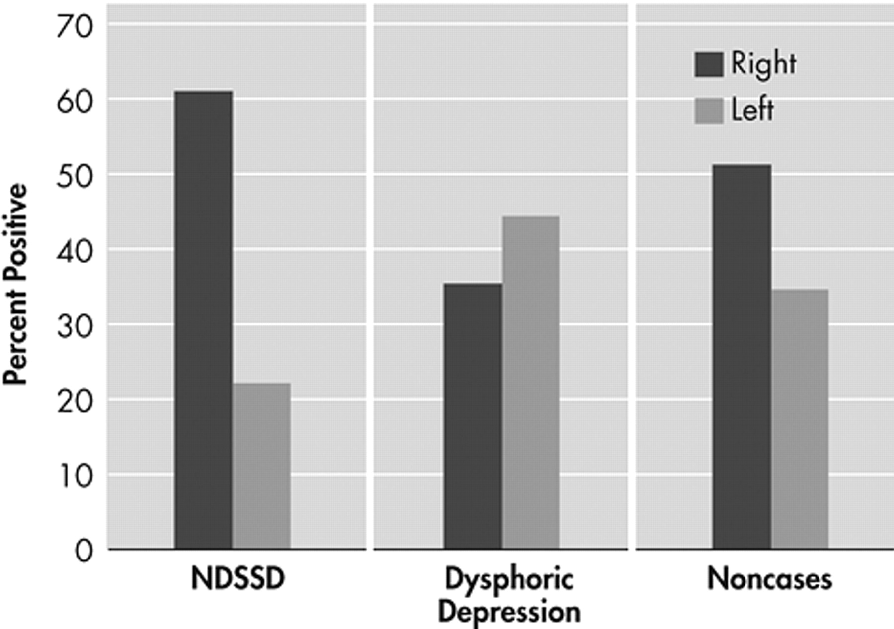
Our main hypothesis of a significant association between nondysphoric subsyndromal depression and right hemisphere lesion was substantiated (i.e., 61% of the patients with this condition, 35% of the dysphoric subjects, and 51% of controls had a right hemisphere lesion (chi-square [6]=18.1, p<0.006) ( Table 3 ). Comparing only dysphoric and nondysphoric depressed subjects with single unilateral lesions revealed an effect of hemispheric side of lesion (chi-square [1]=4.4, p<0.04) ( Figure 1 ).
TABLE 3. Location and Volume of Damage
FIGURE 1. Percent of Subjects per Diagnostic Group with CT-Ascertained Right or Left Hemisphere Lesions
NDSSD=nondysphoric subsyndromal depression. Significantly more nondysphoric subsyndromal depression subjects had right hemisphere damage compared with persons with dysphoric depression (chi-square [1]=4.4, p<0.04).
While the distance of the anterior margin of the damaged area to the frontal pole was comparable across the three groups ( F [2, 140]=0.56, p>0.5), the distance of the posterior margin showed a significant group effect ( F [2, 16]=4.4, p<0.02), demonstrating that the damage in dysphoric and control patients extended significantly more posteriorly compared to nondysphoric subsyndromal depression patients (p<0.05 in both cases). There was no significant group association with volume ( F [2, 144]=2.1, p=0.12), or damage to cortical or subcortical regions (chi-square [4]=3.7, p>0.4). In summary, analyses of the location of the damage support our hypothesis that nondysphoric depression is associated with right hemisphere damage localized to more anterior structures than dysphoric depression.
DISCUSSION

The main goal of this study was to begin to understand the brain mechanisms of nondysphoric subsyndromal depression using a large database of stroke subjects systematically assessed for psychopathology and lesion location. Despite reports of influential clinicians 1317 and empirical publications addressing construct validity, 11, 13 the brain correlates of this condition had not been examined until now. The present study found that nondysphoric depression is a moderately frequent and potentially significant psychopathological condition in the poststroke period. Overall, nondysphoric depressed patients had greater psychosocial impairment and impairment in daily living than comparison patients but less than dysphoric depressed patients following stroke, with the only statistically significant differences between dysphoric and nondepressed subjects. Notably, more persons with nondysphoric depression had a negative head CT than persons with dysphoric depression, but for subjects with a positive head CT, a right hemisphere lesion was found more frequently in nondysphoric than dysphoric depression.
Before discussing these findings, we must acknowledge some limitations of the present study. A relatively large portion of persons were lower socioeconomic class and African-American. Because subsyndromal depression has been found to be twice as frequent in Caucasians compared to African Americans, 44 our study may have underestimated the frequency of nondysphoric subsyndromal depression. Patients with significant comprehension deficits or markedly decreased levels of consciousness were excluded. Because patients were studied during the first few weeks after the onset of neurological symptoms, the results may not generalize beyond the acute stroke period. In addition, not all subjects received a radiographic examination to determine the location of their brain lesion. No significant group difference in demographic and impairment variables (except for gender) was found between patients who did and did not receive a brain scan, but this should be considered in the interpretation of the data.

Frequency and Clinical Variables

Nondysphoric subsyndromal depression appears to be a moderately frequent clinical condition in the acute poststroke period for it was present in almost 7% of acute stroke patients. A surprising finding was that the dysphoric and nondysphoric conditions were not significantly different in terms of many demographic and clinical variables including race, handedness, education, socioeconomic and marital status, history of prior stroke, psychiatric family history, and alcohol consumption or abuse. The age of patients with dysphoric depression was significantly younger than nondepressed controls but this difference did not reach significance in the dysphoric/nondysphoric comparison. Also, the female to male ratio was similar in both dysphoric and nondysphoric depression, but compared with dysphoric depression significantly more people with nondysphoric depression had been diagnosed with a psychiatric disorder or treated with psychotropic medications, including antidepressants, before they suffered a stroke.
The difference in the phenomenological presentation between dysphoric and nondysphoric depression goes beyond what would be expected based on the inclusion criteria. In addition to the absence of endorsement of depressed mood and anhedonia, nondysphoric depression was characterized by greater psychomotor slowing and self-depreciation. If confirmed by future studies, these psychopathological differences may be useful in diagnosing this elusive depressive disorder in stroke patients. 45

Mechanisms

Brain scan data suggest possible differences in the mechanisms of dysphoric and nondysphoric depression following stroke. Unipolar depression is the most common psychiatric disorder that follows brain damage of any etiology. 4647 Worldwide pooled data demonstrate that 41% of patients develop a depressive disorder after stroke (21.7% major and 19.5% minor depression). 18 Frequencies for other psychiatric disorders that follow an insult to the brain such as mania or psychosis are much lower. Episodes of depression before stroke increase the vulnerability to develop depression following stroke. 48
Because depression following brain injury is so common, a central question in neuropsychiatry concerns what prevents the development of a dysphoric depressive disorder as a sequelae of stroke. The significant association linking nondysphoric subsyndromal depression with damage to the right anterior region of the brain may help to clarify this phenomenon. A general concept key to understanding the mechanisms leading to nondysphoric subsyndromal depression in brain injury is that the specific location of the damage may have either or both etiological and pathoplastic (i.e., influencing the presentation of the illness) effects. This view is consistent with research showing that the right hemisphere predominantly subserves emotion processing, 4950 and damage to the right hemisphere may alter the (correct) identification of personal emotional states. 31, 5152 Hence, destruction of lateralized brain regions supporting perception of emotional material may lead persons at risk of depression to develop cognitive and vegetative but not emotional symptoms (sadness or anhedonia) because of impairment in perception of personal affective states. 51 In this model, brain damage and vulnerability to develop a depressive disorder interact with the strategic location of the lesion, shaping the phenomenological presentation of depression after stroke. This finding is consistent with the notion that major depression in the acute poststroke period is more likely after a stroke to the left anterior hemisphere 18 —which is not thought to be involved in assessment of personal affective states.
The model also helps to explain the lack of significant difference in terms of association with right hemisphere damage between nondysphoric subsyndromal depression and nondepressed comparison subjects. Considering the negative effects of right hemisphere damage on the capacity to perceive and/or endorse depressed emotion and mood, this finding was not totally surprising. In fact, as a consequence of their more frequent history of depression prior to stroke, nondysphoric subsyndromal depression subjects would be expected to have a considerably elevated vulnerability to develop a depressive relapse after stroke. They, however, developed a nondysphoric presentation following strategic damage to the right anterior brain.

Emotion Psychopathology

Irrespective of brain damage, significantly more patients with dysphoric than nondysphoric depression endorsed emotional emptiness and poor reactivity to emotional events, and significantly more persons with nondysphoric depression were assessed as displaying severe sad affect than dysphoric depression or comparison subjects. Sad affect in absence of endorsed sadness and anhedonia is consistent with reduced conscious access to emotions. Impaired conscious access to emotions should not reduce the physical manifestations of the depressive illness which will continue to be characterized by depressive ideation, vegetative symptoms and sad facial expression and posture (affect).
While the mechanisms of nondysphoric depression in the absence of gross brain lesion are not understood, it is plausible that high levels of alexithymia (i.e., reduced emotional awareness and verbalization) prevent reporting sadness. It is noteworthy that greater alexithymic features have been found to be associated with smaller right cingulate gyrus gray matter 31 and that right hemisphere stroke leads to high levels of alexithymia. 51 Thus, a parsimonious interpretation of the findings in this study is that nondysphoric depression may be due to reduced access to personal emotions that may follow either right hemisphere stroke damage or high levels of alexithymia, or both. While in stroke, direct damage to structures subserving emotional awareness may lead to “acquired alexithymia” and impair the capacity to endorse sadness or anhedonia; 30 nondysphoric depression not obviously due to large strokes 3 may be ascribed to dysfunction of brain structures found to be associated with high alexithymic features, including the right inferior, middle and orbital frontal cortex 28 and the anterior cingulate gyrus. 29
Underreporting of sadness and anhedonia may be responsible for underdiagnosing depression and lack of appropriate treatment in primary care. 53 Nondysphoric depression leads to increased need of welfare benefits, 11 medical illness and psychosocial distress, 18, 10 cognitive impairment, and mortality. 3 For these reasons, it is important to systematically examine the extent to which poststroke nondysphoric subsyndromal depression contributes a negative prognostic factor and is less than optimally identified and treated.
There have been no specific treatment trials of nondysphoric depression although data are beginning to emerge on treatment of subsyndromal depressive conditions. 5456 The importance of identifying and treating persons with reduced emotional awareness in the poststroke period and the validation of nondysphoric subsyndromal depression as a particular presentation of depressive disorder was underscored in a recent study in which 8 weeks of treatment with sertraline or fluoxetine significantly improved the ability to identify and describe emotions in patients with right hemisphere stroke. 52
In summary, the present study found that poststroke nondysphoric subsyndromal depression is a moderately common clinical condition associated with right hemisphere anterior brain damage and poor ability to report emotion-related symptoms. Future studies should examine nondysphoric subsyndromal depression in other populations, which may help to illuminate the cause of this potentially significant clinical condition and foster treatment research.

Acknowledgments

This work was supported by the Edward J. Mallinckrodt Jr. and Dana Foundations and an NIH-sponsored Mentored Research Scholarship (K12) (Dr. Paradiso).

Footnote

Received December 12, 2006; revised February 9, 2007; accepted February 12, 2007.
The authors are affiliated with The University of Iowa, Department of Psychiatry, Department of Neurology, Roy J. and Lucille A. Carver College of Medicine, Iowa City, Iowa. Address correspondence to Sergio Paradiso, M.D., Ph.D., University of Iowa, Department of Psychiatry, 200 Hawkins Dr., W278 GH, Iowa City, IA 52242; [email protected] (e-mail).
Copyright © 2008 American Psychiatric Publishing, Inc.

References

1.
Broadhead WE, Blazer DG, George LK, et al: Depression, disability days, and days lost from work in a prospective epidemiology survey. JAMA 1990; 264:2524–2528
2.
Bruce ML, Seeman TE, Merrill SS, et al: The impact of depressive symptomatology on physical disability. MacArthur studies of successful aging. Am J Public Health 1994; 84:1796–1799
3.
Gallo JJ, Rabins PV, Lyketsos CG, et al: Depression without sadness: functional outcomes of nondysphoric depression in later life. J Am Geriatr Soc 1997; 45:570–578
4.
Horwath E, Johnson J, Klerman GL, et al: Depressive symptoms as relative and attributable risk factors for first-onset major depression. Arch Gen Psychiatry 1992; 49:817–823
5.
Judd LL, Paulus MP, Wells KB, et al: Socioeconomic burden of subsyndromal depressive symptoms and major depression in a sample of the general population. Am J Psychiatry 1996; 153:1411–1417
6.
Olfson M, Broadhead WE, Weissman MM, et al: Subthreshold psychiatric symptoms in a primary care group practice. Arch Gen Psychiatry 1996; 53:880–886
7.
Penninx BW, Guralnik JM, Ferrucci L, et al: Depressive symptoms and physical decline in community-dwelling older persons. JAMA 1998; 279:1720–1726
8.
Penninx BW, Leveille S, Ferrucci L, et al: Exploring the effect of depression on physical disability: longitudinal evidence from the established populations for epidemiologic studies of the elderly. Am J Public Health 1999; 89:1346–1352
9.
Sadek N, Bona J: Subsyndromal symptomatic depression: a new concept. Depress Anxiety 2000; 12:30–39
10.
Wells KB, Stewart A, Hays RD, et al: The functioning and well being of depressed patients: results from the medical outcomes study. JAMA 1989; 262:914–919
11.
Judd LL, Rapaport MH, Paulus MP, et al: Subsyndromal symptomatic depression: a new mood disorder. J Clin Psychiatry 1994; 55:18–28
12.
Paradiso S, Robinson RG: Minor depression after stroke: an initial validation of the DSM-IV construct. Am J Geriatr Psychiatry 1999; 7:244–251
13.
Keilholz P: Masked Depression. Berne, Hans Huber Publishers, 1973, pp 61–76
14.
Asnis GM, McGinn LK, Sanderson WC: Atypical depression: clinical aspects and noradrenergic function. Am J Psychiatry 1995; 152:31–36
15.
Klerman GL, Budman S, Berwick D, et al: Efficacy of a brief psychosocial intervention for symptoms of stress and distress among patients in primary care. Med Care 1987; 25:1078–1088
16.
Sherbourne CD, Wells KB, Hays RD, et al: Subthreshold depression and depressive disorder: clinical characteristics of general medical and mental health specialty outpatients. Am J Psychiatry 1994; 151:1777–1784
17.
Mossey JM, Knott KA, Higgins M, et al: Effectiveness of a psychosocial intervention, interpersonal counseling for subdysthymic depression in medically ill elderly. J Gerontol A Biol Sci Med Sci 1996; 51:M172–178
18.
Robinson RG: The Clinical Neuropsychiatry of Stroke . Cambridge, UK, Cambridge University Press, 2006
19.
Martinot JL, Hardy P, Feline A, et al: Left prefrontal glucose hypometabolism in the depressed state: a confirmation. Am J Psychiatry 1990; 147:1313–1317
20.
Kozel FA, George MS: Meta-analysis of left prefrontal repetitive transcranial magnetic stimulation (rTMS) to treat depression. J Psychiatr Pract 2002; 8:270–275
21.
Hughlings-Jackson J: Selected Writings. London, Hodder and Stoughton, 1931
22.
Denny-Brown D, Meyer JS, Horenstein S: The significance of perceptual rivalry resulting from parietal lesion. Brain 1952; 75:433–471
23.
Hecaen H: The clinical symptomatology of right and left hemispheric lesions. Cah Coll Med Hop Paris 1962; 3:259–270
24.
Hecaen H, Albert M: Disorders of mental functioning related to frontal lobe pathology, in Psychiatric Aspects of Neurological Disease. Edited by Benson DF, Blumer D. New York, Gruen and Stratton, 1975
25.
Sifneos PE: Short-term Psychotherapy and Emotional Crisis. Cambridge, MA, Harvard University Press, 1972
26.
Lane RD, Ahern GL, Schwartz GE, et al: Is alexithymia the emotion equivalent of blindsight? Biol Psychiatry 1997; 42:834–844
27.
Lumley MA: Alexithymia and negative emotional conditions. J Psychosom Res 2000; 49:51–54
28.
Kano M, Fukudo S, Gyoba J, et al: Specific brain processing of facial expression in people with alexithymia: an H2 150-PET study. Brain 2003; 126:1474–1484
29.
Berthoz S, Artiges E, Van De Moortele PF, et al: Effect of impaired recognition and expression of emotions on fronto cingulate cortices: An fMRI study of men with alexithymia. Am J Psychiatry 2002; 159:961–967
30.
Lane RD, Fink GR, Chau PM, et al: Neural activation during selective attention to subjective emotional responses. Neuroreport 1997; 8:3969–3972
31.
Jones AM, Paradiso S, Fiedorowicz J, et al: Sub regions of the human anterior cingulate cortex and alexithymia. Presented at the 17 th annual meeting of the American Neuropsychiatric Association. La Jolla, CA, February 18–21, 2006
32.
Castillo CS, Starkstein SE, Fedoroff JP, et al: Generalized anxiety disorder after stroke. J Nerv Ment Dis 1993; 181:100–106
33.
Downhill JE, Robinson RG: Longitudinal assessment of depression and cognitive impairment following stroke. J Nerv Ment Dis 1994; 182:425–431
34.
Wing JK, Cooper JE, Sartorius N: Measurement and classification of psychiatric symptoms: an instruction manual for the PSE and Catego Program. London, Cambridge University Press, 1974
35.
Robinson RG, Benson DF: Depression in aphasic patients: frequency, severity, and clinical-pathology correlations. Brain Lang 1981; 14:282–291
36.
Robinson RG, Szetela B: Mood change following left hemispheric brain injury. Ann Neurol 1981; 8:447–453
37.
Levine DN, Grek A: The anatomic basis of delusions after right cerebral infarction. Neurol 1984; 34:577–582
38.
Starkstein SE, Robinson RG, Price TR: Comparison of cortical and subcortical lesions in the production of post-stroke mood disorders. Brain 1987; 110:1045–1059
39.
Robinson RG, Bolla-Wilson K, Kaplan E, et al: Depression influences intellectual impairment in stroke patients. Br J Psychiatry 1986; 148:541–547
40.
Robinson RG, Kubos KL, Starr LB, et al: Mood disorders in stroke patients. importance of location and lesion. Brain 1984; 107:81–93
41.
Starr LB, Robinson RG, Price TR: Reliability, validity, and clinical utility of the social functioning exam in the assessment of stroke patients. Exp Aging Res 1983; 9:101–106
42.
Robinson RG, Starr LB, Kubos KL, et al: A two-year longitudinal study of post-stroke mood disorders: findings during the initial evaluation. Stroke 1983; 14:736–741
43.
Hamilton M: Development of a rating scale for primary depressive illness. Br J Soc Clin Psychol 1967; 6:278–296
44.
Cohen CI, Magai C, Yaffee R, et al: Comparison of users and non-users of mental health services among depressed, older, urban African Americans. Am J Geriatr Psychiatry 2005; 13:545–553
45.
Carota A, Staub F, Bogousslavsky J: Emotions, behaviors, and mood changes in stroke. Curr Opin Neurol 2002; 15:57–69
46.
Benbadis SR, Sila CA, Christea RL: Mental status changes and stroke. J Gen Intern Med 1994; 9:485–487
47.
Koponen S, Taiminen T, Portin R, et al: Axis I and II psychiatric disorders after traumatic brain injury: a 30-year follow-up study. Am J Psychiatry 2002; 159:1315–1321
48.
Paradiso S, Robinson RG: Gender differences in post-stroke depression. J Neuropsychiatry Clin Neurosci 1998; 10:41–47
49.
Borod JC, Koff E, Perlman Lorch M, et al: The expression and perception of facial emotion in brain-damaged patients. Neuropsychologia 1986; 24:169–180
50.
Bryden MP, Ley RG: Right-hemisphere involvement in the perception and expression of emotion in normal humans, in Neuropsychology of Human Emotion. Edited by Heilman K, Satz P. New York, Guilford Press, 1983
51.
Spalletta G, Pasini A, Costa A, et al: Alexithymic features in stroke: effects of laterality and gender. Psychosom Med 2001; 63:944–950
52.
Spalletta G, Ripa A, Bria P, et al: Response of emotional unawareness after stroke to antidepressant treatment. Am J Geriatr Psychiatry 2006; 14:220–227
53.
Charney DS, Reynolds CF III, Lewis L, et al: Depression and bipolar alliance support consensus statement on the unmet needs in diagnosis and treatment of mood disorders in late life. Arch Gen Psychiatry 2003; 60:664–672
54.
Dietrich AJ, Oxman TE, Williams JW, et al: Re-engineering systems for the treatment of depression in primary care: cluster randomized controlled trial. Br Med J 2004; 329:602
55.
Lyness JM: Treatment of depressive conditions in later life: real-world light for dark (or dim) tunnels. JAMA 2004; 291:1626–1628
56.
Williams JW, Barrett J, Oxman T, et al: Treatment of dysthymia and minor depression in primary care: a randomized controlled trial in older adults. JAMA 2000; 284:1519–1526

Information & Authors

Information

Published In

Go to The Journal of Neuropsychiatry and Clinical Neurosciences
The Journal of Neuropsychiatry and Clinical Neurosciences
Pages: 52 - 61
PubMed: 18305284

History

Published online: 1 January 2008
Published in print: Winter, 2008

Authors

Details

Sergio Paradiso, M.D., Ph.D.
Robert G. Robinson, M.D.

Metrics & Citations

Metrics

Citations

Export Citations

If you have the appropriate software installed, you can download article citation data to the citation manager of your choice. Simply select your manager software from the list below and click Download.

For more information or tips please see 'Downloading to a citation manager' in the Help menu.

Format
Citation style
Style
Copy to clipboard

View Options

View options

PDF/EPUB

View PDF/EPUB

Get Access

Login options

Already a subscriber? Access your subscription through your login credentials or your institution for full access to this article.

Personal login Institutional Login Open Athens login
Purchase Options

Purchase this article to access the full text.

PPV Articles - Journal of Neuropsychiatry and Clinical Neurosciences

PPV Articles - Journal of Neuropsychiatry and Clinical Neurosciences

Not a subscriber?

Subscribe Now / Learn More

PsychiatryOnline subscription options offer access to the DSM-5-TR® library, books, journals, CME, and patient resources. This all-in-one virtual library provides psychiatrists and mental health professionals with key resources for diagnosis, treatment, research, and professional development.

Need more help? PsychiatryOnline Customer Service may be reached by emailing [email protected] or by calling 800-368-5777 (in the U.S.) or 703-907-7322 (outside the U.S.).

Media

Figures

Other

Tables

Share

Share

Share article link

Share