Full access
REGULAR
Published Online: 1 October 2010

Increased Cortisol Levels and Anticholinergic Activity in Cognitively Unimpaired Patients

Publication: The Journal of Neuropsychiatry and Clinical Neurosciences
T he neurotransmitter acetylcholine plays an important role in the processes of learning and memory. Consequently, anticholinergic serum levels might be associated with disturbances in cognitive function such as delirium, depression, or dementia. 1
Elderly patients scheduled for a surgery because of urological reasons are often characterized by multimorbidity, changed neurotransmitter balance, and/or polypharmacy. Additionally, medications with anticholinergic properties (e.g., sedatives, narcotics, and antibiotics) are regularly applied in perioperative conditions and may lead to an increased anticholinergic burden. 2 The concept of an anticholinergic burden addresses a situation in which a variety of medications with anticholinergic actions are prescribed, leading to anticholinergic toxicity which can be detected as increased serum anticholinergic activity (SAA) by a muscarinic anticholinergic radioreceptor assay, 3 independently of the origin of anticholinergic burden.
Using this assay technique, previous work has suggested a link between SAA and cognitive decline. Thus, several studies reported a correlation of SAA with cognitive impairment during depression 4 or dementia 1, 5 as well as with delirium in intensive care patients. 6, 7 Recently, particular interest has been directed to the anticholinergic burden in surgical patients due to its possible relationship with an increased risk of transient and persistent postoperative cognitive dysfunction. 2, 6 Cerebral cholinergic deficit is considered one of the main reasons for abrupt cognitive decline and postoperative dysfunctions including delirium. 8, 9 Postoperative cognitive impairment is of particular significance as it is associated with worsened outcome in hospitalized patients, including prolonged hospital stay and increased mortality. 10, 11 Because SAA reflects the cumulative binding capacity of muscarinic receptors to acetylcholinergic substances, 3 most of these studies support the hypothesis that increased SAA is related to an anticholinergic burden caused by anticholinergic medications. 1214
However, the question of the relationship between endogenous and exogenous factors modifying SAA levels is controversially discussed due to the finding of elevated SAA in patients taking no known anticholinergic medications. 15 Therefore, this work raised the possibility that in addition to medications, an endogenous source of anticholinergic activity may exist. Pre- and postsurgical stress could be such a factor. Stress is mainly mediated through high blood cortisol levels. In parallel with neurotransmitters including acetylcholine, it is well known that steroid hormones influence different functions of the nervous system. 16 Whether elevated cortisol or cholinergic deficits are causally, or as an epiphenomenon, related to cognitive impairment is not clear. However, it is shown that sustained high glucocorticoid levels may be associated with the progression of CNS diseases such as stroke, depression, and neurodegenerative diseases. 17 Furthermore, a growing body of evidence showed that the basal tonus of the hypothalamic-pituitary-adrenal (HPA) axis also increases with aging, promoting hypercortisolemia, which is of interest since aging is recognized as a risk factor for cognitive disorders. 18 Although it is well known that, in addition to acetylcholine, glucocorticoids play an important role in learning and memory functions, 1719 the role of cortisol during transient delirium is not clear. Likewise, the interaction between acetylcholinergic neurotransmission and glucocorticoid actions in relation to cognitive processes is still unknown, in particular under the conditions of perioperative surgical stress.
Thus, the central question of the present study is to investigate whether acute stress expressed by increased blood cortisol is associated with patients’ acetylcholinergic levels and cognition in perioperative surgical settings for urological reasons.

METHODS

Patients

We recruited patients between April and December 2007 at the Department of Anesthesiology at University of Heidelberg in Germany after obtaining written informed consent. This study was approved by the local ethics committee and institutional review board and was performed in accordance with the ethical standards of the Declaration of Helsinki.
To exclude the effect of sexual hormones on glucocorticoids we included only male patients. Patients older than 18 admitted for elective surgery in urology were included with the exception of patients with psychiatric or neurological disorders, such as dementia, depression, or schizophrenia, and stroke patients or those with a history of alcohol abuse. Furthermore, we excluded patients with pronounced hearing and/or visual impairment problems and non-German-speaking people because the neuropsychological tests were in German. In all, five patients were not included in the present study because of the exclusion criteria. Therefore, in accordance to power analysis (see statistics), we included 30 patients in the present study.

Characterization of the Patients

For preoperative evaluation a structural study tool was employed that included the patients’ age, sex, body weight and height, American Society of Anesthesiologists classification, and hospital admission diagnosis. Lengths of hospital stay and hospital mortality were also recorded. In addition, preoperative and postoperative medication as the number of drugs with potential and known anticholinergic properties according to previous studies 19, 20 was recorded. We included the number of preoperative anticholinergic medications that had been administered continuously for longer than 4 weeks before patients’ inclusion in our present study (long-term effect of medication).

Blood and CSF Sampling and Spinal Anesthesia

Blood samples were taken three times: (I) 1 day before surgery, (II) shortly before spinal anesthesia, and (III) on the first postoperative day between 9 and 11 a.m. Additionally, patients’ physiological (blood pressure, heart rate, and body temperature) and arterial blood parameters (pO 2, pCO 2, glucose, pH, base excess, standard bicarbonate, potassium, calcium, sodium, hematocrit, and hemoglobin levels) were determined at the end of surgery (IV).
CSF samples were taken once between 8 a.m. and 10 a.m., shortly before spinal anesthesia (respectively to II) strictly by the same investigator (PT) in a standardized manner. All patients were premedicated orally with 7.5 mg midazolam at 24 hours and 1 hour before the planned surgery. Two milliliters of venous blood and 1 ml of CSF were taken under local anesthesia (subcutaneous: mepivaciane 1%) and stored at 4°C for a maximum of 1 hour before further processing.

Scoring Tests

The investigator involved in neuropsychological testing (JM) was trained by a psychologist in scoring procedures; cross-center observation and cross-scoring of test protocols were used to assure data quality before the beginning of the study.
Delirium was assessed in every patient by the same investigator using the German translation of the Abbreviated Mental Test, the confusion assessment method, and the Delirium Index 2123 on the basis of ICD-10 and DSM-IV criteria by evaluating four items: acute onset of cognitive changes with a fluctuating course, inattention, disorganized thinking, and altered level of consciousness.
Neuropsychological testing included the Mini-Mental State Examination (MMSE). 24 Working memory capacities were determined using the Auditory Verbal Learning Test (word list: immediate and delayed) and the Digit Span Task (forward and backward) from the German Nuernberger Alters-Inventar. 25 The duration of the MMSE and time for performing the whole battery of tests was recorded.
The testing was performed 1 day before surgery and on the first postoperative day immediately before blood was taken for biochemical analysis.

Blood Analysis

The venous blood samples (2 ml) were centrifuged at 7,000 rpm for 10 minutes. Thereafter, the supernatant was taken and stored together with vials containing the CSF for a maximum of 1 month at −80°C until anticholinergic activities were determined.
An investigator blinded to all clinical data assessed the plasma anticholinergic activities by competitive radioreceptor binding assay as described by Tune and Coyle. 3
For measurement of total cortisol levels, 100 μL of plasma or CSF was extracted with 1 ml of diethyl ether. The organic phase was evaporated in a stream of nitrogen. The residue was analyzed applying a commercial cortisol enzyme immunoassay kit, which operates on the basis of competition between an HRP-cortisol conjugate and the cortisol in the sample (Oxford Biomedical Research, Rochester Hills, Michigan). Intra-assay and inter-assay coefficients of variation were 7.1% and 3.4%, respectively.

Statistical Analysis

Power analysis, assuming a clinically important difference of 15% in cortisol levels between the groups, suggested that 30 patients were required for the study (α=0.05; 1−β=0.8). Comparisons of normally distributed data were made using the unpaired two-tailed t test (subgroup comparison). The data for descriptive variables were analyzed by chi-square test; the data from blood analyses were compared for statistical significance using ANOVA followed by the Tukey’s post hoc test. Bivariate correlation analysis was done according to Pearson.
Statistical analyses were performed on SPSS, version 16.0 (SPSS Inc., Chicago). The results are expressed in Table 1 and Table 2 . A p value of <0.05 was considered to be statistically significant.
TABLE 1. Patient Characteristics
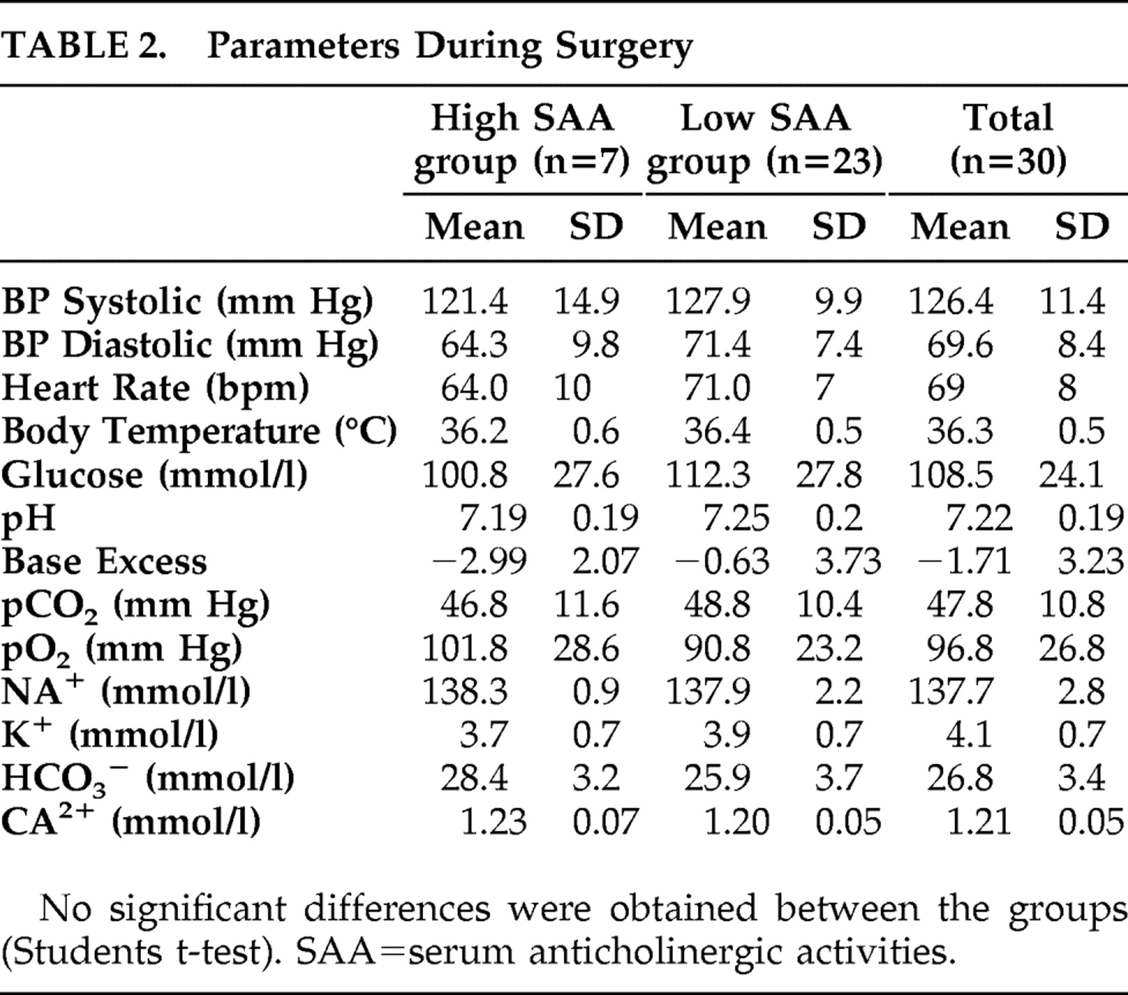
TABLE 2. Parameters During Surgery

RESULTS

Patient Characteristics

All male participants underwent elective surgery for underlying urological conditions (benign prostate hyperplasia [n=15], bladder carcinoma [n=6], urethra dysplasia [n=5], urethra constriction [n=4]).
The mean age of patients was 64.5 years old (SD=13.0), the mean body weight was 85 kg (SD=12), and the mean duration of hospital stay was 4.4 days (SD=2.0). All patients corresponded to American Society of Anesthesiologists (ASA) classification I–III. None of the patients died during their hospital stay.
In order to generate an SAA baseline in an age-related population (60 years old [SD=15]), SAA was measured in an earlier investigation including 50 ASA classification I patients in which SAA was found to be less than 4 pmol/ml in all cases (baseline data not shown). Previous studies 26, 27 suggest using SAA=4 pmol/ml as a cutoff point for an increased anticholinergic load. Based on this recommendation the present study population was divided in two subgroups, those with SAA values ≥4 pmol/ml (high SAA group, n=7) and those with SAA values <4 pmol/ml (low SAA group, n=23). Patient characteristics in subgroups are shown in Table 1 . Patients with high SAA levels received about two times more anticholinergic medication before surgery than the low SAA subgroup.

Time Course Investigations of Serum Anticholinergic Activities and Cortisol

Serum Anticholinergic Activities (SAA)

One day before surgery, the mean anticholinergic activity in all patients (n=30) was found to be 3.1 pmol/ml (SD=1.9). A significant transient increase in SAA to 6.0 pmol/ml (SD=3.7) was recorded after patients received premedication, as measured immediately before spinal anesthesia. At the first postoperative day, SAA levels were still significantly increased to 4.9 pmol/ml (SD=2.2) compared with preoperative SAA levels. No further medication without known anticholinergic action, including systemic analgesics, was applied on the first postoperative day except for antibiotics.
When analyzing both subgroups by using the cutoff value of 4 pmol/ml, the difference between the high SAA group and the low SAA group was significant at all time points ( Figure 1 ).
FIGURE 1. Subgroup Analysis of Serum Anticholinergic Activities (SAA)
Mean blood SAA levels ± SD, Student’s t-test
*p<0.05 between subgroups

Cortisol Level

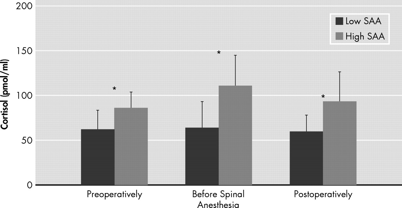
One day before surgery, the mean serum cortisol level of all patients (n=30) was found to be 70.0 ng/ml (SD=32.0). Serum cortisol levels increased to 74.4 ng/ml (SD=36.0) (p>0.05) immediately before spinal anesthesia, before a decrease to 64.6 ng/ml (SD=11.0) was obtained at the first postoperative day (p>0.05).
Results of subgroup analysis show a significant difference in serum cortisol levels between the high SAA group and the low SAA group before spinal anesthesia and after surgery ( Figure 2 ).
FIGURE 2. Subgroup Analysis of Blood Cortisol Levels
Mean blood SAA levels ± SD, Student’s t-test
*p<0.05 between subgroups
SAA=serum anticholinergic activity

Parameters During Surgery

For the parameters obtained during surgery, no significant differences were found between patients with high and with low SAA levels ( Table 2 ), nor was the duration of surgery significantly different between the subgroups (mean=56.7 minutes [SD=31.1] compared with mean=55.0 minutes [SD=26.3]). Hemoglobin concentrations amounted to 13.4 g/dl (SD=2.1) in patients with high SAA compared with 13.1 g/dl (SD=2.1) in patients with low SAA levels (p>0.05). Therefore, distinctions in SAA or cortisol are not associated with intraoperative differences.

CSF Analysis

Anticholinergic activity in all patients (n=30) was found to be 7.6 pmol/ml (SD=1.8) when measured immediately before spinal anesthesia (time point II). Results of subgroup analysis showed a marked difference (p<0.001) in SAA levels between the high SAA group (mean=9.5 pmol/ml [SD=2.3]) and the low SAA group (mean=4.6 pmol/ml [SD=1.7]).
Cortisol amounted to 9.3 ng/ml (SD=5.5) in the CSF (n=30). Results of subgroup analysis showed increased CSF cortisol levels in the high SAA group (mean=12.4 ng/ml [SD=9.3]) and diminished CSF cortisol levels in the low SAA group (mean=8.6 ng/ml [SD=3.9]; p>0.05).

Neuropsychological Tests

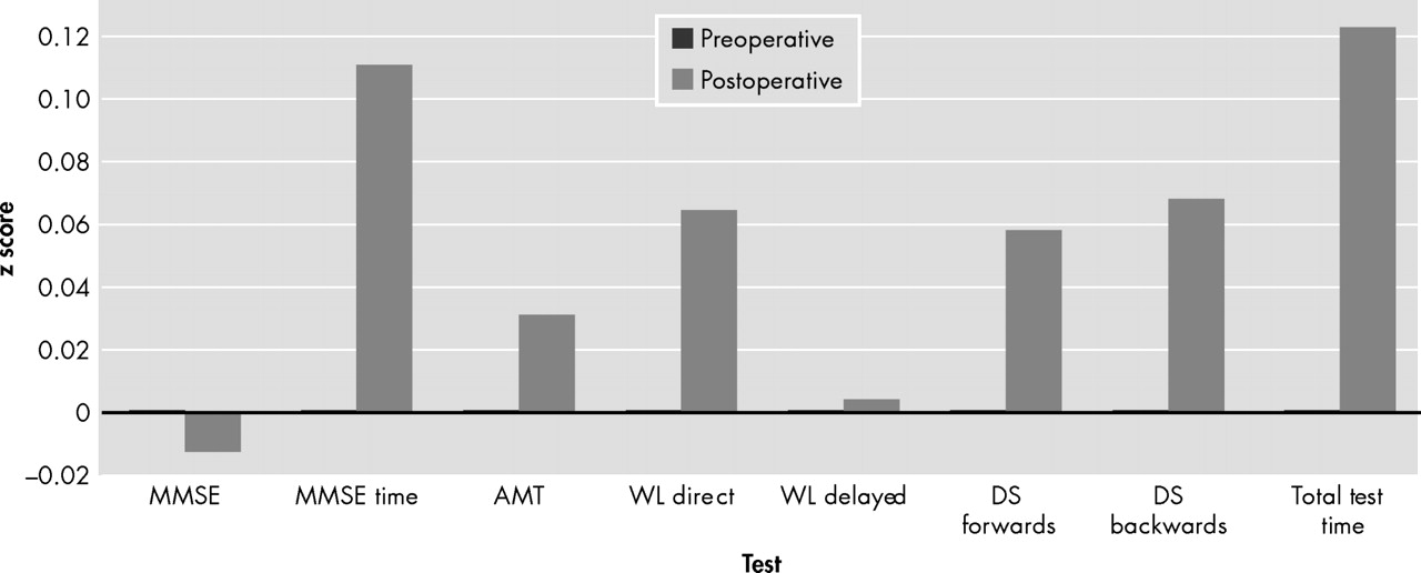
None of the study patients was delirious either under the preoperative or postoperative condition. Additionally, only a slight tendency for deterioration was observed in nearly all neuropsychological tests when preoperative levels were compared with the postoperative data in these patients; however, these differences were not statistically significant ( Figure 3 ). Furthermore, no marked differences in the neuropsychological data were detected when comparing the two subgroups with high and low preoperative SAA levels (data not shown).
FIGURE 3. Neuropsychological Examination
z score (subtracting the population mean from an individual raw score and then dividing the difference by the population standard deviation) was calculated. The differences between the pre- (=0) and postoperative data are illustrated.
MMSE=Mini-Mental State Examination (maximum 30 points); AMT=Abbreviated Mental Test (maximum 10 points); WL=word list (maximum 8 points); DS=digit span (maximum 9 points).
No significant differences were observed between preoperative and postoperative data obtained from neuropsychological testing.

Correlation Analyses

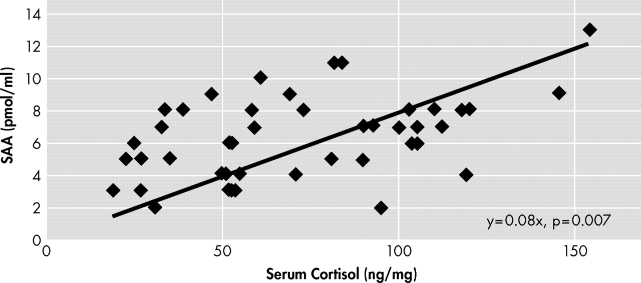
Data from all patients were correlated using the Pearson correlation test to analyze the effects between SAA levels and following preoperative parameters: anticholinergic medications (r=0.614, p=0.003), classification of the patients in relation to kinds of illnesses (American Society of Anesthesiologists classification; r=0.237, p=0.216), patients’ age (r=0.183, p=0.331), and serum cortisol (r=0.654, p<0.001). A significant positive linear correlation between SAA and serum cortisol levels was also detected if pre- and postoperative measurements were included ( Figure 4 ). In addition, significant correlation (p=0.015) (r=0.464) between anticholinergic activities and cortisol was also obtained in CSF.
FIGURE 4. Interrelation Between Serum Anticholinergic Activities (SAA) and Cortisol Levels
A significant (p=0.007) linear correlation (r=0.44) between plasma anticholinergic activity (pmol/ml atropine equivalents) and cortisol (ng/mg) was detected by including pre- and postoperative measurements.
Furthermore, a highly significant linear correlation of 0.866 between SAA and anticholinergic activities in CSF could be shown (p<0.001); the correlation between respective cortisol levels in CSF and serum was just within the significant interval (p=0.048, r=0.462). With neuropsychological data, none of these parameters showed significant association.

DISCUSSION

Significant correlations between peripheral (serum) and central (CSF) compartments for cortisol and anticholinergic activities are one precondition that both peripheral measured markers are adequately reflecting the CNS situation.
We show for the first time in our knowledge that there is a linear positive correlation between cortisol levels and SAA in patients scheduled for surgery for urological reasons. Furthermore, we have shown that the number of preoperative anticholinergic drugs in patients’ history is also clearly associated with higher SAA levels. This result is consistent with most other studies 1214 and supports the hypothesis that increased SAA is related to the anticholinergic burden caused by anticholinergic medications. In addition, the results confirm our findings of a previous study with a smaller sample that the relationship between serum and CSF anticholinergic activities helps validate SAA. 26
As stated above, however, endogenous sources of SAA related to fever, acute infection, or stress have been postulated independently of anticholinergic medication. 15, 27 We did not find a correlation between SAA and patients’ medical history as defined by the American Society of Anesthesiologists (ASA) guidelines. However, only patients with ASA classification I to III were included in this investigation. From endogenous factors, serum cortisol levels seem to play an important role for interpretation of SAA because high serum cortisol concentrations were associated with increased SAA levels ( Figure 4 ). Thus, the present data also support the hypothesis that some individuals may have anticholinergic activity in the absence of anticholinergic drugs. 15, 28
Taking both results together, our data confirm the hypothesis that both endogenous as well as exogenous factors had an influence on patients’ SAA levels. Thus, sources of the anticholinergic burden can be highly individual, and therefore the SAA may be considered as a conglomerate of anticholinergic properties of exogenous and endogenous origin. However, the generalizability of our findings needs to be estimated carefully. Our study was done only with urological patients. Further investigations on other patient populations should be conducted to justify our conclusion in relation to the interpretation of the role of SAA in general.
In a chronic state, the deleterious role of high serum cortisol levels leading to cognitive impairment is shown. 1618 Chronic stress, which can have a physiological and a psychological source, causes a permanent activation of the sympathetic nervous system, which in turn activates the HPA axis and causes elevation in serum cortisol. The role of acute stress in the development of postoperative delirium and cognitive dysfunction, however, is controversially discussed. 2932 Elevated serum cortisol levels have been associated with delirium, for instance, in Cushing’s disease and high-dose steroid treatment. 29 Only a few small studies have examined the association between serum cortisol levels and delirium in general medical and surgical settings, and the results are controversial. 30, 31 One further report suggests that individuals who fail to suppress in the dexamethasone suppression test appear to be at increased risk for delirium. 32 Thus, the lack of suppression in the dexamethasone suppression test is a sign of an overactive HPA axis. Therefore, the results of the study seem to be fully consistent with our present findings. In line with other biomarkers, however, careful control of patient characteristics should be evaluated, and further studies will be required to confirm the importance of hypercortisolism as a biomarker for delirium.
If stress, as mediated by elevated cortisol, may be an endogenous source of anticholinergic activity, an interrelation between glucocorticoids and cholinergic system is assumed. Therefore, we used this SAA assay according to Tune and Coyle 3 to detect a direct effect of cortisol in a preliminary study. Our results (data not shown) showed that exogenous applied cortisol had no direct effect on muscarinic receptors under in vitro conditions. Therefore, another—likely an indirect—interaction between cortisol and cholinergic system seems likely. Little is known about the interaction of glucocorticoids and acetylcholine. In the periphery, glucocorticoids may interfere with the storage and inactivation of acetylcholine (e.g., cholinesterase activity may be up-regulated by glucocorticoids). 33 Furthermore, long-term treatment with glucocorticoids increases synthesis and stability of junctional acetylcholine receptors on innervated cultured human muscle. 34 The human acetylcholine esterase gene was demonstrated to include a glucocorticoid-responsive element. Furthermore, evidence exists that alternative splicing forms of the acetylcholine esterase may be associated with stressful events. 35
Diverging results have been reported on the question of whether SAA correlates with cognitive dysfunction. Although most of the studies suggest that there is a clear association between SAA levels and cognitive impairment, 47 we could not confirm this effect in our present study; despite some high SAA levels in our included patients, no postoperative cognitive dysfunctions were detected. To investigate the role of SAA in relation to cognition, other groups of patients undergoing surgery under general anesthesia or with a longer in-hospital stays should be investigated with a more detailed battery of neuropsychological tests. Furthermore, a regression analysis is now planned to perform a risk factor analysis in undergoing studies. Additionally, the severity of illness in our middle-aged patients was moderate and the duration of the surgical procedures was relatively short (<1 hour). Thus, the risk of postoperative cognitive dysfunction in the presented study design was low. However, our previous studies on critically ill intensive care patients could also not detect an association between high SAA levels and delirium in postoperative delirium. 36 Therefore, the association between SAA and patients’ cognitive function still remains controversial and should be further investigated in larger patient populations.
It can be concluded from the present study that sources of the anticholinergic burden seem to be individual, and SAA therefore may be considered as a conglomerate of anticholinergic properties of endogenous and exogenous origin. The results of the present study pointed out that for interpretation of diagnostic benefit of SAA, different factors should be taken into account as we have shown for anticholinergic medication and cortisol.
Further studies are necessary for investigation of the role of SAA as a diagnostic marker for surgical patients with a high stress level or a high anticholinergic burden caused by polypharmacy with anticholinergic medication.

Acknowledgments

This study was registered at http://www.clinicaltrials.gov, NCT00463333. The work was carried out at the Department of Anesthesiology, University of Heidelberg, in Germany. This work was supported in part by the Else Kroener-Fresenius-Stiftung, Bad Homburg v.d.H., in Germany. All other funding was from within-departmental resources. We want to thank Ms. S. Himmelsbach (Department of Pathology, University of Heidelberg, D-69120 Heidelberg, Germany) for excellent technical assistance and Ms. Manuela Schwegler and the colleagues from the central patients’ management (Department of Anesthesiology and Urology, University of Heidelberg, D-69120 Heidelberg, Germany).

Footnote

Received July 17, 2009; revised September 29 and November 13, 2009; accepted November 28, 2009. Dr. Plaschke, Mr. Mattern, Dr. Martin, and Dr. Teschendorf are affiliated with the Department of Anesthesiology at the University of Heidelberg in Germany; Dr. Kopitz is affiliated with the Department of Pathology at the University of Heidelberg in Germany; Address correspondence to Konstanze Plaschke, Ph.D., Department of Anesthesiology, University of Heidelberg, Im Neuenheimer Feld 110, D-69120 Heidelberg, Germany; [email protected] (e-mail).
Copyright © 2010 American Psychiatric Publishing, Inc.

References

1.
Thienhaus OJ, Allen A, Bennett JA, et al: Anticholinergic serum levels and cognitive performance. Eur Arch Psychiatry Clin Neurosci 1990; 240:28–33
2.
Lechevallier-Michel N, Molimard M, Dartigues JF, et al: Drugs with anticholinergic properties and cognitive performance in the elderly: results from the Paquid Study. Br J Clin Pharmacol 2005; 59:143–151
3.
Tune L, Coyle JT: Serum levels of anticholinergic drugs in treatment of acute extrapyramidal side effects. Arch Gen Psychiatry 1980; 37:293–297
4.
Nebes RD, Pollock BG, Mulsant BH, et al: Low-level serum anticholinergicity as a source of baseline cognitive heterogeneity in geriatric depressed patients. Psychopharmacol Bull 1997; 33:715–720
5.
Chew ML, Mulsant BH, Pollock BG: Serum anticholinergic activity and cognition in patients with moderate-to-severe dementia. Am J Geriatr Psychiatry 2005; 13:535–538
6.
Golinger RC, Peet T, Tune LE: Association of elevated plasma anticholinergic activity with delirium in surgical patients. Am J Psychiatry 1987; 144:1218–1220
7.
Tune LE, Damlouji NF, Holland A, et al: Association of postoperative delirium with raised serum levels of anticholinergic drugs. Lancet 1981; 2:651–653
8.
Hall R, Popkin M, Henry L: Angel’s trumpet psychosis: a central nervous system anticholinergic syndrome. Am J Psychiatry 1977; 134:312–314
9.
Trzepacz P: The neuropathogenesis of delirium: the clinical observation that delirium is one manifestation of anticholinergic toxicity. Psychosomatics 1994; 4:374–391
10.
Ely EW, Shintani A, Truman B, et al: Delirium as a predictor of mortality in mechanically ventilated patients in the intensive care unit. JAMA 2004; 291:1753–1762
11.
Milbrandt EB, Deppen S, Harrison PL, et al: Costs associated with delirium in mechanically ventilated patients. Crit Care Med 2004; 32:955–962
12.
Moore AR, O’Keeffe ST: Drug-induced cognitive impairments in the elderly. Drugs Aging 1999; 15:15–28
13.
Tune LE: Serum anticholinergic activity levels and delirium in the elderly. Semin Clin Neuropsychiatry 2000; 5:149–153
14.
Kay GG, Abou-Donia MB, Messer WS Jr, et al: Antimuscarinic drugs for overactive bladder and their potential effects on cognitive function in older patients. J Am Geriatr Soc 2005; 53:2195–2201
15.
Flacker JM, Wei JY: Endogenous anticholinergic substances may exist during acute illness in elderly medical patients. J Gerontol A Biol Sci Med Sci 2001; 56:M353–M355
16.
Melcangi RC, Panzica G: Steroids and the nervous system: introduction. Ann N Y Acad Sci 2003; 1007:1–5
17.
de Quervain DJ, Aerni A, Schelling G, et al: Glucocorticoids and the regulation of memory in health and disease. Front Neuroendocrinol 2009; 30:358–370
18.
Lupien SJ, Fiocco A, Wan N, et al: Stress hormones and human memory function across the lifespan. Psychoneuroendocrinology 2005; 30:225–242
19.
Tune LE, Egeli S: Acetylcholine and delirium. Dement Geriatr Cogn Disord 1999; 10:342–344
20.
Lu CJ, Tune LE: Chronic exposure to anticholinergic medications adversely affects the course of Alzheimer disease. Am J Geriatr Psychiatry 2003; 11:458–461
21.
Linstedt U, Berkau A, Meyer O, et al: The Abbreviated Mental Test in a German version for detection of postoperative delirium. Anaesthesiol Intensivmed Notfallmed Schmerzther 2002; 37:205–208
22.
Inouye SK, van Dyck CH: Clarifying confusion: the confusion assessment method. A new method for detection of delirium. Ann Intern Med 1990; 113:941–948
23.
McCusker J, Cole MG, Dendukuri N, et al: The Delirium Index, a measure of the severity of delirium: new findings on reliability, validity, and responsiveness. J Am Geriatr Soc 2004; 52:1744–1749
24.
Folstein MF, Folstein SE, McHugh PR: “Mini-mental state”: a practical method for grading the cognitive state of patients for the clinician. J Psychiatr Res 1975; 12:189–198
25.
Oswald WD, Fleischmann UM: Nuernberger-Alters-Inventar (NAI), 4th ed. Bern, Toronto, Seattle, Hofgrefe Verlag Goettingen, 1999, pp 1–202
26.
Plaschke K, Thomas C, Engelhardt R, et al: Significant correlation between plasma and CSF anticholinergic activity in presurgical patients. Neurosci Lett 2007; 417:16–20
27.
Mulsant BH, Pollock BG, Kirshner M, et al: Serum anticholinergic activity in a community-based sample of older adults: relationship with cognitive performance. Arch Gen Psychiatry 2003; 60:198–203
28.
Hori K, Funaba Y, Konishi K, et al: Assessment of pharmacological toxicity using serum anticholinergic activity in a patient with dementia. Psychiatry Clin Neurosci 2005; 59:508–510
29.
Flacker JM, Lipsitz LA: Serum anticholinergic activity changes with acute illness in elderly medical patients. J Gerontol A Biol Sci Med Sci 1999; 54:M12–M16
30.
McIntosh TK, Bush HL, Yetson NS, et al: Beta-endorphin, cortisol, and postoperative delirium: a preliminary report. Psychoneuroendocrinology 1985; 30:303–313
31.
Gustafson Y, Olsson T, Asplund K, et al: Acute confusional state (delirium) soon after stroke is associated with hypercortisolism. Cerebrovasc Dis 1993; 3:33–38
32.
O’Keeffe ST, Devine JG: Delirium and the dexamethasone suppression test in the elderly. Neuropsychobiology 1994; 30:153–156
33.
Goto K, Chiba Y, Sakai H, et al: Glucocorticoids inhibited airway hyperresponsiveness through downregulation of CPI-17 in bronchial smooth muscle. Eur J Pharmacol 2008; 591:231–236
34.
Braun S, Askanas V, Engel WK, et al: Long-term treatment with glucocorticoids increases synthesis and stability of junctional acetylcholine receptors on innervated cultured human muscle. J Neurochem 1993; 60:1929–1935
35.
Shaked I, Zimmerman G, Soreq H: Stress-induced alternative splicing modulations in brain and periphery: acetylcholinesterase as a case study. Ann N Y Acad Sci 2008; 1148:269–281
36.
Plaschke K, Hill H, Engelhardt R, et al: EEG changes and serum anticholinergic activity measured in patients with delirium in the intensive care unit. Anesthesia 2007; 62:1217–1223

Information & Authors

Information

Published In

Go to The Journal of Neuropsychiatry and Clinical Neurosciences
The Journal of Neuropsychiatry and Clinical Neurosciences
Pages: 433 - 441
PubMed: 21037129

History

Published online: 1 October 2010
Published in print: Fall, 2010

Authors

Details

Konstanze Plaschke, Ph.D.
Peter Teschendorf, M.D.

Metrics & Citations

Metrics

Citations

Export Citations

If you have the appropriate software installed, you can download article citation data to the citation manager of your choice. Simply select your manager software from the list below and click Download.

For more information or tips please see 'Downloading to a citation manager' in the Help menu.

Format
Citation style
Style
Copy to clipboard

View Options

View options

PDF/EPUB

View PDF/EPUB

Get Access

Login options

Already a subscriber? Access your subscription through your login credentials or your institution for full access to this article.

Personal login Institutional Login Open Athens login
Purchase Options

Purchase this article to access the full text.

PPV Articles - Journal of Neuropsychiatry and Clinical Neurosciences

PPV Articles - Journal of Neuropsychiatry and Clinical Neurosciences

Not a subscriber?

Subscribe Now / Learn More

PsychiatryOnline subscription options offer access to the DSM-5-TR® library, books, journals, CME, and patient resources. This all-in-one virtual library provides psychiatrists and mental health professionals with key resources for diagnosis, treatment, research, and professional development.

Need more help? PsychiatryOnline Customer Service may be reached by emailing [email protected] or by calling 800-368-5777 (in the U.S.) or 703-907-7322 (outside the U.S.).

Media

Figures

Other

Tables

Share

Share

Share article link

Share