The continuing increase in human life expectancy places a sense of urgency on understanding the causes of disability in the elderly so that therapeutic interventions can be developed to improve their health and quality of life
(1). Among the major causes of disability in the elderly are those pertaining to changes in brain function
(2). The brain dopamine system, which plays a pivotal role in motor activity, motivation, and reward, is particularly vulnerable to age
(3). In the human brain, significant losses over a normal lifespan have been reported for dopamine D
2 receptors
(4), dopamine D
1 receptors
(5), and dopamine transporters
(6). Studies of dopamine metabolism have been less consistent, showing decreases as well as no changes with aging
(7). However, very little is known about the functional consequences of the age-related dopamine decline. Part of the difficulty is that most of the studies in humans have been done postmortem, and thus there is limited information on the functional state of the brain of these subjects.
With imaging techniques such as positron emission tomography (PET), it is now possible to measure dopamine-specific parameters and parameters that reflect brain function (e.g., regional brain glucose metabolism) in living human subjects
(8). For instance, through PET we recently documented that the age-related decline in dopamine D
2 receptors was associated not only with impaired motor performance but also with impaired performance in neurocognitive tasks that involve the frontal cortex
(9). We interpreted these findings as suggesting that the neurocognitive deficits were due to a dysregulation of the frontal regions from the loss in dopamine brain activity. Although multiple studies have revealed a significant decline in frontal and cingulate metabolism with age
(10), no study has evaluated directly whether the changes in dopamine brain activity are, in fact, associated with the changes in frontal and cingulate metabolism.
Dopamine could modulate frontal activity indirectly through the striatal-pallidal-thalamic circuit
(11) or directly through projections from mesencephalic dopamine cells
(12). The importance of dopamine in frontal activity has been suggested by imaging studies, which have shown that dopamine-enhancing agents increase frontal activation in response to task stimulation
(13,
14).
The purpose of this study was to map the effects of dopamine receptor availability on regional cerebral metabolism and investigate the association between age-related decrements in D2 receptors and regional brain glucose metabolism.
RESULTS
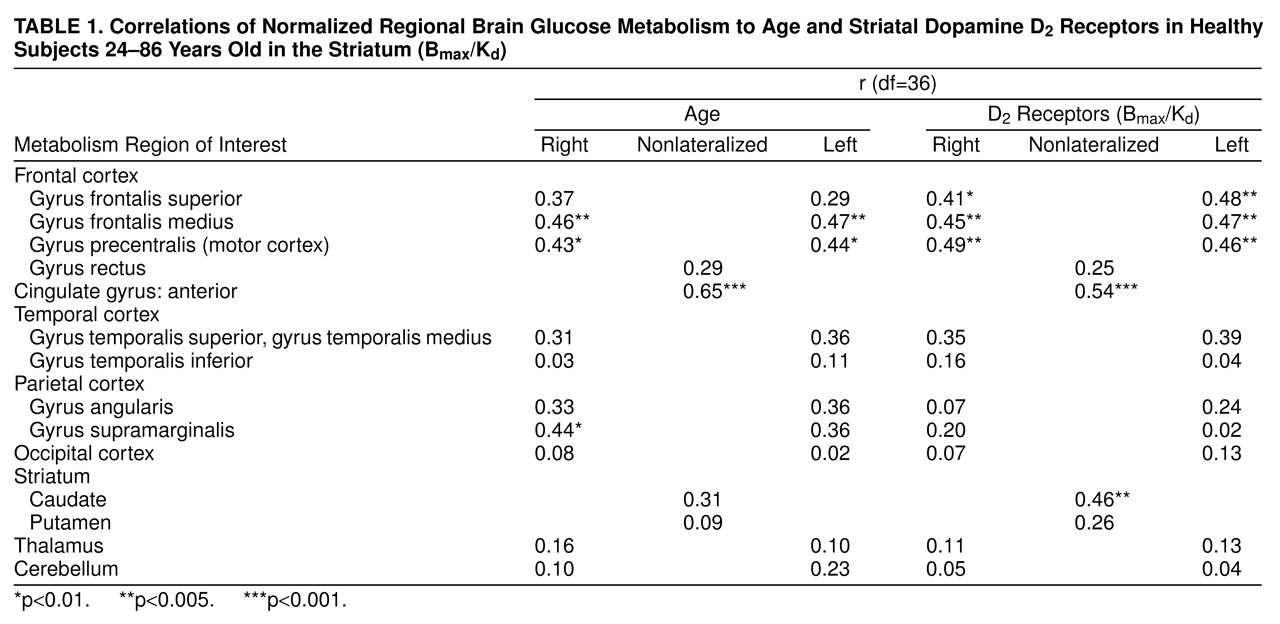
Global absolute metabolism ranged between 26 and 41 µmol/100g per minute and was not correlated with age or with D
2 receptor availability. However, the regional measures for metabolism declined significantly with age in several frontal brain regions and in the anterior cingulate as well as in the right supramarginal gyrus (
table 1). Age was also associated with a significant decline in striatal dopamine D
2 receptors (r=0.55, df=36, p<0.0001). We consequently used partial correlations to remove the effects of age from all subsequent analyses, which were found to be robust to this procedure. Therefore, the raw, uncorrected results will be reported.
The correlations between regional glucose metabolism and dopamine D
2 receptor availability were significant mostly for cortical brain regions.
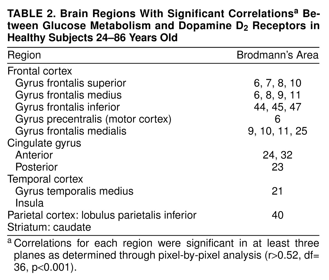
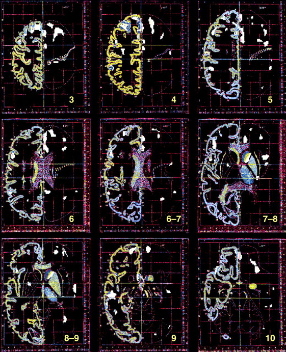
figure 1 shows (superimposed on the Talairach-Tournoux atlas) the pixels with significant (r<0.52, df=36, p<0.001) correlations between D
2 receptors and metabolism. Most of the significant correlations were located in the anterior cingulate gyrus and the frontal cortex, including areas in the prefrontal, motor, and orbitofrontal cortex (
table 2). Significant correlations were also located in association areas in the temporal cortex and in the caudate.
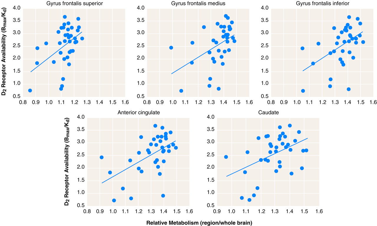
This pattern of correlations was independently corroborated when the analysis was done by using the relative metabolic measures obtained in preselected regions of interest (
table 1,
figure 2). Because the variance from relative measures can result either from changes in the region or in the global measure, we also corroborated whether the significant correlations were also observed for the absolute measures. The correlations with the absolute measures were significant for the cingulate gyrus (r=0.47, df=36, p<0.005) and left caudate (r=0.53, df=36, p<0.001) and showed a trend for significance in the frontal and superior temporal gyrus (r>0.33, df=36, 0.01<p>0.05).
DISCUSSION
In this group of healthy participants, glucose metabolism was found to decrease with age in frontal brain regions and anterior cingulate gyrus. Reductions in frontal and anterior cingulate metabolism with age have been consistently reported by imaging studies of the aging human brain
(10). The present study indicates that these age-linked metabolic reductions were coupled with the decline in dopamine D
2 receptors. We documented a similar association in a group of cocaine abusers in whom the reductions in D
2 receptors were associated with metabolic decrements in the prefrontal cortex and in the anterior cingulate gyrus
(23). However, this is the first demonstration in healthy adults of a direct association between the levels of D
2 receptors in brain and cerebral metabolic activity.
It is likely that dopamine dysfunction underlies motor impairment in the elderly (dyskinesias and rigidity), but the present results suggest that it may also underlie some of the cognitive changes seen in older individuals. Note that the cognitive deficits associated with age involve frontal lobe function
(24). Moreover, in healthy elderly people the decline in D
2 receptors is associated with disrupted performance in tasks related to frontal lobe function, such as executive function (Wisconsin Card Sorting Test) and response inhibition (Stroop Interference)
(9). The significant association between D
2 receptors and metabolism in prefrontal and anterior cingulate cortices provides an explanation of how decline in dopamine activity would result in impairment in these tasks, since these brain regions are involved in their execution
(25,
26)). Although there was a significant association between striatal D
2 receptors and metabolism in caudate, the correlation in putamen was not significant. The reason for this differential correlation is unclear.
It is noteworthy that some brain regions showing correlations between metabolism and D2 receptors did not decline with age (i.e., caudate), while metabolism that declined with age in some areas was not correlated with D2 receptors (supramarginal gyrus). This most likely reflects age-related changes in multiple neurotransmitter systems, which, in turn, affect the activity of brain regions they modulate. Moreover, the function of a given brain region is likely to be regulated by more than one neurotransmitter. Thus, it is unlikely that complete correspondence would be found between degeneration of the dopamine system and the function of a given brain region.
For this study the relationship between D
2 receptors and metabolism was obtained during baseline conditions (no active stimulation). This would imply that dopamine’s modulation of projection regions under nonstimulation conditions, and hence presumably for tonic dopamine release
(27), could be, in part, dependent on the availability of D
2 receptors. In fact, increases in dopamine concentration induced by methylphenidate, a drug that blocks dopamine reuptake into the terminal, changed regional brain metabolism in proportion to the availability of D
2 receptors
(28). Although this study supports the role of dopamine in regulating frontal activity through its interaction with D
2 receptors, a different pattern of interactions may emerge if the analysis is done for other dopamine receptors such as D
1.
A limitation for this study was that the metabolic measures were not corrected for brain atrophy. However, it is unlikely that the correlations were driven by atrophy, since atrophy accounts for only a small percent of the variance in the regional metabolic measures seen with age
(29). Another limitation is the concern of chance findings when performing multiple correlations. However, the fact that the two methods of analysis used to corroborate the findings (pixel by pixel and preselected regions of interest) gave an almost identical pattern of correlations suggests that the findings are not due to chance. Even though the correlation between frontal metabolism and D
2 receptors remained significant after removing age effects, future studies in which this relationship is assessed in elderly subjects would serve to solidify this finding. Finally, although regional brain glucose metabolism, as assessed with PET, serves as a good marker of brain function, it does not allow one to determine the cellular mechanisms underlying the changes in metabolic activity. Studies in laboratory animals are required to determine which molecular targets are responsible for the association between the decline in dopamine receptors and the reduction in frontal metabolism.
This study provides direct evidence in humans that age-related losses in D2 receptors are functionally significant and predominantly affect the frontal and cingulate brain regions. Understanding the role that the degeneration of the dopamine system has on the function of the human brain in the elderly is a crucial step in the development of interventions targeted toward ameliorating these changes and retarding their presentation.