Full access
Article
Published Online: 1 November 2000

Neural Correlates of Imaginal Aggressive Behavior Assessed by Positron Emission Tomography in Healthy Subjects

Abstract

OBJECTIVE: Neurodegenerative or traumatic lesions of the frontal lobes often lead to abnormally aggressive behavior. The authors hypothesized that the imaginal evoking of scenarios involving aggressive behavior would be associated with a modulation of the functional activity in the human frontal cortex. METHOD: Regional cerebral blood flow (rCBF) determinations by positron emission tomography and psychophysiological measures of emotional responsivity were obtained in a group of 15 young healthy volunteers with good visual imagery abilities and no history of abnormal behavior while they imagined the same scenario with four variations involving emotionally neutral behavior and aggressive behavior. RESULTS: Compared to the imagined neutral scenario, the imagined scenarios involving aggressive behavior were associated with significant emotional reactivity and rCBF reductions in the ventromedial prefrontal cortex, suggesting that a functional deactivation of this cortical area occurs when individuals respond to the eliciting of imagined aggressive behavior. CONCLUSIONS: These results in healthy subjects further expand previous findings from animal and human studies by providing an in vivo functional demonstration of the involvement of the orbitofrontal cortex in the expression of aggressive behavior. They are also consistent with the hypothesis that a functional alteration of this cortical region may be present in individuals with pathological aggressive behavior.
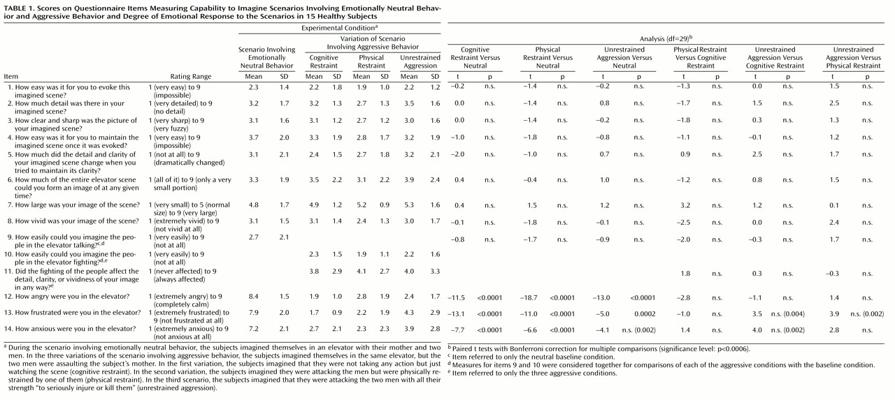
The orbitofrontal and the ventromedial frontal cortex are thought to help inhibit inappropriate social decision making and aggressive behavior (18). In humans and monkeys, stimulation of the orbital and ventromedial prefrontal cortex leads to inhibition of anger and aggression, while neurodegenerative, traumatic, or neoplastic lesions of these structures facilitate the expression of aggressive behavior and behavioral disinhibition (5, 7, 8). In humans, positron emission tomography (PET) studies at rest have shown a lower cerebral glucose metabolism in frontal cortical regions in murderers than in nonviolent comparison subjects (911). PET studies have also shown a correlation between lower glucose utilization in the orbitofrontal cortex and history of aggressive behavior in subjects with personality disorders (12). Although these findings have suggested a relation between dysregulation of aggressive behavior and prefrontal dysfunction, they have not informed us about the changes in brain activity that occur during the expression of aggressive behavior in healthy human subjects. We conducted a study to determine the functional role of the frontal lobes and the potential involvement of additional brain areas in the modulation of aggressive behavior. Functional brain imaging methods, including PET, have been successfully employed to explore the in vivo neural correlates of emotional behavior (1318); these methods have also been used in combination with visual imagery strategies in such research (1924). In the study reported here, functional neuroimaging was combined with qualitative and quantitative measures to examine brain activity and behavioral and autonomic responsiveness during the imagined evocation of aggressive behaviors in healthy volunteers with good visual imagery capabilities and without a history of mental disorders or aggression.

Method

Subjects

Fifteen right-handed healthy volunteers (eight men, seven women; mean age=22 years, SD=2; mean education=16 years, SD=1), were screened to ensure good to excellent imagery abilities (Gordon Visual Imagery Test [25] score mean=22.9, SD=1.7, range=18–24, minimum score=0 [worst imagery], maximum score=24 [best]; Betts Questionnaire of Mental Imagery [26] vividness score mean=71.5, SD=19.5, range=42–110, minimum score=35 [best imagery], maximum score 245 [worst]) and underwent clinical, neurological, and psychiatric examination (including assessment with personality rating scales) and a magnetic resonance imaging (MRI) scan of the brain. History or presence of any medical, neurological, and psychiatric illness, including head trauma with prolonged loss of consciousness, exposure to toxic substances, disruptive behavior disorders, or abnormalities on  MRI were exclusionary criteria. Furthermore, subjects who had deceased parent(s) or had experienced a violent episode during their life similar to the scenario used for the experimental paradigms (described below) were excluded. All subjects were medication-free, including over-the-counter medications and painkillers, for at least 2 weeks before entering the study and refrained from alcohol for 24 hours before the PET scan (27). All participants gave their written informed consent after the purpose of the study and risks were explained (as approved by the institutional review board of the National Institute of Neurological Disorders and Stroke; protocol 93-N-0077).

PET Scanning

rCBF was measured by using a PC2048-15B positron emission tomographer (Scanditronix, Uppsala, Sweden) in a sequence of eight scans with 37 mCi of [15O]H2O that alternated among the four different experimental conditions described below (see reference 28 for details of PET methods). Total time for each run was approximately 90 seconds, and each run was separated from the next one by 12 minutes to allow for radiation decay, administration of the rating scales, and the return to baseline emotional and autonomic state (see below).

Imagery Conditions

The four experimental conditions required the subjects to visually imagine the same scenario with one variation that involved emotionally neutral behavior and three variations that involved aggressive behavior. Specifically, in the scenario involving emotionally neutral behavior, subjects imagined themselves in an elevator with their mother and two men taking a typical elevator ride. In the three variations that involved aggressive behavior, the elevator context was identical but the two men would assault the subject’s mother. In the first of the three variations, subjects had to pretend that they would not take any action but just watch the scene (cognitive restraint). In the second variation, subjects would attack the men but would be physically restrained by one of them, and despite their efforts to free themselves and continue their counterattack, they would be unsuccessful (physical restraint). In the third variation, subjects had to pretend that they were able to attack the two men and hit them with all their strength “to seriously injure or kill them” (unrestrained aggression). A standard description of each scenario was read to the subjects immediately before the radioisotope injection; subjects were instructed to keep their eyes closed, to listen carefully to the script, and to focus on the evoked situation until they were told to stop. Each imagined condition lasted for approximately 100 seconds. Presentation of the four distinct scenarios was varied across subjects to control for sequence effect. Before the scan, subjects were instructed that they would have to evoke imagined situations during the study, but, to maintain novelty, no details about the nature of the scenarios were provided.
In conjunction with each scan and experimental condition, qualitative and quantitative measures of the subjects’ ability to evoke the imaginal situation and of the associated emotional response and indices of peripheral nervous system activation were obtained. Changes in rCBF were determined by comparing the results associated with each of the three aggressive scenarios with the results associated with the neutral scenario.

Behavioral and Physiological Measures

Immediately before the instructions for each condition were read and immediately after each condition was completed, subjects’ blood pressure and heart rate were measured by using a Dinamap Plus automatic blood pressure machine (Critikon, Tampa, Fla.). The capability to imagine the different scenarios and the degree of the emotional responses were assessed with a 14-item questionnaire with answers on a Likert-type scale ranging from 1 (extremely) to 9 (not at all). Table 1 lists the questionnaire items and the results for each experimental condition. Data from the first and the second scan for the same condition were averaged together. Behavioral data and changes in blood pressure and heart rate were compared across conditions by using paired t tests.

PET-rCBF Data Analysis

PET image data were analyzed by using statistical parametric mapping software package (SPM 95) (29). Data were coregistered to the mean scan, spatially normalized to a standard brain (30) and smoothed with a 20 × 20 × 12 mm Gaussian filter. In addition, high-resolution MRI scans of the brain were obtained by using a 1.5-T scanner (GE Medical Systems, Milwaukee) with a T1-weighted three-dimensional spoiled gradient recalled at steady-state sequence (TE=5.4 msec, TR=14 msec, flip angle=20°, number of excitations=1, slice thickness=1 mm, field of view=24 × 24, acquisition matrix=256 × 192). The structural MRI scans were normalized into the same standardized space as the PET scans to allow for superimposition of PET activations and deactivations onto an averaged structural image. Within-group comparisons were performed on rCBF data that were adjusted with analysis of covariance to correct for differences in whole brain CBF. Mean rCBF increases and decreases were calculated by comparing each of the three experimental conditions involving aggressive situations to the neutral condition by using t tests (29). Our a priori hypothesis predicted that ventromedial and orbital frontal cortical regions would show decreased activity in association with the imaginal evoking of situations involving aggressive behavior, compared to the neutral condition. Therefore, given these strong predictions, we reported foci in frontal cortex showing rCBF decreases for which z>3.29 (approximately equivalent to p<0.001, two-tailed, uncorrected for multiple comparisons). A p<0.001 threshold is frequently used in pixel-based analyses of PET data and has been used in previous PET-rCBF studies of emotional states induced through script-driven imagery (20); however, less conservative thresholds have been adopted by other PET studies of emotions (e.g., reference 16). In addition, to minimize the risk of type I error, we repeated the analyses of frontal rCBF decreases with a correction for spatial extent (p<0.05) (31), which confirmed the results. Furthermore, as this functional study is the first we are aware of to assess brain response to imaginal evoking of aggressive behavior, for the sake of completeness and to minimize the possibility of neglecting the potential involvement of other brain structures, we also report on brain areas that were not part of our a priori hypothesis but that showed significant rCBF changes (increases) during each of the three aggressive conditions, compared with the neutral condition.

Results

Behavioral Measures

All subjects were able to complete the study and provided similarly detailed and clear mental representations for each experimental condition, as indicated by the scores in Table 1. Moreover, no significant differences in measures of mental imagery abilities were found between each of the conditions involving aggressive behavior and the neutral condition. The measures of emotional response indicated that subjects experienced significantly greater anger, frustration, and anxiety during the aggressive conditions compared to the neutral condition. Furthermore, the subjects experienced more frustration and anger during the scenarios involving cognitive restraint and physical restraint than when they were free to carry out their aggressive response in the scenario involving unrestrained aggression. However, these comparisons did not maintain statistical significance in analyses that used a more conservative p value (with a Bonferroni correction for multiple comparisons) (Table 1).

rCBF

Table 2 shows the brain areas with significant rCBF changes in the distinct contrasts. During each of the three conditions involving aggressive content, the subjects showed significant rCBF decreases in the medial orbitofrontal cortex compared to the neutral condition (Figure 1 shows the rCBF decreases during the scenario involving unrestrained aggression). No decrease in rCBF was seen in any other region. The distribution of rCBF changes was similar across the three aggressive conditions, although the largest changes were associated with the scenario involving unrestrained aggression.
During the scenario involving unrestrained aggression, the peak deactivation was seen in the left medial frontal gyrus (Brodmann’s area 11). The area of deactivation extended to include the right medial frontal gyrus (Brodmann’s area 10) and the rostral portion of the right superior frontal gyrus (Brodmann’s area 10). During the scenario involving cognitive restraint, decreases in rCBF were seen in the medial frontal gyrus (Brodmann’s area 10) and in the left anterior cingulate gyrus (Brodmann’s area 24/25/32). During the scenario involving physical restraint, rCBF decreases were present in the right medial frontal gyrus (Brodmann’s area 10) and in the left anterior cingulate gyrus (Brodmann’s area 32).
In a comparison of the results associated with the scenarios involving cognitive restraint and unrestrained aggression, there was significantly greater rCBF in the medial frontal gyrus bilaterally (Brodmann’s area 10/11) during the scenario involving cognitive restraint, indicating that the functional deactivation of these frontal cortical areas was strongest when subjects were instructed to express rather than to try to inhibit their aggressive behavior. Additional areas of significantly greater rCBF were found in the right middle (Brodmann’s area 11) and inferior frontal (Brodmann’s area 47) gyri during the scenario involving cognitive restraint, as compared to the unrestrained aggression scenario.
Furthermore, during the scenario involving unrestrained aggression, increases in diastolic blood pressure were significantly greater than in the neutral condition (t=2.16, df=29, p<0.04). Increases in heart rate were also greater during the scenario involving unrestrained aggression; but the difference did not reach statistical significance (t=1.91, df=27, p<0.07). Increases in heart rate and in systolic blood pressure were inversely correlated with rCBF in the medial frontal gyrus (Brodmann’s area 10/11) (Talairach coordinates for local maxima: 2, 60, 12 [for heart rate, r=0.41, df=1, 14, p<0.03] and –4, 32, –12 [for blood pressure, r=0.43, df=1, 14, p<0.02]). No other significant correlations were found.
The scenario involving unrestrained aggression also elicited statistically significant rCBF increases in primary and association visual cortical areas, limbic regions, and the cerebellum (Table 2 and Figure 2). Changes in rCBF in these regions during the scenarios involving cognitive restraint and physical restraint did not reach statistical significance.

Discussion

Clinical observations in humans and experimental reports in primates have consistently indicated that the prefrontal cortex, and in particular the orbitofrontal region, plays a pivotal role in the regulation of social and aggressive behavior (3237). Furthermore, PET studies have shown abnormal glucose utilization in prefrontal cortical areas in the resting state in violent murderers (9, 10), in psychiatric patients with violent behavior (12), and in association with aggressive behavior in individuals with personality disorders (11). Recently, an average 11% reduction in gray matter of the prefrontal cortex was shown in subjects with antisocial personality disorder compared to healthy subjects (38), further supporting the evidence that prefrontal dysfunction is associated with abnormal aggressive and violent behavior (412, 36, 39).
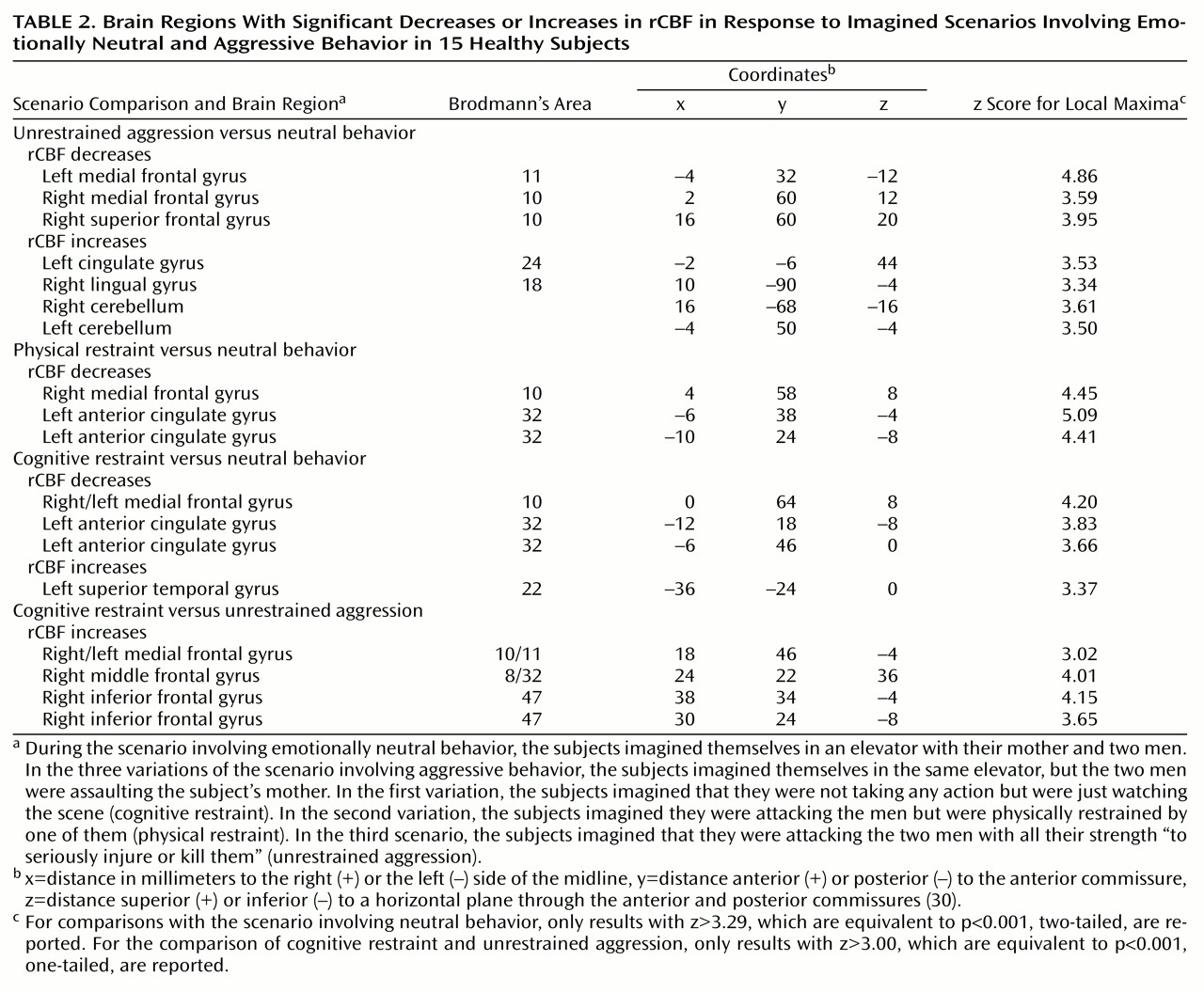
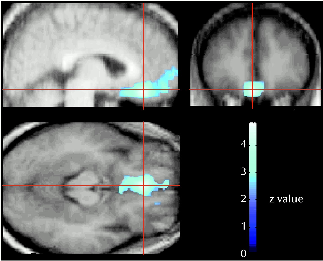
In this study, we assessed the functional brain response when healthy individuals are confronted with situations involving aggressive behavior. Consistent with our hypothesis, the results indicate that a functional decrease in the activity of the medial orbitofrontal cortex accompanies the expression of aggressive behavior even at an imaginal level. The medial orbitofrontal cortex is considered the “limbic” portion of the frontal association cortex (32) and is intimately connected with the amygdala and the limbic system (35, 4042). It is thought to play a role in integrating motivational and emotional processes (43). It has long been known that the human orbitofrontal cortex plays a crucial role in the modulation of the expression of social and emotional behavior (37). In rats, lesions of the orbitofrontal cortex lead to increased aggression (44). In humans, lesions of the medial orbitofrontal cortex disrupt inhibitory and emotional mechanisms and lead to impulsive and socially inappropriate behavior (1, 2, 33, 34, 39, 45). In a study by our group of a large cohort of patients who had received penetrating head injuries during the Vietnam War (7), the patients with lesions of the ventromedial frontal cortex consistently showed significantly greater scores on scales and questionnaires assessing aggressive and violent behavior than did healthy comparison subjects or patients with lesions in other parts of the brain. Frontal lobe seizures are often associated with rage and aggression (46); disinhibition of behavior, including verbal and physical aggressiveness and failure to observe social and moral rules, often is among the prominent features of degenerative processes of the frontal lobes (47), whereas personality and behavior are virtually intact until the end stages of dementia in degenerative disorders that spare the frontal lobes (27). Besides guiding our everyday social interactions and mediating behavior consistent with rules of socialization (35), the modulation exercised by the orbitofrontal cortex enables humans to suppress inappropriate reactions (such as physical violence) to environmental provocations (37). Our results are in agreement with the findings from recent PET studies that demonstrated involvement of the orbitofrontal cortex in response to anger (22, 48). In one of those studies (22), induction of anger in healthy male subjects through script-driven imagery resulted in activation of the left orbitofrontal cortex (including the orbital gyrus of Brodmann’s area 11 and the medial inferior frontal gyrus of Brodmann’s areas 10 and 47), compared to an emotionally neutral baseline. The authors proposed that this part of the orbitofrontal cortex was activated to inhibit an aggressive behavioral response elicited by the induction of anger. In the other study, Blair and colleagues (48) showed enhanced activity in the right orbitofrontal cortex in response to angry facial expressions that correlated with expression intensity. Considering that angry expressions are displayed to curtail the socially inappropriate behavior of others and that the orbitofrontal cortex is involved in the mediation of behavioral extinction (49), the authors suggested that the orbitofrontal cortex activation in response to stimuli featuring angry expressions acts to suppress current inappropriate behavior. This suppression may occur either through an inhibition of current behavior or by activation of alternative behavioral patterns (48).
The role of orbitofrontal and prefrontal cortical areas in the evaluation of distinct negative and positive emotional situations, including sadness, happiness, fear, disgust, aversive gustatory stimulation, anticipatory anxiety, and recollection of autobiographic traumatic events, has been supported by several functional brain studies in both healthy subjects and patients with psychiatric disorders (e.g., references (1324, 5054). Further, the identification of emotional expression is impaired in patients with lesions of subcallosal and other ventromedial prefrontal regions (55). In particular, the ventromedial prefrontal cortex is involved in making judgments on the basis of the emotional valence of stimuli (56, 57). Thus, the medial prefrontal cortical regions could be involved in the conscious experience of emotion, including modulation and inhibition of expression of emotions or of emotionally driven behavior (16, 22, 48). A proper modulatory role of this cortical region in the subject’s emotional state and consequent behavioral response may be needed to make personally relevant decisions (16, 34, 58).
The experimental paradigm designed for this study included three distinct scenarios involving aggressive behavior to control for variables that could have affected the results. Specifically, the scenario involving cognitive restraint was meant to control for the potential effects of the (imagined) motor response component, which is absent in this scenario but is present in the scenario involving physical restraint and even more so in the scenario involving unrestrained aggression. Although the specific features of the three scenarios differed in some regards, all of them involved aggressive behavior. We argue that the reduced ventromedial frontal cortex activation associated with these scenarios compared to the scenario involving emotionally neutral behavior is selectively linked to the aggressive component, as indicated by the fact that reduced rCBF in the ventromedial cortex was associated with all three aggressive scenarios. Even when the subjects were instructed to inhibit the expression of aggressive behavior (cognitive restraint), they were indeed exposed to aggressive behavior and experienced an emotionally arousing response similar to that elicited by the other two aggressive scenarios (as indicated by the scale scores). However, they had to prevent themselves from reacting. This was not an easy task because it was somewhat “unnatural” and went against the natural aggressivity of the subject. On the other hand, the rCBF associated with cognitive restraint was higher than the rCBF associated with the unrestrained expression of aggression, which suggests a less profound inactivation of this cortical area when aggression is manifested in others but must be inhibited in the observer.
The functional meaning of rCBF decreases observed in PET studies may require some additional comments. rCBF in neocortical regions reflects the balance between local excitatory and inhibitory synapses. The accepted explanation for decreases in rCBF is that they represent functional deactivations and are due to a net decrease in regional synaptic metabolic demand. This interpretation is supported by both PET studies in humans and autoradiographic studies in animals that demonstrated a decrease in local cerebral glucose metabolism and blood flow in relation to the inhibition of cortical neurons (5961). For instance, the intracarotid injection of the GABAA agonist 4,5,6,7-tetrahydroisoxazolo(5,4-c) pyridin-3-ol (THIP), which induces postsynaptic neuronal inhibition by increasing the membrane conductance to Cl– and thereby enhancing the negative membrane resting potential, is associated with a dose-dependent decrease in rCBF in the cortical areas of the affected hemisphere (61). These deactivations may be due to a decreased excitation (i.e., the deactivated area may receive less input from another brain structure) or to a local net increase in synaptic inhibition (62, 63). Therefore, whether due to direct inhibition or a reduced excitatory input, the decreased rCBF in the frontal cortical regions shown by our subjects during the scenarios involving aggressive behavior compared to the neutral scenario can be regarded as a true index of reduction of the functional activity in these cortical areas.
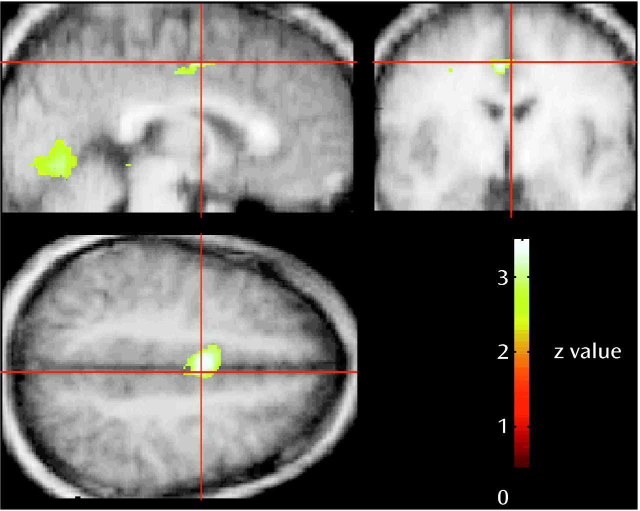
In addition to the rCBF reductions in the orbitofrontal cortex, activation of visual and limbic cortical areas and the cerebellum occurred during the scenario involving unrestrained aggression. The involvement of these regions can be explained by the distinctive features of the unrestrained aggression scenario compared to the neutral scenario. In previous studies, increased rCBF in cortical areas 17 and 18 was found while subjects visualized aversive versus neutral stimuli and during the actual viewing of emotionally aversive versus neutral pictures (13, 14, 19, 34, 35). Thus, the higher blood flow in the visual cortical areas during unrestrained aggression may have been due to the aversive content and to greater emotional arousal and visual attention associated with this situation compared with the neutral content of the baseline condition, although no significant rCBF increase in visual cortical areas was present in the other aggressive conditions, which had similarly aversive and arousing content. Another possibility is that activation of the visual area, along with activation of the anterior cingulate cortex (Brodmann’s area 24) and of the cerebellum, may be related to the subjects’ imagining themselves actively moving while planning their “counterattacks” as well as to change in their visual perspective during the scenario involving unrestrained aggression compared with the neutral scenario or the aggressive scenarios in which they were restrained (6669). Motor imagery, which can be regarded as an incomplete motor program terminated before execution (69), led to activation of cerebellar hemispheres in several studies that used different stimulation paradigms (66, 6971). It is of interest that lateral regions in the cerebellar hemispheres also were activated during imagery but not during actual motion (69). This additional functionality during imagery may reflect inhibitory processes that terminate the motor program before execution (72), sustained motor representations when the motor program awaits execution (73), or elaboration of visually imagined motor actions (71, 74). Finally, cerebellar activation may be linked to emotional processing. A growing body of evidence in the last decade has supported a cognitive and affective role for the cerebellum, in addition to its more classic role in motor function (75, 76). Behavioral changes, including disinhibited and inappropriate behavior, are part of the so-called cerebellar cognitive affective syndrome that follows lesions of the posterior part of the cerebellum (77). As in the case of the visual cortex, however, this interpretation cannot explain why no significant cerebellar activation was present in association with the remaining two scenarios with aggressive content, given that subjects reported a similar emotional and arousal response.
Possible gender differences in brain response are another factor that may help explain the lack of statistically significant rCBF increases in the remaining two conditions with aggressive content (18, 78). Preliminary analyses of gender by condition in our study group showed that while rCBF decreased in relation to the aggressive scenarios in both male and female subjects, the distribution of increases in rCBF differed significantly between the two groups. Male subjects showed a significantly greater activation in visual cortical areas than female subjects, and female subjects showed a significantly greater activation in limbic structures than male subjects (79, 80). A significantly larger activation of limbic system areas in female subjects than in male subjects has also been found during the self-induction of transient sadness (78), despite the observation that men and women had similar abilities in self-inducing sadness. In-progress studies of gender differences in personality traits and in behavioral and brain functional response to imaginal evoking of aggressive behavior (79, 80) will likely clarify this issue.
The magnitude and type of aggressive behavior response that can be induced during functional brain imaging studies is subject to both practical and ethical constraints. Mental imagery has been successfully employed to evaluate cognitive, behavioral, and emotional responses in previous neuropsychological and imaging studies (18, 2023, 27, 65, 8183). Although it is impossible to determine what subjects actually do during a visual imagery scan, in this study we obtained multiple measures, in addition to rCBF changes, that indicated that subjects were engaging in mental imagery. For example, subjects’ blood pressure was significantly higher and their heart rate was higher, although not significantly so, during aggressive scenarios compared to the neutral scenario. Furthermore, all participants in this study were selected on the basis of good or excellent visual imagery abilities and were trained in the experimental procedures to minimize anxiety or discomfort associated with PET scanning and thereby to enhance compliance during the study (84). Indeed, if the 15 subjects in the study had not engaged in the cognitive/emotional task of the distinct experimental conditions as requested, we would have had progressively increased noise in the signal and we would have not observed the significant physiological, behavioral, and rCBF results that are consistent with specific imagery that varied with experimental instructions.
In summary, although the results of this study are preliminary and should be replicated in different experimental conditions, these findings provide some initial in vivo evidence in healthy humans to support the crucial role of the orbitofrontal cortical regions in the modulation of aggressive behavior. When aggressive behavior was evoked through script-driven mental imagery, subjects showed a significant decrease in the neural activity of the orbitofrontal cortex, which is known to control the expression of aggression. These results, along with the findings from previous PET studies of baseline brain glucose utilization in subjects with abnormal behavior (912) and with data from recent structural (38) and functional brain studies on the prefrontal cortex (22, 48), suggest that abnormal expressions of aggressive behavior in violent individuals are accompanied by a functional “shut-down” of the orbitofrontal cortex (37). As more sophisticated experimental paradigms are developed in conjunction with functional brain imaging methods, specific hypotheses can be tested in the investigation of the neural correlates of normal and pathological expressions of human emotions and behavior (1324, 5054).
TABLE 1
TABLE 2
Figure 1. Decreases in rCBF in Response to an Imagined Scenario Involving Unrestrained Aggression Compared With an Imagined Scenario Involving Emotionally Neutral Behavior in 15 Healthy Subjectsa
aPET data are superimposed onto the averaged MRI scan of the 15 subjects and displayed at the voxel of peak deactivation in the transverse, sagittal, and coronal planes. The color scale indicates the z scores for comparisons of rCBF in the neutral and aggressive conditions, with the peak of deactivation in white (z>3.29, equivalent to p<0.001, two-tailed, is considered significant). For the peak deactivation in the left medial frontal gyrus (Brodmann’s area 11), the coordinates in the Talairach space (30) are x=–4, y=32, z=–12, and the z score is 4.86.
Figure 2. Increases in rCBF in Response to an Imagined Scenario Involving Unrestrained Aggression Compared With an Imagined Scenario Involving Emotionally Neutral Behavior in 15 Healthy Subjectsa
aPET data are superimposed onto the averaged brain MRI scan of the 15 subjects and displayed at the voxel of peak activation for the cingulate gyrus in the transverse, sagittal, and coronal planes. The color scale indicates the z scores for comparisons of rCBF in the neutral and aggressive conditions, with the peak of activation in white (z>3.29, equivalent to p<0.001, two-tailed, is considered significant). For the peak activation in the left cingulate gyrus (Brodmann’s area 24), the coordinates in the Talairach space (30) are x=–2, y=–6, z=44, and the z score is 3.53.

Footnote

Presented in part at the 28th annual meeting of the Society for Neuroscience, Los Angeles, Nov. 7–12, 1998, and at the European Regional Congress of the World Federation Societies of Biological Psychiatry, Florence, Italy, April 8–11, 1999. Received July 26, 1999; revisions received March 28, 2000 and May 16, 2000; accepted May 17, 2000. From the Cognitive Neuroscience Section, NINDS, NIH, Bethesda, Md; and the Departments of Human and Environmental Sciences, Psychiatry, Pharmacology, and Neurobiology and Biotechnologies, University of Pisa, Pisa, Italy. Address reprint requests to Dr. Grafman, Cognitive Neuroscience Section, National Institute of Neurological Disorders and Stroke, NIH, Bldg. 10, Rm. 5C205, 10 Center Dr., MSC 1440, Bethesda, MD 20892-1440; [email protected] (e-mail).Supported by an intramural NINDS/NIH grant.The authors thank Neel Sandhu for assistance with PET scanning and data entry; the NIH PET technologists headed by Paul Baldwin; and Drs. James V. Haxby, Etienne Koechlin, Paolo Nichelli, Giulio Tononi, and Tiziana Zalla for an internal review of an earlier version of this manuscript.

References

1.
Bechara A, Damasio AR, Damasio H, Anderson SW: Insensitivity to future consequences following damage to human prefrontal cortex. Cognition 1994; 50:7–12
2.
Fuster JM: The Prefrontal Cortex: Anatomy, Physiology and Neuropsychology of the Frontal Lobe. New York, Raven Press, 1989
3.
Butter CM, Snyder DR, McDonald JA: Effects of orbital frontal lesions on aversive and aggressive behaviors in rhesus monkeys. J Comp Physiol Psychol 1970; 72:132–144
4.
Damasio AR, Tranel D, Damasio H: Individuals with sociopathic behavior caused by frontal damage fail to respond autonomically to social stimuli. Behav Brain Res 1990; 41:81–94
5.
Elliott FA: Neurology of aggression and episodic dyscontrol. Semin Neurol 1990; 10:303–312
6.
Giancola PR, Zeichner A: Neuropsychological performance on tests of frontal-lobe functioning and aggressive behavior in men. J Abnorm Psychol 1994; 103:832–835
7.
Grafman J, Schwab K, Warden D, Pridgen A, Brown HR, Salazar AM: Frontal lobe injuries, violence, and aggression: a report of the Vietnam Head Injury Study. Neurology 1996; 46:1231–1238
8.
Lapierre D, Braun CMJ, Hodgings S: Ventral frontal deficits in psychopathy: neuropsychological test findings. Neuropsychologia 1995; 33:139–151
9.
Raine A, Buchsbaum MS, Stanley J, Lottenberg S, Abel L, Stoddard J: Selective reductions in prefrontal glucose metabolism in murderers. Biol Psychiatry 1994; 36:365–373
10.
Raine A, Stoddard J, Bihrle S, Buchsbaum M: Prefrontal glucose deficits in murderers lacking psychosocial deprivation. Neuropsychiatry Neuropsychol Behav Neurol 1998; 11:1–7
11.
Volkow ND, Tancredi L: Neural substrates of violent behavior: a study with positron emission tomography. Br J Psychiatry 1987; 151:668–673
12.
Goyer PF, Andreason PJ, Semple WE, Clayton AH, King AC, Compton-Toth BA, Schulz SC, Cohen RM: Positron-emission tomography and personality disorders. Neuropsychopharmacology 1994; 10:21–28
13.
Paradiso S, Robinson RG, Andreasen NC, Downhill JE, Davidson RJ, Kirchner PT, Watkins GL, Boles Ponto LL, Hichwa RD: Emotional activation of the limbic circuitry in elderly normal subjects in a PET study. Am J Psychiatry 1997; 154:384–389
14.
Paradiso S, Johnson DL, Andreasen NC, O’Leary DS, Watkins GL, Boles Ponto LL, Hichwa RD: Cerebral blood flow changes associated with attribution of emotional valence to pleasant, unpleasant, and neutral visual stimuli in a PET study of normal subjects. Am J Psychiatry 1999; 156:1618–1629
15.
Davidson RJ, Irwin W: The functional neuroanatomy of emotion and affective style. Trends Cogn Sci 1999; 3:11–21
16.
Reiman EM, Lane RD, Ahern GL, Schwartz GE, Davidson RJ, Friston KJ, Yun L-S, Chen K: Neuroanatomical correlates of externally and internally generated human emotion. Am J Psychiatry 1997; 154:918–925
17.
Pietrini P, Furey ML, Guazzelli M: In vivo biochemistry of the brain in understanding human cognition and emotions: towards a molecular psychology. Brain Res Bull 1999; 50:417–418
18.
George MS, Ketter TA, Parekh PI, Horwitz B, Herscovitch P, Post RM: Brain activity during transient sadness and happiness in healthy women. Am J Psychiatry 1995; 152:341–351
19.
Kosslyn SM, Shin LM, Thompson WL, McNally RJ, Rauch SL, Pitman RK, Alpert NM: Neural effects of visualizing and perceiving aversive stimuli: a PET investigation. Neuroreport 1996; 7:1569–1576
20.
Shin LM, McNally RJ, Kosslyn SM, Thompson WL, Rauch SL, Alpert NM, Metzger LJ, Lasko NB, Orr SP, Pitman RK: Regional cerebral blood flow during script-driven imagery in childhood sexual abuse-related PTSD: a PET investigation. Am J Psychiatry 1999; 156:575–584
21.
Shin LM, Kosslyn SM, McNally RJ, Alpert NM, Thompson WL, Rauch SL, Macklin ML, Pitman RK: Visual imagery and perception in post-traumatic stress disorder: a positron emission tomographic investigation. Arch Gen Psychiatry 1997; 54:233–241
22.
Dougherty DD, Shin LM, Alpert NM, Pitman RK, Orr SP, Lasko M, Macklin ML, Fischman AJ, Rauch SL: Anger in healthy men: a PET study using script-driven imagery. Biol Psychiatry 1999; 46:466–472
23.
Kimbrell TA, George MS, Parekh PI, Ketter TA, Podell DM, Danielson AL, Repella JD, Benson BE, Willis MW, Herscovitch P, Post RM: Regional brain activity during transient self-induced anxiety and anger in healthy adults. Biol Psychiatry 1999; 46:454–465
24.
Partiot A, Grafman J, Sadato N, Wachs J, Hallett M: Brain activation during the generation of non-emotional and emotional plans. Neuroreport 1995; 6:1397–1400
25.
Richardson A: Mental Imagery. New York, Springer, 1969
26.
Sheehan PW: A shortened form of Betts’ questionnaire upon mental imagery. J Clin Psychol 1967; 23:386–389
27.
Pietrini P, Furey ML, Graff-Radford NR, Freo U, Alexander GE, Grady CL, Dani A, Mentis MJ, Schapiro MB: Preferential metabolic involvement of visual cortical areas in a subtype of Alzheimer’s disease: clinical implications. Am J Psychiatry 1996; 153:1261–1268
28.
Grady CL, Maisog JM, Horwitz B, Ungerleider LG, Mentis MJ, Salerno JA, Pietrini P, Wagner E, Haxby JV: Age-related changes in cortical blood flow activation during visual processing of faces and location. J Neurosci 1994; 14:1450–1462
29.
Friston KJ, Holmes AP, Worsley KJ, Poline J-B, Frith CD, Frackowiak RSJ: Statistical parametric maps in functional imaging: a general linear approach. Hum Brain Mapp 1995; 2:189–210
30.
Talairach J, Tournoux P: Co-Planar Stereotaxic Atlas of the Human Brain. New York, Thieme Medical, 1988
31.
Friston KJ, Worsley KJ, Frackowiak RSJ, Mazziotta JC, Evans AC: Assessing the significance of focal activations using their spatial extent. Hum Brain Mapp 1994; 1:210–220
32.
Mesulam M-M: Frontal cortex and behavior. Ann Neurol 1986; 19:320–325
33.
Damasio AR: On some functions of the human prefrontal cortex. Ann NY Acad Sci 1995; 769:241–251
34.
Damasio AR: Descartes’ Error: Emotion, Rationality and the Human Brain. New York, Putnam, 1994
35.
Zald DH, Kim SW: The anatomy and function of the orbito-frontal cortex, I: anatomy, neurocircuitry, and obsessive-compulsive disorder. J Neuropsychiatry Clin Neurosci 1996; 8:125–138
36.
Dolan RJ: On the neurology of morals. Nat Neurosci 1999; 11:927–929
37.
Grafman J, Litvan I: Importance of deficits in executive functions. Lancet 1999; 354:1921–1923
38.
Raine A, Lencz T, Bihrle S, LaCasse L, Colletti P: Reduced prefrontal gray matter volume and reduced autonomic activity in antisocial personality disorder. Arch Gen Psychiatry 2000; 57:119–127
39.
Anderson SW, Bechara A, Damasio H, Tranel D, Damasio AR: Impairment of social and moral behavior related to early damage in human prefrontal cortex. Nat Neurosci 1999; 11:1032–1037
40.
Porrino LJ, Crane AM, Goldman-Rakic PS: Direct and indirect pathways from the amygdala to the frontal lobe in rhesus monkeys. J Comp Neurol 1981; 198:121–136
41.
Price JL, Carmichael ST, Drevets WC: Networks related to the orbital and the medial prefrontal cortex: a substrate for emotional behavior. Prog Brain Res 1996; 107:523–536
42.
Barbas H: Organization of cortical afferent input to the orbitofrontal area in the rhesus monkey. Neuroscience 1993; 56:841–864
43.
Malloy P, Bihrle A, Duffy J: The orbitomedial frontal syndrome. Arch Clin Neuropsychol 1993; 8:185–201
44.
de Bruin JP, van Oyen HG, Van de Poll N: Behavioural changes following lesions of the orbital prefrontal cortex in male rats. Behav Brain Res 1983; 10:209–232
45.
Grafman J: Similarities and distinctions among current models of prefrontal cortical functions. Ann NY Acad Sci 1995; 769:337–368
46.
Gedye A: Episodic rage and aggression attributed to frontal lobe seizures. J Ment Defic Res 1989; 33:369–379
47.
Duffy JD, Campbell JJ: The regional prefrontal syndromes: a theoretical and clinical overview. J Neuropsychiatry Clin Neurosci 1994; 6:379–387
48.
Blair RJR, Morris JS, Frith CD, Perett DI, Dolan RJ: Dissociable neural responses to facial expressions of sadness and anger. Brain 1999; 122:883–893
49.
Rolls ET: The orbitofrontal cortex. Philos Trans R Soc Lond B Biol Sci 1996; 351:1433–1443
50.
Pardo JV, Pardo PJ, Raichle ME: Neural correlates of self-induced dysphoria. Am J Psychiatry 1993; 150:713–719
51.
Lane RD, Chua PM, Dolan RJ: Common effects of emotional valence, arousal and attention on neural activation during visual processing in pictures. Neuropsychologia 1999; 37:989–997
52.
Zald DH, Lee JT, Fluegel KW, Pardo JV: Aversive gustatory stimulation activates limbic circuits in humans. Brain 1998; 121:1143–1154
53.
Rauch SL, Jenike MA, Alpert NM, Baer L, Breiter HCR, Savage CR, Fishman AJ: Regional cerebral blood flow measured during symptom provocation in obsessive-compulsive disorder using oxygen 15-labeled carbon dioxide and positron emission tomography. Arch Gen Psychiatry 1994; 51:62–70
54.
Rauch SL, Savage CR, Alpert NM, Miguel EC, Baer L, Breiter HCR, Fishman AJ, Manzo PA, Moretti C, Jenike MA: A positron emission tomographic study of simple phobic symptom provocation. Arch Gen Psychiatry 1995; 52:20–28
55.
Dias R, Robbins TW, Roberts AC: Dissociation in prefrontal cortex of affective and attentional shifts. Nature 1996; 380:69–72
56.
Bechara A, Tranel D, Damasio H, Damasio AR: Failure to respond autonomically to anticipated future outcomes following damage to the prefrontal cortex. Cereb Cortex 1996; 6:215–225
57.
Damasio AR: The somatic marker hypothesis and the possible functions of the prefrontal cortex. Philos Trans R Soc Lond B Biol Sci 1996; 351:1413–1420
58.
Damasio H, Grabowski T, Frank R, Galaburda A, Damasio A: The return of Phineas Gage: clues about the brain from the skull of a famous patient. Science 1994; 264:1102–1105
59.
Kelly PA, McCulloch J: Effects of the putative GABAergic agonists, muscimol and THIP, upon local cerebral glucose utilization. J Neurochem 1982; 39:613–624
60.
Kelly PA, McCulloch J: The effects of the GABAergic agonist muscimol upon the relationship between local cerebral blood flow and glucose utilization. Brain Res 1993; 258:338–342
61.
Roland PE, Friberg L: The effect of the GABA-A agonist THIP on regional cortical blood flow in humans: a new test of hemispheric dominance. J Cereb Blood Flow Metab 1988; 8:314–323
62.
Kawashima R, O’Sullivan BT, Roland PE: Positron-emission tomography studies of cross-modality inhibition in selective attentional tasks: closing the “mind’s eye.” Proc Natl Acad Sci USA 1995; 92:5969–5972
63.
Haxby JV, Horwitz B, Ungerleider LG, Maisog JM, Pietrini P, Grady CL: The functional organization of human extrastriate cortex: a PET-rCBF study of selective attention to faces and locations. J Neurosci 1994; 14:6336–6353
64.
Lang PJ: Emotional arousal and activation of the visual cortex: an fMRI analysis. Psychophysiology 1998; 2:199–210
65.
Rauch SL, van der Kolk BA, Fisler RE, Alpert MN, Orr SP, Savage CR, Fishman AJ, Jenike MA, Pitman RK: A symptom provocation study of posttraumatic stress disorder using positron emission tomography and script-driven imagery. Arch Gen Psychiatry 1996; 53:380–387
66.
Decety J, Perani D, Jeannerod M, Bettinardi V, Tadary B, Woods R, Mazziotta JC, Fazio F: Mapping motor representations with positron emission tomography. Nature 1994; 371:600–602
67.
Roland PE, Skinhoj E, Lassen NA, Larsen B: Different cortical areas in man in organization of voluntary movements in extrapersonal space. J Neurophysiol 1980; 43:137–150
68.
Ryding E, Decety J, Sjolhom H, Stenberg G, Ingvar DH: Motor imagery activates the cerebellum regionally: a SPECT rCBF study with 99m Tc-HMPAO. Brain Res Cogn Brain Res 1993; 1:94–99
69.
Luft AR, Skalej M, Stefanou A, Klose U, Voigt K: Comparing motion- and imagery-related activation in the human cerebellum: a functional MRI study. Hum Brain Mapp 1998; 6:105–113
70.
Jueptner M, Ottinger S, Fellows SJ, Adamschewski J, Flerich L, Mueller SP, Diener HC, Thilmann AF, Weiller C: The relevance of sensory input for the cerebellar control of movements. Neuroimage 1997; 5:41–48
71.
Parsons LM, Fox PT, Downs JH, Glass T, Hirsch TB, Martin CC, Jerabek PA, Lancaster JL: Use of implicit motor imagery for visual shape discrimination as revealed by PET. Nature 1995; 375:54–58
72.
Berthoz A: The role of inhibition in the hierarchical gating of executed and imagined movements. Brain Res Cogn Brain Res 1996; 3:101–113
73.
Jeannerod M: Mental imagery in the motor context. Neuropsychologia 1995; 33:1419–1432
74.
Decety J, Ingvar DH: Brain structures participating in mental simulation of motor behavior: a neuropsychological interpretation. Acta Psychol 1990; 73:13–34
75.
Dolan RJ: A cognitive affective role for the cerebellum. Brain 1998; 121:545–546
76.
Schmahmann JD, Pandya DN: Prefrontal cortex projections to the basilar pons in rhesus monkey: implications for cerebellar contribution to higher function. Neurosci Lett 1995; 199:175–178
77.
Schmahmann JD, Sherman JC: The cerebellar cognitive affective syndrome. Brain 1998; 121:561–579
78.
George MS, Ketter TA, Parekh PI, Herscovitch P, Post RM: Gender differences in regional cerebral blood flow during transient self-induced sadness or happiness. Biol Psychiatry 1996; 40:859–871
79.
Pietrini P, Guazzelli M, Grafman J: Gender differences in brain activation during aggressive imageries (abstract). Int J Psychophysiol 1998; 30:33
80.
Pietrini P, Guazzelli M, Jaffe K, Sandhu N, Grafman J: Brain response during imaginal aggressive behavior in healthy young male and female subjects: a PET-rCBF study. Abstracts of the Society for Neuroscience 1998; 24(part 1):781
81.
Kossylin SM, Ochsner KN: In search of occipital activation during visual mental imagery. Trends Neurosci 1994; 17:290–292
82.
Kossylin SM, Pascual-Leone A, Felician O, Camposano S, Keenan JP, Thompson WL, Ganis G, Sukel KE, Alpert NM: The role of area 17 in visual imagery: convergent evidence from PET and rTMS. Science 1999; 284:167–170
83.
Kossylin SM: Image and the Brain: The Resolution of the Imagery Debate. Cambridge, Mass, MIT Press, 1994
84.
Pietrini P, Dani A, Furey ML, Alexander GE, Freo U, Grady CL, Mentis MJ, Mangot D, Simon EW, Horwitz B, Haxby JV, Schapiro MB: Low glucose metabolism during brain stimulation in older Down’s syndrome subjects at risk for Alzheimer’s disease prior to dementia. Am J Psychiatry 1997; 154:1063–1069

Information & Authors

Information

Published In

Go to American Journal of Psychiatry
American Journal of Psychiatry
Pages: 1772 - 1781
PubMed: 11058474

History

Published online: 1 November 2000
Published in print: November 2000

Authors

Affiliations

Pietro Pietrini, M.D., Ph.D.

Metrics & Citations

Metrics

Citations

Export Citations

If you have the appropriate software installed, you can download article citation data to the citation manager of your choice. Simply select your manager software from the list below and click Download.

For more information or tips please see 'Downloading to a citation manager' in the Help menu.

Format
Citation style
Style
Copy to clipboard

There are no citations for this item

View Options

View options

PDF/ePub

View PDF/ePub

Get Access

Login options

Already a subscriber? Access your subscription through your login credentials or your institution for full access to this article.

Personal login Institutional Login Open Athens login
Purchase Options

Purchase this article to access the full text.

PPV Articles - American Journal of Psychiatry

PPV Articles - American Journal of Psychiatry

Not a subscriber?

Subscribe Now / Learn More

PsychiatryOnline subscription options offer access to the DSM-5-TR® library, books, journals, CME, and patient resources. This all-in-one virtual library provides psychiatrists and mental health professionals with key resources for diagnosis, treatment, research, and professional development.

Need more help? PsychiatryOnline Customer Service may be reached by emailing [email protected] or by calling 800-368-5777 (in the U.S.) or 703-907-7322 (outside the U.S.).

Media

Figures

Other

Tables

Share

Share

Share article link

Share