Full access
Images in Neuroscience
Published Online: 1 February 2001

Neural Networks: Neural Systems VI: Basal Ganglia
Publication: American Journal of Psychiatry
Neural systems in the human brain not only mediate cognitive, motor, and affective functions but can also distribute psychotropic drug actions. The basal ganglia thalamocortical pathways course from the frontal cortex through the striatum, globus pallidus, substantia nigra, and thalamus in parallel, segregated fashion before returning to the frontal cortex. Because the basal ganglia is rich in diverse neurotransmitters and modulators, it is a plausible target for drug action. Moreover, because the striatum is richly interconnected with the thalamus as well as the frontal and limbic cortex, drug actions in the striatum can have distant, transmitted effects. Regional changes in neuronal activity in the human frontal cortex that are associated with therapeutic change can be a transmitted response to a primary drug-induced perturbation in the striatum. In a set of functional brain imaging studies to identify the CNS regions involved in antipsychotic drug action, the changes in neuronal activity before and after administration of haloperidol implicate the basal ganglia thalamocortical system in the dissemination of antipsychotic drug action from the basal ganglia to the limbic cortex and neocortex.
Haloperidol is useful for this type of study because of its predominant antidopaminergic actions in the brain with potent D2 dopamine family receptor blockade. Since the highest concentration of these receptors is in the striatum, this is the area where investigators have looked first for the mechanism of this drug’s antipsychotic action.
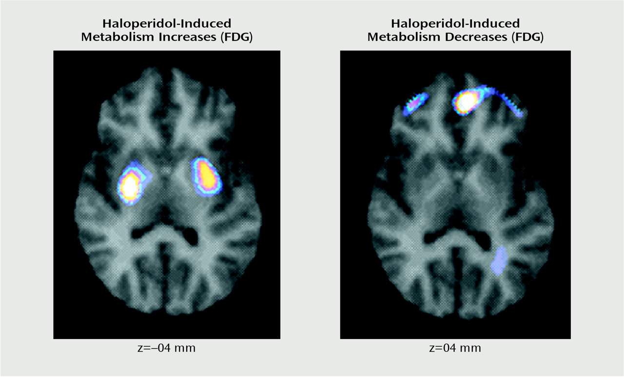
The left image shows the brain regions where haloperidol produces an elevation in rCBF, including the caudate, putamen, and thalamus (the latter is not shown). The right image shows brain regions where haloperidol produces a reduction in metabolism, including the middle frontal cortex and the anterior cingulate and medial frontal regions. A parsimonious interpretation of these results is that haloperidol has its primary effect in the caudate and putamen at the D2 dopamine receptor to block an inhibitory receptor, thereby increasing neuronal activity (hence metabolism) in this region. This activation of the striatum is transmitted through the medial and lateral globus pallidus to the thalamus, where it causes an activation in thalamic activity. Since the afferent pathway to the thalamus is γ-aminobutyric-acid-ergic, the increased synaptic activity in the thalamus produces an inhibitory effect, thus inhibiting thalamic efferent pathways to the cortex. This neuronal inhibition could be represented by the reduced metabolism found in the middle frontal and anterior cingulate cortex. This example provides evidence that the brain’s own intrinsic neuronal pathways can participate in the distribution of therapeutic drug action.
Figure
These images are subtraction images from a haloperidol treatment study where volunteers with schizophrenia were treated with haloperidol for 4 weeks (0.3 mg/kg per day) followed by placebo for 4 weeks and scanned after each treatment period using [ 18F]fluorodeoxyglucose (FDG) and positron emission tomography. The image on the left shows the group average off-haloperidol scan subtracted from the group average on-haloperidol scan to illustrate brain regions where neuronal activity is increased by antipsychotic drug action. The caudate and putamen show regional activation; the thalamic activation can be visualized in an inferior slice. The image on the right shows the group average on-haloperidol scan subtracted from the group average off-haloperidol scan to illustrate brain regions where activity is decreased by antipsychotic drug action. The middle frontal and anterior cingulate regions show reduced metabolism.

Footnote

Address reprint requests to Dr. Tamminga, Maryland Psychiatric Research Center, University of Maryland, P.O. Box 21247, Baltimore, MD 21228. Images are courtesy of Dr. Holcomb.

Information & Authors

Information

Published In

Go to American Journal of Psychiatry
American Journal of Psychiatry
Pages: 185
PubMed: 11156797

History

Published online: 1 February 2001
Published in print: February 2001

Authors

Affiliations

Carol A. Tamminga, M.D.
Henry H. Holcomb, M.D.

Metrics & Citations

Metrics

Citations

Export Citations

If you have the appropriate software installed, you can download article citation data to the citation manager of your choice. Simply select your manager software from the list below and click Download.

For more information or tips please see 'Downloading to a citation manager' in the Help menu.

Format
Citation style
Style
Copy to clipboard

There are no citations for this item

View Options

View options

PDF/ePub

View PDF/ePub

Get Access

Login options

Already a subscriber? Access your subscription through your login credentials or your institution for full access to this article.

Personal login Institutional Login Open Athens login
Purchase Options

Purchase this article to access the full text.

PPV Articles - American Journal of Psychiatry

PPV Articles - American Journal of Psychiatry

Not a subscriber?

Subscribe Now / Learn More

PsychiatryOnline subscription options offer access to the DSM-5-TR® library, books, journals, CME, and patient resources. This all-in-one virtual library provides psychiatrists and mental health professionals with key resources for diagnosis, treatment, research, and professional development.

Need more help? PsychiatryOnline Customer Service may be reached by emailing [email protected] or by calling 800-368-5777 (in the U.S.) or 703-907-7322 (outside the U.S.).

Media

Figures

Other

Tables

Share

Share

Share article link

Share