Patients with borderline personality disorder are characterized by impulsive aggressive behaviors, repeated self-injury, suicidal behavior, affective lability, and disturbed, chaotic relationships. Across diagnostic categories, these behaviors have been associated with indices of low serotonin (or 5-hydroxytryptamine [5-HT]) neurotransmission: low CSF concentrations of 5-hydroxyindoleacetic acid
(1–
3), blunted prolactin responses to 5-HT agonists
(4), and disturbances to markers on platelets and in plasma
(5,
6). Acute tryptophan depletion, a procedure that transiently decreases 5-HT neurotransmission, has been reported to increase impulsive
(7) and aggressive
(8,
9) behaviors. Together, these studies support the hypothesis that low serotonergic tone plays an etiological role in the pathophysiology of behavioral disinhibition/impulsivity. However, an important caveat to most of these studies is that they relied on peripheral indices of 5-HT function, which at best represent crude measures of neurotransmission in the brain.
Recent developments in functional neuroimaging have enabled researchers to reassess the 5-HT hypothesis of impulsivity by more directly measuring aspects of 5-HT neurotransmission in the brains of living humans. One such method is positron emission tomography (PET) with the tracer α-[
11C]methyl-
l-tryptophan (α-[
11C]MTrp). α-MTrp is a synthetic analog of the 5-HT precursor
l-tryptophan. The methyl group prevents the tracer’s incorporation into protein
(10). Autoradiography studies indicate that α-MTrp is taken up by 5-HT neurons
(11), where it is trapped in the 5-HT synthesis precursor pool and/or metabolized into α-methylserotonin
(11,
12). It has been proposed that the rate of this irreversible trapping of α-[
11C]MTrp (K*) provides an index of 5-HT synthesis capacity
(12).
Method
Subjects
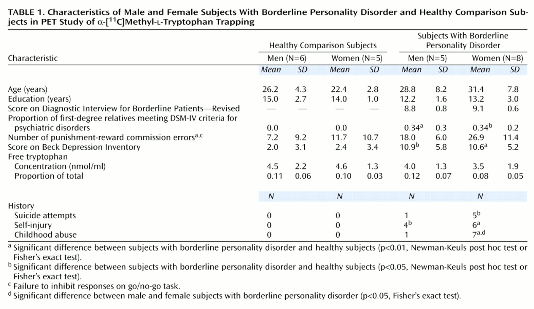
The primary entry criteria for the subjects with borderline personality disorder were satisfaction of the criteria for borderline personality disorder of both DSM-IV and the Diagnostic Interview for Borderline Patients—Revised (DIB-R)
(13) (for the latter, we used the recommended cutoff of at least eight out of 10), no current major depression or substance abuse, never having used the putative 5-HT neurotoxins 3,4-methylenedioxymethamphetamine (MDMA) or 3,4-methylenedioxyamphetamine (MDA), and being right-handed. On the go/no-go task, all but one of the subjects with borderline personality disorder scored at least 1 standard deviation above the mean of our normative data.
Volunteers were recruited by newspaper advertisements. The advertisement to recruit subjects with borderline personality disorder described four features: moodiness, temper dysregulation, self-injury, and relationship problems. Approximately 331 respondents were assessed on the telephone, 35 were assessed in person, and 17 were accepted into the study, 14 of whom were scanned. All of the borderline personality disorder subjects were interviewed by one of us (M.L.) with the Structured Clinical Interview for DSM-IV (SCID) and the DIB-R. Family pedigrees were constructed, and histories of psychiatric disorders were determined in first-degree relatives by using DSM-IV criteria. For each relative, psychiatric disorders were judged to be absent, possible, probable, or definite.
All 14 subjects reported extensive histories of impulsive, affectively labile behavior and disturbed interpersonal relationships compatible with moderate to severe cluster B personality disorders, and the features were consistent with borderline personality disorder. Each subject met repeatedly with a research psychologist (M.L.); the total contact time ranged from 12 to 20 hours. Diagnoses were assigned on the basis of all available information, including interviews with the subject and, when deemed possible, other family members and previous treating clinicians. Written reports based on the assessments were reviewed independently by two psychiatrists (C.B., J.P.); all three investigators had to agree that the volunteer met the entry criteria. Seven subjects were recontacted at 1-year follow-up, and the persistence of borderline personality disorder symptoms was confirmed; the other seven could not be reached.
All of the subjects with borderline personality disorder were medication free at testing, and the shortest interval since last treatment was 6 months. The PET data from one subject with borderline personality disorder were lost because of technical difficulties.
Healthy comparison subjects were also recruited through advertisement; 92 respondents to that advertisement were assessed on the telephone, 18 were assessed in person, and 13 were accepted into the study, 11 of whom were scanned. All healthy subjects were interviewed by one of us (M.L.) with the nonpatient version of the SCID, and the reports were reviewed by another (C.B.). Acceptance into the study depended on agreement by both investigators. The exclusion criteria included a history of psychiatric disorders in the subject or a first-degree relative, a score on the Beck Depression Inventory higher than 8, and past use of MDMA or MDA.
All subjects were physically healthy, as determined by a physical examination and standard laboratory tests. On the day of the PET scan, all tested negative on a urine drug screen sensitive to cocaine, opiates, phencyclidine, LSD, tetrahydrocannabinol, and amphetamines (triage panel for drugs of abuse). Women were scanned during the follicular phase of the menstrual cycle.
Written informed consent was obtained from all subjects. The study was carried out in accordance with the Declaration of Helsinki and was approved by the Research Ethics Committee of the Montreal Neurological Institute.
Impulsivity
Impulsivity was operationally defined as the number of commission errors (responding when one should not) in the punishment-reward condition of a computerized go/no-go task
(14). Of the four go/no-go conditions, punishment-reward is unique in that it is necessary to delay responding (pushing a button) to learn the task. High numbers of commission errors might reflect difficulty delaying and inhibiting behavior. In a larger study group
(15), punishment-reward was the go/no-go condition that best discriminated subjects with borderline personality disorder from the healthy comparison group.
Tryptophan Measurement
Three 2-ml venous blood samples were drawn from each subject during the PET study. The samples were centrifuged, and the ultrafiltrate was stored at –80°C for measurement of plasma free tryptophan with high-performance liquid chromatography.
PET and MRI
α-[
11C]MTrp was prepared as described previously
(16). The studies were carried out in the late morning or early afternoon (scan start time, 11:00 a.m. to 3:00 p.m.) after an overnight fast. The day before the PET study, the subjects received low-protein meals to reduce variability in plasma amino acid concentrations attributable to differences in diet. All subjects were scanned with an ECAT HR+ tomograph (CTI/Siemens). Three-dimensional dynamic images were blurred to 8.1 mm full width at half maximum in the transaxial direction by using a Hanning filter. Between 200 and 800 MBq of α-[
11C]MTrp were administered intravenously over 2 minutes. All studies consisted of 60-minute dynamic PET scans, starting at the onset of tracer injection. Thirteen blood samples were drawn from an antecubital vein to plot plasma time activity curves. Before tracer injection, transmission scans were performed by using gallium-68 for attenuation correction.
Each subject underwent a magnetic resonance imaging (MRI) scan (160 slices, 1 mm thick) obtained with a 1.5-T Philips Gyroscan. Coregistration of the individual PET and MRI images was performed by using an automatic procedure
(17), and the PET images were then transferred into Talairach space
(18).
Calculation of α-MTrp Trapping
Input functions were estimated with the venous-sinus-normalized venous plasma radioactivity curve
(19). To calculate α-MTrp trapping, distribution volumes (milliliters per gram) were fitted as a function of exposure time (minutes)
(20). The unidirectional trapping of the tracer was then determined by using data points obtained from 20 to 60 minutes, during which apparent steady state is achieved
(19). Functional images of α-MTrp trapping were then calculated pixel by pixel
(21).
Statistical Parametric Mapping Analysis
The functional PET data were analyzed with SPM96
(22). Each functional image in Talairach space was smoothed by using a Gaussian filter (14 mm full width at half maximum) to reduce the effect of individual variability in cortical gyral anatomy. Proportional scaling was applied to remove the effect of global differences on regional values among subjects. Functional images were normalized by the mean global values. Statistical parametric mapping (SPM) comparisons were made at a threshold of 80% of the peak value, the usual cut-off in SPM studies of regional cerebral blood flow (rCBF). The t test was applied pixel by pixel to determine regional differences between groups. The t values were then converted to a normal standard distribution (z values), independent of the degree of freedom of the error, and this distribution constituted the statistical parametric map. The errors associated with the pixel values have been shown to approximate a Gaussian distribution
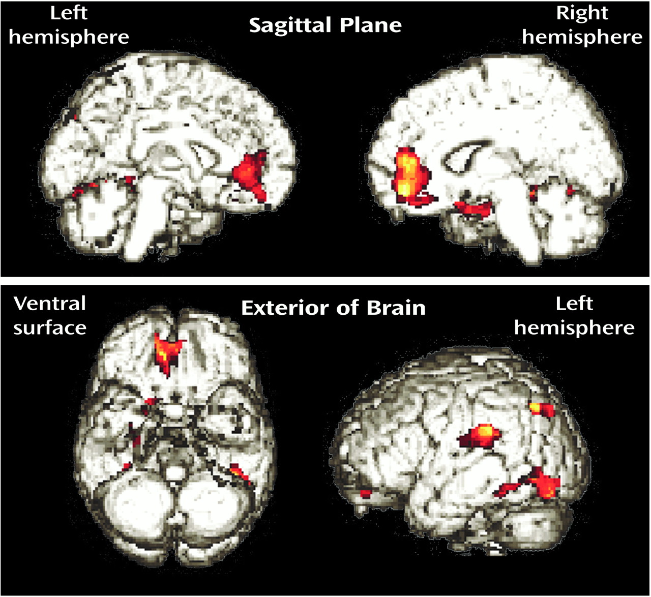
(21). Statistically significant regional differences were identified by using dual criteria. First, the height threshold (u) used to interpret the t test in terms of probability level was set at z=2.58 (p=0.01, two-tailed), with each cluster requiring a peak of z≥3.0. Second, the extent threshold (k) was set as 100 voxels, suitable for the 14-mm full width at half maximum filter and sufficient to remove small noisy clusters, which may reach significance by chance. The distribution of significant clusters (SPM{t}) was depicted by rendering significant pixel clusters onto three-dimensional images of the brain. Since sex-related differences in regional α-[
11C]MTrp trapping have been reported
(23), men and women were analyzed separately in the group comparisons.
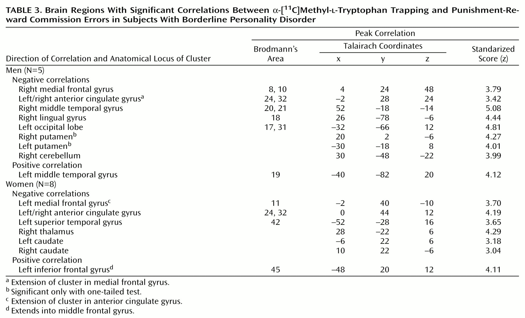
We also identified regions where α-MTrp trapping correlated with the number of punishment-reward commission errors. Using SPM96, we generated pixel-by-pixel correlational maps and identified clusters that met the preceding criteria for statistical significance.
Statistical Analyses
Unless otherwise specified, the data were analyzed with 1) analyses of variance followed by Newman-Keuls post hoc tests or 2) Fisher’s exact tests.
Discussion
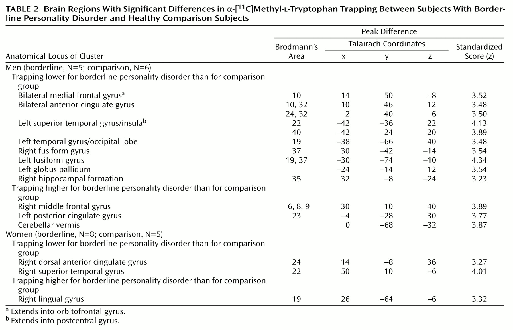
To our knowledge, the present study is the first investigation in vivo of an index of presynaptic 5-HT neurotransmission in the brains of unmedicated subjects with borderline personality disorder. The results suggest that 1) as a group, borderline personality disorder subjects exhibit disturbed α-[11C]MTrp trapping in both the cortex and basal ganglia and 2) α-[11C]MTrp trapping correlates in selective brain pathways with a measure of impulsivity.
Compared to healthy subjects, subjects with borderline personality disorder have higher levels of α-[11C]MTrp trapping in some brain regions and less in others. Both men and women with borderline personality disorder exhibited low α-[11C]MTrp trapping in aspects of the superior temporal gyrus (Brodmann’s area 22) and anterior cingulate (Brodmann’s area 24). In the male subjects, low α-[11C]MTrp trapping was also observed in the medial frontal cortex (Brodmann’s area 10) extending into the orbitofrontal cortex, as well as in the corpus striatum. These sites correspond to regions thought to have important roles mediating the planning, initiation, and inhibition of goal-directed behaviors (frontal cortex, orbitofrontal cortex, anterior cingulate gyrus, striatum), working memory (medial frontal cortex, anterior cingulate, and superior temporal gyrus), and affect (temporal cortex, limbic areas).
The regions identified in the present study have also been implicated in previous functional neuroimaging studies of patients with a wide range of clinically impulsive disorders. Glucose metabolism and rCBF appear to be low in the frontal cortex and their subcortical connections, including the prefrontal and premotor cortex, anterior cingulate, thalamus, and caudate-putamen
(24,
25). Lifetime histories of impulsive, aggressive acts have been reported to correlate negatively with glucose metabolism in the medial frontal cortex, anterior frontal cortex, orbitofrontal cortex, and temporal cortex
(24). This hypocortical activity might be related to low 5-HT neurotransmission. A recent report
(26) suggested that in patients with cluster B personality disorders, rCBF responses to a fenfluramine challenge—an indirect measure of serotonergic responsivity—are blunted in the prefrontal, cingulate, and orbitofrontal cortices.
The statistical parametric maps of the differences between the borderline personality disorder and comparison groups differed for men and women. A sex difference in α-[11C]MTrp trapping might be related to gender differences in the behavioral phenotype. As seen in larger study groups, more of the men than women met the criteria for antisocial personality disorder (two of five versus zero of eight), narcissistic personality disorder (two of five versus zero of eight), and a past substance use disorder (five of five versus four of eight), and fewer had a history of binge eating (one of five versus five of eight).
The subjects with borderline personality disorder made significantly more punishment-reward commission errors on the go/no-go task than did the healthy subjects. This finding is consistent with the clinical observation that patients with borderline personality disorder have difficulty inhibiting behavior and/or delaying responses. Among our subjects with borderline personality disorder, punishment-reward commission errors correlated with α-[11C]MTrp trapping. These correlations were observed within or near brain regions that differentiated these subjects from the healthy comparison group. In both the men and women with borderline personality disorder, punishment-reward commission errors were negatively correlated with α-[11C]MTrp trapping in the medial frontal gyrus, anterior cingulate gyrus, temporal cortex, and striatum. These results suggest that 1) the degrees of dysregulated impulse control and α-[11C]MTrp trapping are related and 2) female subjects with borderline personality disorder who have high impulsivity scores have serotonergic disturbances in the same regions as men with borderline personality disorder.
The validity of these findings rests on the following considerations. The α-MTrp method is a new technique, and concerns have been expressed regarding the physiological significance of tracer trapping in the brain. In particular, it has been argued that the α-MTrp method images only transport of tryptophan through the blood-brain barrier
(27,
28) and/or its incorporation into the protein precursor pool
(29). Some of these concerns have been addressed in detail elsewhere
(30,
31). To further characterize the kinetics of α-MTrp and tryptophan in the brain, the brain regional uptake and trapping constants for labeled tryptophan and α-MTrp were recently compared in rats for the two metabolic pathways, the 5-HT metabolic pathway and tryptophan incorporation into proteins
(32). The results indicate that, in the 5-HT metabolic pathway, the brain uptake and trapping constants of labeled tryptophan and α-MTrp are significantly correlated (p=0.0007). In comparison, the tissue uptake and trapping constants of α-MTrp do not correlate with the incorporation of tryptophan into proteins or the transport of either tryptophan or α-MTrp across the blood-brain barrier, as assessed by the permeability surface area product
(32–
34). Moreover, the regional α-[
11C]MTrp distribution volume dynamic curves in the brain are consistent with trapping of the tracer. A two-compartment fit of distribution volume data would be expected to show that, over time, the distribution volume would plateau, whereas in the case of a three-compartment fit, the distribution volume would be expected to increase, as a reflection of trapping. As recently noted by others
(28), adding a constant for irreversible trapping of the tracer significantly improves fit. This observation, together with the finding that α-MTrp trapping correlates with tryptophan metabolism in the 5-HT pathway, supports the contention that α-[
11C]MTrp brain uptake and trapping, termed 5-HT synthesis capacity
(12), relates directly to regional 5-HT synthesis.
There are several other issues for consideration. First, the size of the study group was modest (N=13). Second, the subjects met the criteria not only for borderline personality disorder but also, on average, for two to three additional diagnoses, typically other cluster B disorders, past substance abuse, or past mood disorders. The possible relation between abnormal α-[
11C]MTrp trapping and these other disorders is unclear. Preliminary results
(35) suggest that major depression is associated with disturbances in α-[
11C]MTrp trapping that overlap, although are not identical to, those seen in the present study. Third, the female subjects with borderline personality disorder tended to be older than the healthy women, but most were in their 20s and 30s.
In conclusion, the PET/α-[
11C]MTrp method is relatively new, and the present results should be seen as exploratory. However, the study suggests that, in at least some patients with borderline personality disorder, α-[
11C]MTrp trapping is disturbed in neocortical and limbic structures, and these alterations could contribute to decreasing the threshold for behavioral disinhibition. Neither impulsivity nor disturbances to 5-HT neurotransmission are likely to account for the entire clinical picture of borderline personality disorder, though
(36). Instead, the etiology of personality disorders is undoubtedly complex and multifactorial, including various genetic, neurobiological, developmental, psychodynamic, and psychosocial factors, all thought to contribute to the diverse clinical expressions of borderline personality disorder.