Full access
Clinical Case Conference
Published Online: 1 September 2002

Prompt Identification of Alzheimer’s Disease With Brain PET Imaging of a Woman With Multiple Previous Diagnoses of Other Neuropsychiatric Conditions

Publication: American Journal of Psychiatry
Clinical identification and differential diagnosis of dementia is especially challenging in its early stages, partly because of the difficulty in distinguishing it from the mild decline in memory that can occur with normal aging and from mild cognitive manifestations of other neuropsychiatric conditions, such as depression. As reported by the U.S. Agency for Health Care Policy and Research in 1996 (1), “Early recognition of the condition has important benefits,” which is even more the case today, now that several medications for the treatment of Alzheimer’s disease are available by prescription; however, “early-stage dementia is often unrecognized or misdiagnosed.” This is especially unfortunate for dementias like Alzheimer’s due to neurodegenerative disease, since patients potentially have the most to gain from effective therapies that intervene as early as possible in the course of progressive irreversible damage to brain tissue.
The University of California, Los Angeles, Memory Clinic is primarily directed toward serving patients with mild memory changes who frequently have not experienced sufficient cognitive losses to meet diagnostic criteria for dementia. The mission of the clinic is driven by clinicopathological studies of recent years indicating that a substantial portion of such individuals are actually suffering from the earliest manifestations of Alzheimer’s disease and related dementias, while conventional methods for evaluating those patients are inadequate for making reliable diagnostic and prognostic assessments. During the same period, advances in dementia research have identified genetic and environmental factors that significantly increase the risk of developing neurodegenerative disease; moreover, it has become increasingly evident that measurement of regional cerebral metabolism through positron emission tomography (PET) sensitively detects such disease at the time of its earliest symptomatic expression, or even preclinically. Although new PET technologies (2) are being developed to measure the concentration of amyloid plaques and neurofibrillary tangles, the pathognomonic lesions of Alzheimer’s disease, this technology is as yet available in clinical settings. By contrast, PET measurements of cerebral glucose metabolism are clinically available; considerable evidence indicates their usefulness in the early diagnosis of dementia. The clinical case described illustrates the typical outcomes of a conventional evaluation process and the dementia evaluation process as it is carried out in our memory clinic.

Case Presentation

Ms. A was a 53-year-old woman who came to the memory clinic with the chief complaint that it was “hard to put thoughts together.” She reported a progressive decline in her memory abilities over the previous 4 years, for which she had undergone general medical, neurological, psychiatric, and formal neuropsychological evaluations. She had been variously diagnosed as having depression, migraines, fibromyalgia, attention deficit disorder, and posttraumatic stress disorder stemming from childhood abuse. Her medications included the selective serotonin reuptake inhibitor citalopram (40 mg/day for the previous 18 months), levothyroxine for hypothyroidism secondary to Hashimoto’s thyroiditis, and estrogen-replacement therapy.

Past History

A review of Ms. A’s records from an outside health organization revealed that her internist first noted a cognitive problem 2 years earlier when he recorded “memory loss” of 1 years’ duration. At that time he also diagnosed depression, obtained laboratory screening test results, referred Ms. A to a neurologist, and scheduled her for a return visit in 6 weeks.
The next month, Ms. A, accompanied by her husband, had her first visit with the neurologist. He noted that a computerized tomographic scan of the brain and tests of her CSF, obtained in the remote past for workup of headache, were unremarkable. Her examination was normal, except for her mental status: “She appeared anxious and hyperventilated at times.” Her neurologist concluded, “Most of the patient’s symptoms are likely psychiatrically based. She is currently in therapy and being treated. Nevertheless, because of the indication of deteriorated function at work, documented by her husband [with whom she ran a small restaurant], I would like to get [magnetic resonance imaging] MRI of the brain to rule out any frontal lobe disease.”
An MRI of her brain performed without contrast the next week showed a normal cortex, brainstem, basal ganglia, thalami, and ventricles. A “hazy 1-cm area of abnormal signal in the right parietal deep white matter” without mass effect was observed on fluid-attenuated inversion recovery and T2-weighted images but not on T1-weighted images. A postcontrast MRI, performed 2 weeks later, demonstrated a nonenhancing “vague area of slightly increased signal in the area of the previously noted increased signal” on one axial image. The radiologist concluded, “The finding may represent a small area of ischemic change.”
Two and one-half months after the second MRI, Ms. A returned to her internist, who noted, “Memory lapses persist.” His plan was to follow up with the neurologist, refer Ms. A to the behavioral health unit, and have her return in 6 weeks. When she returned 6 weeks later, the internist again noted that “Memory lapses persist,” and his assessment was “depression” and “memory loss.”
Two weeks later, Ms. A had her second visit with the neurologist. He noted “a small area of T2 change that is unchanged on serial MRI, consistent with her history of migraine.” His impression stated, “The differential does include the possibility of frontal lobe dementia, but the patient has no change in personality or other frontal lobe symptoms, and she also has lack of atrophy on brain MRI. We still cannot rule this out and will need to observe her over time.”
After another 6 months (1 year after Ms. A’s first visit to the internist for memory loss), she returned to her internist, who again noted, “Memory lapses persist.” His assessment indicated, “1) Hypothyroidism, 2) depression, 3) fibromyalgia.”
Eight and one-half months later, Ms. A’s husband brought her to a licensed psychologist for formal neuropsychological testing, again expressing the concern that his wife was “experiencing cognitive problems.” Ms. A told the psychologist that she was afraid she was losing her mental capacity: “I forget things, I forget people I meet, lose things in my mind.” The psychologist reported that Ms. A’s verbal IQ was in the normal range, but her performance IQ was markedly deficient. The psychologist’s summary stated, “The results suggest neurological difficulties. It can be said that posttraumatic stress disorder factors into the results.…However, neurological problems should be ruled out ASAP,” and he further recommended individual counseling, as well as marriage counseling, because of “her concern for her husband’s feelings about her difficulties.”

Evaluation at the Memory Clinic

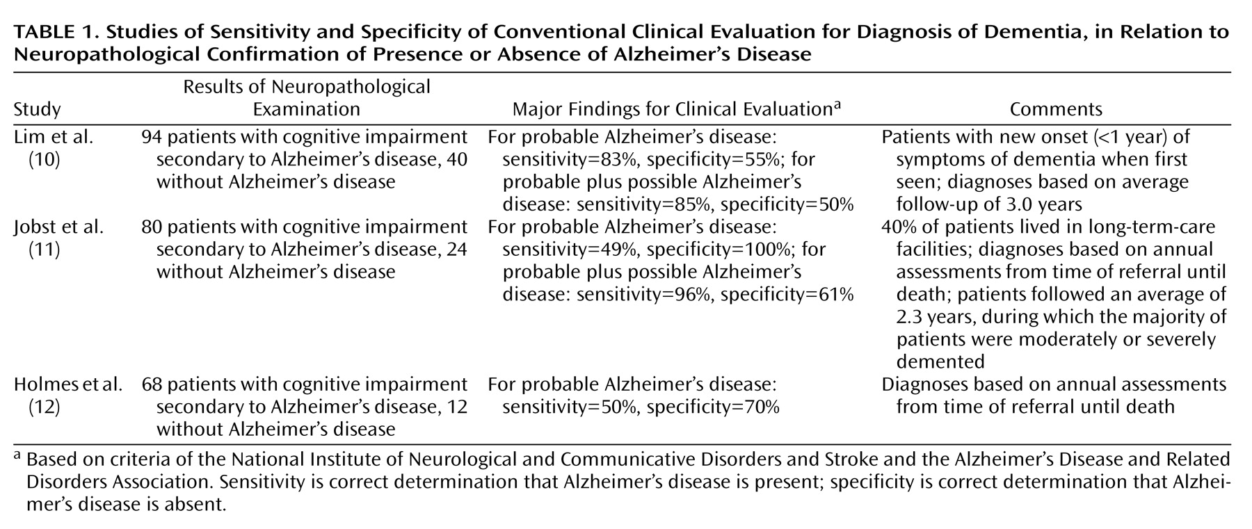
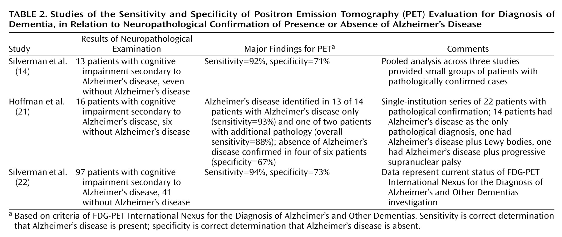
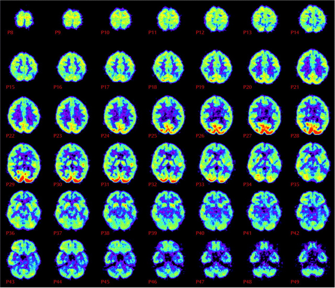
After another 9 months, still without a specific diagnosis for her memory complaints, Ms. A was evaluated for the first time by a geriatric psychiatrist at the memory clinic. At her psychiatric examination, Ms. A was reported to be “alert, mildly anxious, euthymic, friendly, cooperative, with bright affect.” Her performance on the Mini Mental State Examination (MMSE), however, was clearly abnormal; she scored only 18 of a possible 30 points and remembered none of three possible items for short-term recall. Once her previous brain imaging studies had been reviewed, a PET scan, which images regional brain metabolism with the use of [18F]fluorodeoxyglucose (FDG), was obtained. The scan revealed diffuse and moderately severe cortical hypometabolism, especially affecting the bilateral midparietal cortex and left inferior frontal and temporal cortex, but sparing the bilateral sensorimotor and visual cortices (Figure 1). The nuclear medicine physician who interpreted the results noted that this pattern is characteristic of neurodegenerative dementia—most commonly, Alzheimer’s disease. The PET scan was followed by an MRI study with T1-weighted, T2-weighted, and fluid-attenuated inversion recovery sequences, which was normal, with no evidence of infarction or white matter lesions. A week after the PET, a neuropsychologist administered a test battery to Ms. A and concluded that although “other diagnoses that she has received in the past, such as fibromyalgia, attention deficit disorder, posttraumatic stress disorder, and depression could be a factor in her performance, it is emphasized that they would not explain the severity or scope of her current deficits.”

Treatment Plan and Subsequent Course

The psychiatrist who originally saw Ms. A diagnosed probable Alzheimer’s disease, citing the results of PET and neuropsychologic testing, as well as her unremarkable MRI. Treatment was initiated with the cholinesterase inhibitor donepezil at 5 mg/day and increased to 10 mg/day after a month. The family was also referred to the Alzheimer’s Association for education and support and was informed that Ms. A should refrain from driving. She responded well to the treatment: her MMSE score increased from 18 to 21, and she correctly remembered two of three items on the test of short-term recall. Her husband described her as “more engaging with others, more alert.” Ms. A and her husband decided to move to northern California to be closer to their children and to return to Los Angeles every 6 to 12 months for visits to the memory clinic.

Discussion

To summarize Ms. A’s clinical course, 4 years after she had onset of cognitive symptoms, and after 2 years of conventional clinical workup for dementia (including multiple visits with her primary care physician, ongoing care with her psychiatrist, two visits to a neurologist, two MRI scans, and formal neuropsychological testing), she still had no specific diagnosis to explain her memory problems and was accordingly receiving no specific therapy for her dementia symptoms. Furthermore, she and her husband were offered no sense of the prognosis for her disorder or meaningful support for her condition.
By contrast, just 1 month after she came to the memory clinic, a diagnostic PET scan had been acquired, which demonstrated findings characteristic of neurodegenerative dementia, probably Alzheimer’s disease. Neuropsychological testing a week later corroborated this diagnosis, and subsequent MRI ruled out other causes. On this new clinical path, appropriate pharmacotherapy was initiated, and Ms. A showed signs of improvement. In addition, she was linked up with useful community resources and began to formulate, with her husband, meaningful plans for themselves and their family.
Ms. A’s young age may have contributed to some of the diagnostic delay. Most cases of Alzheimer’s disease occur after age 65 years, while frontotemporal dementia more often has an onset in people in their 50s and 60s. A PET scan is useful in differentiating these two dementias: frontotemporal dementia shows a pattern of frontal and temporal hypometabolism (3) in contrast to the posterior deficits observed early in Alzheimer’s disease. Genetic testing was not obtained for this patient because an autosomal-dominant family history was not present. The apolipoprotein E-4 allele is associated with a greater risk for Alzheimer’s disease. Use of apolipoprotein E-4 allele testing may increase diagnostic accuracy in patients with dementia, but it is not recommended as a predictive test in asymptomatic individuals (4). Although neuropsychological testing can be useful in the early differential diagnosis of dementia, the brain has the capacity to compensate for subtle deficits (5). Previous studies (6, 7) have indicated that cerebral metabolic changes may predate cognitive changes observed on neuropsychological testing.
Just how accurate is the conventional diagnostic workup in the context of evaluating early dementia? This is a difficult question to answer for at least two reasons. First, clinical definitions of when dementia begins are necessarily arbitrary, since a long period of gradual neuropathological changes in the brain typically precedes the appearance of cognitive symptoms that are significant enough to clearly fall outside the normal range, making disease onset quite insidious. Second, remarkably few studies have specifically addressed the clinical detection of very mild disease, particularly with regard to the criterion standard of histopathological diagnosis. In one small investigation aimed at doing so (8), patients who initially appeared normal or minimally affected were followed with repeated examinations for an average of 4 years. Even by the end of this longitudinal follow-up period, a neurologist examiner detected Alzheimer’s disease in only 70% of the patients who were histologically positive.
In a recent report of the Quality Standards Subcommittee of the American Academy of Neurology (9), the source of the most comprehensive guidelines and standards for the clinical evaluation of dementia in the last several years, three class I studies (1012) were identified in which the diagnostic value of clinical assessment could be meaningfully measured (Table 1). Class I indicates “a well-designed prospective study in a broad spectrum of persons with the suspected condition, using a ‘gold standard’ for case definition, and enabling the assessment of appropriate tests of diagnostic accuracy” (9). Only one of these (10) focused on evaluating dementia at a relatively early stage. To be included in that investigation, patients were required to have had onset of dementia symptoms within 1 year of entry. All of the 134 patients evaluated underwent a complete standardized diagnostic workup composed of a comprehensive medical history, a physical examination, a neurological examination, neuropsychological testing, laboratory tests, structural neuroimaging, and an average of 3 additional years of clinical follow-up. Sensitivity (accuracy among subjects who actually have the condition) of this assessment for Alzheimer’s disease was 83%–85%, while specificity (accuracy among subjects who do not have the condition) was 50%–55%. It should be emphasized that in these studies, and in most similar studies, the reported sensitivities and specificities represent not the diagnostic accuracy of initial clinical evaluation but of an entire series of evaluations repeated over a period of years.
Conventional MRI or computerized tomography of patients with symptoms of dementia may be useful in identifying unsuspected clinically significant lesions, which are present in approximately 5% of patients (9). However, they are nondiagnostic in patients with Alzheimer’s-related pathophysiologic changes, which are much more common. Such scans are typically read as normal or as demonstrating only the nonspecific finding of cortical atrophy or, worst still, as revealing ischemic changes that are often misinterpreted as pointing to cerebrovascular disease as the primary or sole process responsible for the patient’s cognitive decline. This, in turn, leads to failure to institute appropriate pharmacotherapy (i.e., tacrine, donepezil, rivastigmine, and galantamine—all of which are approved by the Food and Drug Administration only for the indication of “mild to moderate dementia of the Alzheimer’s type”). It is unfortunately not rare for that type of misinterpretation to occur, even among expert clinicians. In a multicenter study involving seven university-affiliated Alzheimer’s Disease Diagnostic and Treatment Centers (13), of the patients who were diagnosed after clinical and structural neuroimaging evaluations as having “vascular dementia” and in whom Alzheimer’s disease and other dementia diagnoses were specifically thought to be absent, only 30% actually had isolated cerebrovascular disease, and the majority (55%) had Alzheimer’s disease, as ascertained by pathological diagnosis.
Over the last two decades, clinicians and researchers have obtained substantial experience in using PET for the identification and differential diagnosis of dementia. Thousands of patients with clinically diagnosed and, in some cases, histopathologically confirmed Alzheimer’s disease from many independent laboratories have been studied by using PET measures of cerebral blood flow and glucose and oxygen use. The resulting findings have been the subject of several reviews (1417). The principal findings that have emerged from that experience are, briefly, as follows: a consistent pattern of focally lower cerebral activity has been identified with PET, which involves neocortical association areas but largely spares the basal ganglia, thalamus, cerebellum, and cortex mediating primary sensory and motor functions; the extent of hypometabolism has been correlated with severity of cognitive impairment (15) and often shows right/left hemispheric asymmetry (18). The classic regional cerebral pattern of biparietotemporal hypometabolism has been found to be associated with a higher likelihood of Alzheimer’s disease being present, and distinct typical patterns have also been identified for many of the other diagnostic entities that may clinically resemble Alzheimer’s disease but are differentiated from one another histopathologically. Blind clinical evaluations of PET scans can differentiate patients with Alzheimer’s disease from patients with other dementias and from cognitively intact persons (15, 19, 20).
Studies comparing neuropathological examination with PET imaging, as with clinical assessment, are most informative in assessing diagnostic value (Table 2). In the largest such single-institution series, Hoffman and co-workers (21) studied 22 patients with various types of dementia (including 64% with Alzheimer’s disease alone and 9% with Alzheimer’s disease plus additional neurological disease, who were identified by pathological diagnosis). Visual interpretation of scans, made by readers who were blinded to clinical information, yielded estimates of sensitivity and specificity for identifying the presence of Alzheimer’s disease (88%–93% and 63%–67%, respectively). More recently, a multicenter study compared dementia diagnosis by using FDG PET with neuropathological diagnosis (22). The investigators collected data from an international consortium of clinical facilities, which had acquired both brain FDG PET and histopathological data for patients undergoing evaluation for dementia. Images and pathological data were independently classified as positive or negative for presence of a progressive neurodegenerative disease in general and Alzheimer’s disease specifically. The PET results identified patients with Alzheimer’s disease and patients with any neurodegenerative disease with a sensitivity of 94% in both cases; specificities of 73% and 78%, respectively; and overall accuracies of 88% and 92%.
As suggested by pertinent literature, diagnostic workups that do not include such assessments of cerebral metabolism tend to be substantially less sensitive in the diagnosis of Alzheimer’s disease. If one accepts the recently affirmed recommendation of the American Academy of Neurology (9) that the criteria of the National Institute of Neurological and Communicative Disorders and Stroke and the Alzheimer’s Disease and Related Disorders Association for probable Alzheimer’s disease rather than the more inclusive criteria for possible Alzheimer’s disease should be used routinely, then clinical sensitivity appears to be 66% (range=49–83), while the sensitivity for the use of PET is around 91% (range=88–94) (Table 1 and Table 1). The sensitivity of clinical evaluation can be increased to 90.5% (range=85.0–96.0) (comparable to that with use of PET) by expanding the diagnosis of Alzheimer’s disease to include patients who meet the criteria of the National Institute of Neurological and Communicative Disorders and Stroke and the Alzheimer’s Disease and Related Disorders Association for possible Alzheimer’s disease but at the expense of specificity (55.5%, range=50.0–61.0) for the class I studies. By contrast, at that level of sensitivity, the specificity of the use of PET is 70% (range=67–73).
The American Academy of Neurology’s subcommittee (9) also reviewed nine additional studies that addressed the clinical diagnostic accuracy of Alzheimer’s disease that they classified as having a lower “quality of evidence” than those described in Table 1. Across all of these studies, they found an average clinical specificity of 70% (as occurs with the use of PET), while average sensitivity in that analysis was 81% (compared with 91%, range=88–94, reported for the use of PET). In the two largest class II studies that uniformly employed the diagnostic criteria of the National Institute of Neurological and Communicative Disorders and Stroke and the Alzheimer’s Disease and Related Disorders Association (13, 23), at a sensitivity of 90% (range=89–91) (achieved by including patients with possible Alzheimer’s disease), specificity fell to 29% (range=21–37).
With a preponderance of evidence clearly pointing to greater accuracy when FDG PET is incorporated into the diagnostic algorithm for evaluation of early dementia, the focus turns to the issue of whether it is worth the cost of getting this added information. Apart from financial considerations, several ramifications of having available the added information provided by PET bear upon this issue. For example, if accurate positive diagnoses were achieved early on, patients and their families could be spared the repeated batteries of diagnostic tests performed over extended periods of time, and they and their physicians would less often experience the frustrations of ambiguous diagnostic conclusions. The information would also enhance the ability of families to plan for issues pertinent to future patient care. This is particularly so in light of recent data (24) indicating that the degree of hypometabolism identified by PET in certain affected brain regions predicts the rate of decline in standardized measures of memory that takes place in the years subsequent to PET examination.
Although PET offers a clear benefit in diagnostic accuracy above the standard clinical examination, errors in specificity still occur, raising concern for overdiagnosis of Alzheimer’s disease. In our previous studies (22), many patients inaccurately diagnosed as having Alzheimer’s disease instead had dementia with Lewy bodies or vascular or mixed dementia. Recent evidence (25, 26) indicates that cholinesterase inhibitor drugs, the currently approved treatment for Alzheimer’s disease, also benefit these other dementias, minimizing the concern for such overdiagnosis. An additional risk is the psychological impact of the family learning of an Alzheimer’s disease diagnosis. The greater sensitivity and benefits of early recognition and treatment offset such risks, and the better specificity with PET than with clinical evaluation alone actually decreases the chance of a false positive diagnosis.
In a critical review of the clinical value of neuroimaging in the diagnosis of dementia (14), it was pointed out that the total charges associated with performance of a dedicated brain PET amount to less than the cost of 1 year of unnecessary pharmacotherapy for treatment of a misdiagnosed patient or to 1 month of lost productivity. We recently completed a test of the extent to which the additional costs of scanning would be offset by the savings of improved diagnostic accuracy, employing the formalized tools of decision analysis (27). On the basis of accuracy estimates of clinical evaluation and PET reported in the literature and conservative estimates of savings realized, it was found that the added diagnostic accuracy obtained by incorporation of FDG PET into the routine clinical evaluation of patients seen with early symptoms of dementia could be achieved in an economically practical manner. In fact, the attendant improvement in accuracy allowed PET scans to essentially pay for themselves. A range-of-cost analysis indicated that the overall expense of management of patients seen with early dementia would be somewhat lower when PET was routinely incorporated, for all reimbursed costs of brain PET lower than approximately $2,700. (The amount that is currently reimbursed for brain PET is typically over $1,000 less than that figure.)
The PET scanning procedure is well tolerated by the vast majority of patients; the risks involve minimal radiation exposure. For identifying small tumors and strokes, the higher spatial resolution of a structural MRI scan would make it the preferred procedure. Recent developments in insurance and Medicare reimbursement policies, instrumentation, and commercial PET radiopharmaceutical distribution are rapidly making the acquisition of FDG PET scans achievable in routine clinical settings wherever general nuclear medicine services are currently provided. In the past, PET scanners were only available in a limited number of academic centers. However, the recent decision for Medicare reimbursement for the costs of performing PET for some clinical oncology and cardiology indications means that in the future PET is likely to be more widely available in the United States. The Academy of Molecular Imaging reports that 500 PET scanners were in operation in the United States in 2001. Increasingly, low-cost systems and the development of mobile PET services are making this technology more available (28). The opportunity has thus arrived for making a significant advance in our current approach to evaluating patients for dementia, with use of the tools of molecular-based medical imaging to help elucidate the underlying pathophysiology occurring in the brains of patients who are at an early stage of cognitive decline.
TABLE 1
TABLE 2
Figure 1. [18F]Fluorodeoxyglucose Positron Emission Tomography Brain Scan of a 52-Year-Old Woman With Cognitive Complaintsa
aContiguous planes of brain tissue with slice thickness of 2.5 mm are displayed from superior to inferior levels. Images are displayed with anterior brain at top and posterior brain at the bottom of each image; the left side of the brain is on the right side of the images (often referred to as “radiological convention”). The scan shows the activity of brain cells in different regions of the brain with use of a “rainbow scale”: red, orange, and yellow areas are the most active regions; green areas have midrange activity; and blue and violet areas are the least active. In Alzheimer’s disease, brain activity is lower, especially in the posterior portion of the brain, in areas important for processing language and memories. Metabolism was lower in this brain in the midparietal lobes bilaterally (see green to blue cortex in posterior portion of planes 16–19) and in the left cortex inferiorly (see cortical asymmetry in planes 40–45, where right cortex is yellow to red and left cortex is green to blue).

Footnote

Received Nov. 20, 2001; revision received April 3, 2002; accepted April 10, 2002. From the Departments of Molecular and Medical Pharmacology and Psychiatry and Biobehavioral Sciences, School of Medicine, University of California, Los Angeles, and the VA Greater Los Angeles Healthcare System. Address reprint requests to Dr. Silverman, Ahmanson Biological Imaging Center, CHS AR-144, UCLA School of Medicine, MC694215, Los Angeles, CA 90095-6942; [email protected] (e-mail). Supported by funds from the Los Angeles Alzheimer’s Association, the Turken Family Foundation Award, the Fran and Ray Stark Foundation Fund for Alzheimer’s Disease Research, the Institute for the Study of Aging, Inc., grant MH-52453 from NIMH, grants AG-10123 and AG-13308 from the National Institute on Aging, and grant IIRG-94101 from the Alzheimer’s Association. The views expressed are those of the authors and do not necessarily represent those of the U.S. Department of Veterans Affairs.

References

1.
Clinical Practice Guideline Number 19: Recognition and Initial Assessment of Alzheimer’s Disease and Related Dementias. Rockville, Md, US Department of Health and Human Services, Public Health Service, Agency for Health Care Policy and Research, 1996
2.
Shoghi-Jadid K, Small GW, Agdeppa ED, Kepe V, Ercoli LM, Siddarth P, Read S, Satyamurthy N, Petric A, Huang SC, Barrio JR: Localization of neurofibrillary tangles (NFTs) and beta-amyloid plaques (APs) in the brains of living patients with Alzheimer’s disease. Am J Geriatr Psychiatry 2002; 10:24-35
3.
Ishii K, Sakamoto S, Sasaki M, Kitagaki H, Yamaji S, Hashimoto M, Imamura T, Shimomura T, Hirono N, Mori E: Cerebral glucose metabolism in patients with frontotemporal dementia. J Nucl Med 1998; 39:1875-1888
4.
Relkin NR, Tanzi R, Breitner J, Farrer L, Gandy S, Haines J, Hyman B, Mullan M, Poirer J, Strittmatter W, Folstein M, Mayeux R, Petersen R, Roses A, Schenk D, Small GW, Vangool W, Cook-Deegan R, Fleck L, Kapp M, Karlinsky H, Pericak-Vance M, Post S, Wolpert C: Apolipoprotein E genotyping in Alzheimer’s disease: position statement of the National Institute on Aging/Alzheimer’s Association Working Group. Lancet 1996; 347:1091-1095
5.
Bookheimer SY, Strojwas MH, Cohen MS, Saunders AM, Pericak-Vance MA, Mazziotta JC, Small GW: Patterns of brain activation in people at genetic risk for Alzheimer’s disease. N Engl J Med 2000; 343:450-456
6.
Kuhl DE, Small GW, Riege WH, Fujikawa DG, Metter EJ, Benson DF, Ashford JW, Mazziotta JC, Maltese A, Dorsey DA: Abnormal PET-FDG scans in early Alzheimer’s disease. J Nucl Med 1987; 28(4 suppl):645
7.
Haxby JV, Grady CL, Koss E, Horwitz B, Heston L, Schapiro M, Friedland RP, Rapoport SI: Longitudinal study of cerebral metabolic asymmetries and associated neuropsychological patterns in early dementia of the Alzheimer type. Arch Neurol 1990; 47:753-760
8.
Morris JC, McKell DW, Storandt M, Rubin EH, Price JL, Grant A, Ball MJ, Berg L: Very mild Alzheimer’s disease: informant-based clinical, psychometric, and pathologic distinction from normal aging. Neurology 1991; 41:469-478
9.
Knopman DS, DeKosky ST, Cummings JL, Chui H, Corey-Bloom J, Relkin N, Small GW, Miller B, Stevens JC: Practice parameter: diagnosis of dementia (an evidence-based review): report of the Quality Standards Subcommittee of the American Academy of Neurology. Neurology 2001; 56:1143-1153
10.
Lim A, Tsuang D, Kukull W, Nochlin D, Leverenz J, McCormick W, Bowen J, Teri L, Thompson J, Peskind ER, Raskind M, Larson EB: Clinico-neuropathological correlation of Alzheimer’s disease in a community-based case series. J Am Geriatr Soc 1999; 47:564-569
11.
Jobst KA, Barnetson LP, Shepstone BJ (Oxford Project to Investigate Memory and Aging): Accurate prediction of histologically confirmed Alzheimer’s disease and the differential diagnosis of dementia: the use of NINCDS-ADRDA and DSM-III-R criteria, SPECT, X-ray, CT, and apo E4 in medial temporal lobe dementias. Int Psychogeriatr 1998; 10:271-302
12.
Holmes C, Cairns N, Lantos P, Mann A: Validity of current clinical criteria for Alzheimer’s disease, vascular dementia and dementia with Lewy bodies. Br J Psychiatry 1999; 174:45-50
13.
Victoroff J, Mack WJ, Lyness SA, Chui HC: Multicenter clinicopathological correlation in dementia. Am J Psychiatry 1995; 152:1476-1484
14.
Silverman DHS, Small GW, Phelps ME: Clinical value of neuroimaging in the diagnosis of dementia: sensitivity and specificity of regional cerebral metabolic and other parameters for early identification of Alzheimer’s disease. Clin Positron Imaging 1999; 2:119-130
15.
Mazziotta JC, Frackowiak RSJ, Phelps ME: The use of positron emission tomography in the clinical assessment of dementia. Semin Nucl Med 1992; 22:233-246
16.
Herholz K: FDG PET and differential diagnosis of dementia. Alzheimer Dis Assoc Disord 1995; 9:6-16
17.
Phelps ME: Positron emission tomography provides molecular imaging of biological processes. Proc Natl Acad Sci USA 2000; 97:9226-9233
18.
Duara R, Grady C, Haxby J, Sundaram M, Cutler NR, Heston L, Moore A, Schlageter N, Larson S, Rapoport SI: Positron emission tomography in Alzheimer’s disease. Neurology 1986; 36:879-887
19.
Salmon E, Sadzot B, Maquet P, Degueldre C, Lemaire C, Rigo P, Comar D, Franck G: Differential diagnosis of Alzheimer’s disease with PET. J Nucl Med 1994; 35:391-398
20.
Tedeschi E, Hasselback SG, Waldemar G, Juhler M, Høgh P, Holm S, Garde L, Knudsen LL, Klinken L, Gjerris F: Heterogeneous cerebral glucose metabolism in normal pressure hydrocephalus. J Neurol Neurosurg Psychiatry 1995; 59:608-615
21.
Hoffman JM, Welsh-Bohmer KA, Hanson M, Crain B, Hulette C, Earl N, Coleman RE: FDG PET imaging in patients with pathologically verified dementia. J Nucl Med 2000; 41:1920-1928
22.
Silverman DHS, Small GW, Chang CY, Cummings JL, Kowell AP, Czernin J, Gambhir SS, Hoh CK, Phelps ME: Positron emission tomography in evaluation of dementia: regional brain metabolism and long-term outcome. JAMA 2001; 286:2120-2127
23.
Galasko D, Hansen LA, Katzman R, Wiederholt W, Masliah E, Terry R, Hill LR, Lessin P, Thal LJ: Clinical-neuropathological correlations in Alzheimer’s disease and related dementias. Arch Neurol 1994; 51:888-895
24.
Small GW, Ercoli LM, Silverman DH, Huang S-C, Komo S, Bookheimer SY, Lavretsky H, Miller K, Siddarth P, Mazziotta JC, Saxena S, Wu HM, Mega MS, Cummings JL, Saunders AM, Pericak-Vance MA, Roses AD, Barrio JR, Phelps ME: Cerebral metabolic and cognitive decline in persons at genetic risk for Alzheimer’s disease. Proc Natl Acad Sci USA 2000; 97:6037-6042
25.
McKeith I, Del Ser T, Spano P, Emre M, Wesnes K, Anand R, Cicin-Sain A, Ferrara R, Spiegel R: Efficacy of rivastigmine in dementia with Lewy bodies: a randomised, double-blind, placebo-controlled international study. Lancet 2000; 356:2031-2036
26.
Erkinjuntti T, Kurz A, Gauthier S, Bullock R, Lilienfeld S, Damaraju CV: Efficacy of galantamine in probable vascular dementia and Alzheimer’s disease combined with cerebrovascular disease: a randomised trial. Lancet 2002; 359:1283-1290
27.
Silverman DHS, Gambhir SS, Huang C, Schwimmer J, Kim S, Small GW, Chodosh J, Phelps ME: Evaluating early dementia with and without assessment of regional cerebral metabolism by PET: a comparison of predicted costs and benefits. J Nucl Med 2002; 43:253-266
28.
Phelps ME, Cherry SR: The changing design of positron imaging systems. Clin Positron Imaging 1998; 1:31-45

Information & Authors

Information

Published In

Go to American Journal of Psychiatry
American Journal of Psychiatry
Pages: 1482 - 1488
PubMed: 12202266

History

Published online: 1 September 2002
Published in print: September 2002

Authors

Affiliations

Daniel H.S. Silverman, M.D., Ph.D.

Metrics & Citations

Metrics

Citations

Export Citations

If you have the appropriate software installed, you can download article citation data to the citation manager of your choice. Simply select your manager software from the list below and click Download.

For more information or tips please see 'Downloading to a citation manager' in the Help menu.

Format
Citation style
Style
Copy to clipboard

There are no citations for this item

View Options

View options

PDF/ePub

View PDF/ePub

Get Access

Login options

Already a subscriber? Access your subscription through your login credentials or your institution for full access to this article.

Personal login Institutional Login Open Athens login
Purchase Options

Purchase this article to access the full text.

PPV Articles - American Journal of Psychiatry

PPV Articles - American Journal of Psychiatry

Not a subscriber?

Subscribe Now / Learn More

PsychiatryOnline subscription options offer access to the DSM-5-TR® library, books, journals, CME, and patient resources. This all-in-one virtual library provides psychiatrists and mental health professionals with key resources for diagnosis, treatment, research, and professional development.

Need more help? PsychiatryOnline Customer Service may be reached by emailing [email protected] or by calling 800-368-5777 (in the U.S.) or 703-907-7322 (outside the U.S.).

Media

Figures

Other

Tables

Share

Share

Share article link

Share