Full access
Images in Neuroscience
Published Online: 1 August 2007

GRIK4 and the Kainate Receptor
GRIK4 codes for the KA1 subunit of kainate receptors (KARs). Kainate receptors are tetrameric assemblies of subunits similar in structure to the other ionotropic glutamate receptors, AMPA and NMDA. Their function is less well understood than that of AMPA and NMDA receptors, but a growing knowledge base suggests they are critical regulators of network activity that act by modifying neuronal excitability, directly and indirectly, through GABA-ergic interneurons. Five subunits can assemble to form KARs; GluR5 (coded by GRIK1), GluR6 (coded by GRIK2), and GluR7 (coded by GRIK3) are the low-affinity subunits, with the GluR5 or GluR6 subunit determining the receptor’s permeability to Ca++, and KA1 and KA2 are the high-affinity subunits. The number of subunits and their mix in KARs add to the variety of KAR properties throughout brain. In the adult brain, KARs are located pre- and postsynaptically on pyramidal cells and on interneurons. Presynaptically, the receptors act as both facilitatory and inhibitory autoreceptors. Some kainate receptors also function heterosynaptically, sensing extrasynaptic glutamate. Kainate receptors on GABA-containing interneurons enhance GABA release and thereby downregulate glutamatergic signaling. KARs have been implicated in human brain diseases. Their involvement in seizure disorders is supported by a high sensitivity of hippocampal tissue to kainate in animal models and the resistance of GluR6 knockout animals to kainate-induced seizures. Topiramate, an anticonvulsant, antagonizes the GluR5 subunit of the KAR and may act there to attenuate seizures. Clinical trials are evaluating the effects of GluR5 antagonists in migraine. GluR5 antagonists also have antinociceptive activity. GRIK1 is located on 21q22.1, the region implicated in Down’s syndrome. GRIK2 has shown significant linkage with autism. GRIK3 has been linked to schizophrenia. The crystal structures of the GluR5 and GluR6 subunits have been solved (figures), but the KA1 and KA2 subtypes have resisted structural analysis.
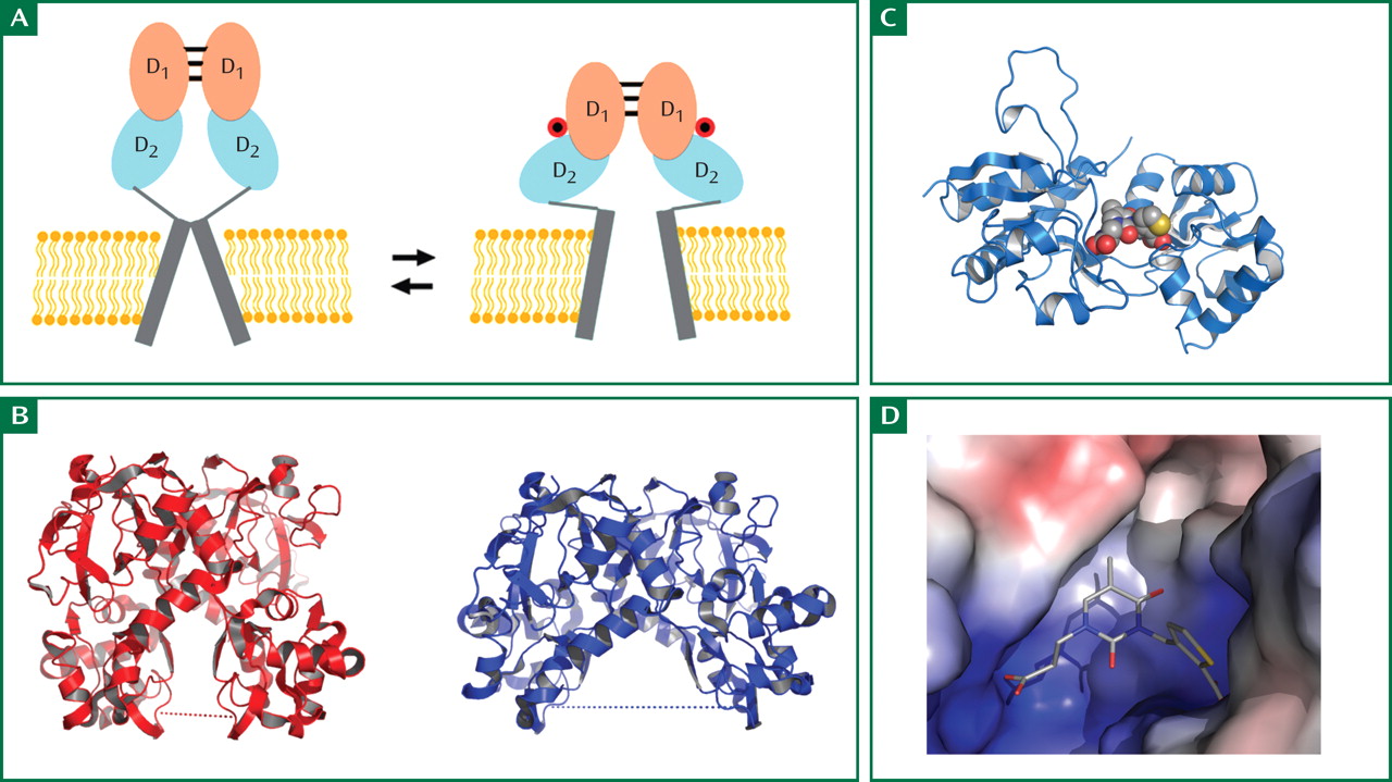
Figure 1. The gating model for activation of kainate receptor ion channels and examples of crystal structures for the ligand-binding domain of the GluR5 subtype trapped in the resting and active conformation. Pairs of subunits assemble as dimers (panel A); the change in “linker” separation, which occurs when the neurotransmitter glutamate binds to the receptor and stabilizes the closed cleft conformation, leads to ion channel gating via a scissors-like motion. Panel B shows crystal structures for the resting and active conformations that led to this model. Panel C shows the crystal structure for a single subunit in complex with the selective GluR5 competitive antagonist UBP310; the antagonist sits in the cleft between the two domains. Panel D shows the molecular surface of the antagonist binding site, colored by surface potential (blue positive and red negative) and reveals that the pair of carboxylate ions in the antagonist bind to positively charged patches in domain 1 and 2.

Footnotes

Address reprint requests to Dr. Tamminga, UT Southwestern Medical Center, Department of Psychiatry, 5323 Harry Hines Blvd., #NE5.110, Dallas, TX 75390-9070; [email protected] (e-mail).
This month’s Image in Neuroscience (doi: 10.1176/appi.ajp.2007.07060996) is drawn from work supported by the Intramural Research Program of the National Institute of Child Health and Human Development, NIH. Figures adapted from data published in Mayer ML, Ghosal A, Dolman NP, Jane DE: Crystal structures of the kainate receptor GluR5 ligand binding core dimer with novel GluR5-selective antagonists. J Neurosci 2006; 26:2852–2861.

Information & Authors

Information

Published In

Go to American Journal of Psychiatry
American Journal of Psychiatry
Pages: 1148
PubMed: 17671275

History

Published online: 1 August 2007
Published in print: August, 2007

Authors

Details

Mark L. Mayer, Ph.D.

Metrics & Citations

Metrics

Citations

Export Citations

If you have the appropriate software installed, you can download article citation data to the citation manager of your choice. Simply select your manager software from the list below and click Download.

For more information or tips please see 'Downloading to a citation manager' in the Help menu.

Format
Citation style
Style
Copy to clipboard

View Options

View options

PDF/EPUB

View PDF/EPUB

Get Access

Login options

Already a subscriber? Access your subscription through your login credentials or your institution for full access to this article.

Personal login Institutional Login Open Athens login
Purchase Options

Purchase this article to access the full text.

PPV Articles - American Journal of Psychiatry

PPV Articles - American Journal of Psychiatry

Not a subscriber?

Subscribe Now / Learn More

PsychiatryOnline subscription options offer access to the DSM-5-TR® library, books, journals, CME, and patient resources. This all-in-one virtual library provides psychiatrists and mental health professionals with key resources for diagnosis, treatment, research, and professional development.

Need more help? PsychiatryOnline Customer Service may be reached by emailing [email protected] or by calling 800-368-5777 (in the U.S.) or 703-907-7322 (outside the U.S.).

Media

Figures

Other

Tables

Share

Share

Share article link

Share