Limitations in technology have prevented the full capture of millisecond-scale neural circuit dynamics in intact tissue from psychiatric disease models. Now, intact circuit function can be mapped and quantified with high temporal- and spatial-resolution optical tools. This approach complements optogenetic technologies used for fast optical control of specific cell types within intact neural circuits.
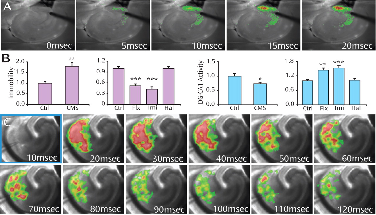
To match the millisecond precision of optogenetics, readout technology that is equally fast is needed. Voltage-sensitive dye imaging (VSDI) captures and quantifies millisecond-scale dynamics of electrically or optically evoked activity propagation while maintaining a full-circuit perspective, thereby allowing circuit-dynamics hypotheses to be tested in animal models of brain disorders. Using high-speed VSDI in living circuits from animals with known behavioral phenotypes, a single measure of activity propagation through specific hippocampal subfields was found to substantially predict both depressed-like behavior and antidepressant treatment response, despite fundamentally different cellular mechanisms. Specifically, in the chronic mild stress (CMS) rodent model of depression, VSDI of slices prepared from the ventral hippocampus revealed a consistent decrease in the relative percolation of evoked electrical activity in the dentate gyrus (DG) versus CA1 subfields, while the opposite effect was seen with antidepressant treatment (panels A, B).
Figure 1. This representative voltage-sensitive dye imaging (VSDI) filmstrip shows the electrically evoked local circuit dynamics of neural activity propagation in the dentate gyrus (DG). Warmer colors indicate greater depolarization (panel A). VSDI quantification can be used to reveal that quantitative differences in the relative dynamics of the DG and CA1 hippocampal subfields substantially account for depression-related behaviors (immobility on the forced swim test) in rodent models of both depressed-like state induction (chronic mild stress [CMS]) and antidepressant treatment (fluoxetine, Flx; imipramine, Imi). The typical antipsychotic haloperidol (Hal) does not affect the depression-related behaviors or circuit-dynamics measures (panel B) (Figure adapted/modified with permission from Airan et al., “High-Speed Imaging Reveals Neurophysiological Links to Behavior in an Animal Model of Depression” [Science 2007; 317:819–823]). Copyright ” American Association for the Advancement of Science. Integrated fast optical control and fast optical imaging reveals circuit dynamics after synchronous activation of a sparse subset of defined neurons in a living slice of mouse hippocampus. Genetically defined (Thy1) hippocampal neurons were modified to express the light-activated cation channel ChR2, and the slice was stained with the voltage-sensitive dye RH-155. The filmstrip reveals the evolving millisecond-scale dynamics of neural activity in the CA1 subfield in response to a single 10 msec optical stimulus pulse of 470nm blue light (frame outlined in blue) to drive an action potential in the targeted cells. Warmer colors indicate greater depolarization. Resulting circuit dynamics in this all-optical experiment can be precisely quantified with regard to total area recruited, time and space constants of propagation, and synchrony and amplitude of activity of individual 25 mm pixels within the intact local circuit (panel C).
This circuit-dynamics approach can be integrated currently with fast optical control of defined cell types. The voltage sensitive dye RH-155 has an absorbance band spectrally separated from the excitation peaks of the optical control tools ChR2, NpHR, and VChR1, which therefore permits smooth integration of fast optical control and recording (panel C). For example, using living brain slices from ChR2 expressing mice, optically evoked hippocampal activity arising from defined cell types can be imaged and quantified on the millisecond time scale (panel C). These fast optical control tools can be used to quantitatively and dynamically determine the causal roles that different cell types play in modulating neural circuit activity in the normal and diseased brain.

Footnote

Address reprint requests to Dr. Tamminga, UT Southwestern Medical Center, Department of Psychiatry, 5323 Harry Hines Blvd., #NE5.110, Dallas, TX 75390-9070; [email protected] (e-mail). Image accepted for publication May 2008 (doi: 10.1176/appi.ajp.2008.08050764).

Information & Authors

Information

Published In

Go to American Journal of Psychiatry
American Journal of Psychiatry
Pages: 800
PubMed: 18593784

History

Published online: 1 July 2008
Published in print: July, 2008

Authors

Details

Karl Deisseroth, M.D., Ph.D.

Metrics & Citations

Metrics

Citations

Export Citations

If you have the appropriate software installed, you can download article citation data to the citation manager of your choice. Simply select your manager software from the list below and click Download.

For more information or tips please see 'Downloading to a citation manager' in the Help menu.

Format
Citation style
Style
Copy to clipboard

View Options

View options

PDF/EPUB

View PDF/EPUB

Get Access

Login options

Already a subscriber? Access your subscription through your login credentials or your institution for full access to this article.

Personal login Institutional Login Open Athens login
Purchase Options

Purchase this article to access the full text.

PPV Articles - American Journal of Psychiatry

PPV Articles - American Journal of Psychiatry

Not a subscriber?

Subscribe Now / Learn More

PsychiatryOnline subscription options offer access to the DSM-5-TR® library, books, journals, CME, and patient resources. This all-in-one virtual library provides psychiatrists and mental health professionals with key resources for diagnosis, treatment, research, and professional development.

Need more help? PsychiatryOnline Customer Service may be reached by emailing [email protected] or by calling 800-368-5777 (in the U.S.) or 703-907-7322 (outside the U.S.).

Media

Figures

Other

Tables

Share

Share

Share article link

Share