A 15-year-old girl of normal intelligence was admitted involuntarily to a local child psychiatric hospital with an acute onset of first-episode psychosis. Despite adequate antipsychotic treatment, her psychiatric and somatic state deteriorated. She was agitated and suffered from severe insomnia, physical exhaustion, and seizure-like episodes. In order to break this vicious circle, she was transferred to the pediatric intensive care unit of our regional tertiary hospital with the need for sedation. Anti-N-methyl-d-aspartate (anti-NMDA) receptor encephalitis was diagnosed, and neuroimmunologic treatment (i.e., methylprednisolone, intravenous immunoglobulins, and rituximab) was commenced. During the disease course her neuropsychiatric state was monitored daily by means of clinical observations and psychometric scales.
After she was weaned off the sedation, her score on the Mini-Mental State Examination (MMSE) (
1,
2) was 15 out of 30 (indicating moderate cognitive impairment), her score on the Pediatric Anesthesia Emergence Delirium Scale (
3) was 9 out of 20 (subsyndromal delirium), and her score on the Positive and Negative Syndrome Scale (PANSS) (
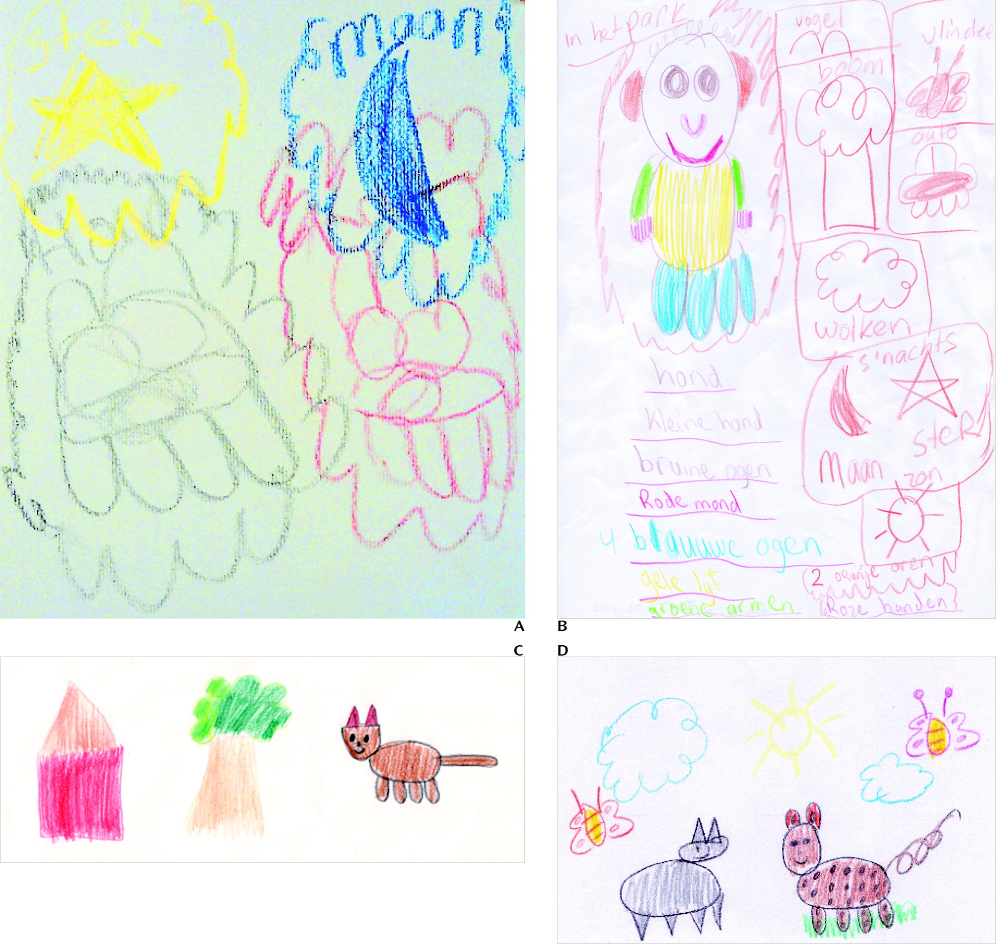
4) was 80 (moderately ill); these values were in line with our clinical examinations. Her medications were lorazepam, trazodone, risperidone, methadone, and methylprednisolone for the insomnia, agitation, and encephalitis. As she gradually recovered we asked her to draw something. She did not know what to draw, so we suggested an animal, such as a dog, but she did not know how to start. When we told her that a dog has four legs, a tail, two ears, two eyes, and a mouth, she drew an abstract figure that consisted of a head with four legs (image A). Her next drawing, of a cat, looked exactly the same, apparently since they share the same basic features. The image showed us pictorial evidence of her higher cortical dysfunctions regarding imaginary representation, praxis, and language.
Two weeks later her MMSE score was 21 (mild cognitive impairment), her score on the Pediatric Anesthesia Emergence Delirium Scale was 2 (no delirium), and her PANSS score was 72 (mildly ill). The medication regimen continued unchanged, and we asked her to draw a dog again (image B). The dog now looked more recognizable but like a human, standing upright, with two arms and four legs. It was drawn with bright colors and not colored in. The surroundings were encircled and monochromatic. The composition was chaotic and inconsistent. All body parts were listed beneath the figure in the same color as they were drawn.
Two months after the patient was transferred to a local rehabilitation center, her MMSE score was 27 (normal), and her PANSS score was 62 (mildly ill). Her medications were reduced to trazodone and hydrocortisone. She was asked to make another drawing (image C). Now the cat was catlike for the first time; it had four legs, was normally proportioned, and was correctly positioned. Colors were used adequately. However, this drawing still looked like one by a primary school child instead of a 15-year-old girl.
Finally, after 5 months of rehabilitation the patient made another drawing. By that time she had largely recovered; her PANSS score was 49 (mildly ill), she was receiving no medication, and no higher cortical dysfunction was present. Her drawing had a normal composition, and it was again colorful and bright (image D). Although she still had the urge to write down what she drew, she did not encircle the figures anymore.
This series of images exemplifies the additional clinical value of chronological drawings in following neuropsychiatric recovery in anti-NMDA receptor encephalitis.


