Full access
New Research
Published Online: 1 June 2013

Decreased Cortical Representation of Genital Somatosensory Field After Childhood Sexual Abuse

Abstract

Objective

Sexual dysfunction is a common clinical symptom in women who were victims of childhood sexual abuse. The precise mechanism that mediates this association remains poorly understood. The authors evaluated the relationship between the experience of childhood abuse and neuroplastic thinning of cortical fields, depending on the nature of the abusive experience.

Method

The authors used MRI-based cortical thickness analysis in 51 medically healthy adult women to test whether different forms of childhood abuse were associated with cortical thinning in areas critical to the perception and processing of specific behavior implicated in the type of abuse.

Results

Exposure to childhood sexual abuse was specifically associated with pronounced cortical thinning in the genital representation field of the primary somatosensory cortex. In contrast, emotional abuse was associated with cortical thinning in regions relevant to self-awareness and self-evaluation.

Conclusions

Neural plasticity during development appears to result in cortical adaptation that may shield a child from the sensory processing of the specific abusive experience by altering cortical representation fields in a regionally highly specific manner. Such plastic reorganization may be protective for the child living under abusive conditions, but it may underlie the development of behavioral problems, such as sexual dysfunction, later in life.
Early adverse experience dramatically increases the risk for developing a wide range of psychiatric disorders and certain medical diseases later in life (1, 2). One common and exceptionally detrimental clinical consequence of childhood sexual abuse is the development of sexual dysfunction, including anorgasmia and inability to experience sexual pleasure, as well as chronic genital or pelvic pain in adulthood (3, 4). The precise mechanism that mediates this association remains poorly understood. A burgeoning number of studies suggest that there is substantial plasticity of the human brain as a function of experience (5). For instance, London taxi drivers have been shown to exhibit navigation-related structural increases in the posterior hippocampus, which is relevant to spatial representation of the environment, suggesting that the brain is capable of adapting its structure in response to environmental demands (6). Another example of experience-induced neural plasticity is observed in limb amputees who exhibit cortical reorganization of the somatosensory cortex after the deprivation of afferent inputs, inasmuch as adjacent cortical representations of intact sensory parts invade the representation field of the lost sensory part (7, 8). It has been suggested that constant cortical competition for space leads to enlargement of areas that are supplied with important information, whereas other areas are narrowed (5). Although the adult brain has the capacity for neural plasticity, the developing brain is clearly more sensitive to the organizing effects of experiences. In classic studies, Wiesel and Hubel (9) demonstrated that visual input during a sensitive period early in life is critical for normal development of the visual cortex and vision. Cortical plasticity during sensitive periods may also account for long-term effects of enriching early experiences. For instance, Elbert et al. (10) reported that earlier onset of musical training during childhood is associated with larger cortical representation of the fingers of the left hand in the primary somatosensory cortex of string players. Extending the cortical competition hypothesis, it can be argued that neuroplastic reorganization may depend on the nature and timing of the experience. More specifically, if an experience is highly aversive and developmentally inappropriate (instead of enriching), the brain may not allocate more resources to the experience but instead narrow its cortical representation to limit detrimental effects.
We hypothesized that the experience of childhood abuse may lead to regionally highly specific neuroplastic thinning of cortical fields, depending on the precise nature of the abusive experience. Such cortical adaptation would serve to immediately shield the child from the experience of the abuse by gating the sensory processing. While initially protective, such cortical thinning may represent a direct biological substrate for the development of behavioral problems later in life, when the behavior in question, such as normal sexual perception, would be developmentally appropriate. Using MRI-based cortical thickness analysis, we demonstrate that different forms of childhood abuse are associated with profound and regionally highly specific cortical thinning, precisely affecting areas that are critical to the perception and/or processing of the specific behavior implicated in the type of abuse.

Method

Participants

Fifty-one healthy adult women (mean age, 27 years [SD=7]; range, 18–45 years; African American, N=24; Caucasian, N=18; and other, N=9) were recruited from the greater Atlanta area to include women with or without reported histories of childhood abuse or neglect that occurred before the onset of puberty and with or without major depression. The women were recruited as part of a larger study of the psychobiology of early-life trauma. All women had regular menstrual cycles and no significant medical illness, as verified by physical examination and standard laboratory testing. All women were free of medication. We excluded women with histories of psychosis, bipolar disorder, substance abuse, or alcohol abuse within 2 years to exclude the effects of these conditions on neural structure. Written informed consent was obtained, and the study was approved by the Emory University institutional review board.

Clinical Assessment

Exposure to childhood maltreatment was quantified using the 28-item form of the Childhood Trauma Questionnaire (CTQ) (11). The CTQ is a self-report questionnaire that assesses five types of maltreatment in separate subscales: emotional abuse, sexual abuse, physical abuse, emotional neglect, and physical neglect. Each subscale includes five items. Examples of questions are: “People in my family called me stupid, lazy, or ugly” (emotional abuse item); “People in my family hit me so hard that it left bruises or marks” (physical abuse item); “Someone threatened to hurt me or tell lies about me unless I did something sexual with them” (sexual abuse item); “I knew there was someone to take care of me and protect me” (emotional neglect inverse item); and “There was someone to take me to the doctor if I needed it” (physical neglect inverse item). Participants rate each item on a five-point Likert scale from 1 (never true) to 5 (very often true). Such Likert-type items create dimensional scales providing quantitative scores that have enhanced reliability and maximized statistical power. In addition, cutoff scores for none to low, low to moderate, moderate to severe, and severe to extreme exposure are provided for each scale. We used the moderate to severe cutoff scores for each subscale to classify participants as positive for a history of childhood trauma category. The cutoff scores are 13 for emotional abuse, 10 for physical abuse, 8 for sexual abuse, 15 for emotional neglect, and 10 for physical neglect. Being identified as positive for a category corresponds with endorsing a substantive number of experiences as often true.
Using this categorization, 28 women (55%) were classified as having moderate to severe maltreatment, and 23 women (45%) were classified as having none to low maltreatment. The CTQ has undergone extensive psychometric testing. The questionnaire demonstrated good internal consistency (Crohnbach's alpha=0.63–0.95) and criterion-related validity (r=0.50–0.75) in clinical and community samples. Convergent reliability with therapist assessments of abuse histories is high. Good specificity and sensitivity of cutoff scores to classify maltreated subjects has been reported as well. We also used a semistructured clinician-administered interview (12) on childhood experiences for cross-validation, and we found high correlations between scores of these two instruments for all subscales (r values, >0.80; p values, <0.001). Information regarding the age at onset and duration of maltreatment was extracted from this interview. Mental health status was assessed using the Structured Clinical Interview for DSM-IV-TR (13). Axis I diagnoses were used to apply exclusion criteria and to control analyses for disorders. A total of 12 women (24%) had current major depression, and nine women (18%) had current PTSD.

MRI Acquisition

All MR images were acquired on a 3-T Siemens Trio (Siemens, Erlangen, Germany). The women were scanned with a high-resolution T1 three-dimensional magnetization-prepared rapid gradient echo sequence (TR=2,600 ms, TE=3.02 ms, flip angle=8°, field of view=256×224 mm, in-plane resolution isotropic, 1 mm3).

Cortical Thickness Analysis

Cortical thickness analysis was performed using the automated analyses pipeline developed at the Montreal Neurological Institute (1416). In brief, anatomical MRIs are corrected for nonuniformities (17), registered into standard stereotaxic space (18), and classified into gray matter, white matter, and CSF using a neural-net classifier (19). At the core of the cortical thickness analysis, a constrained Laplacian anatomical segmentation using proximities is applied to determine white and gray matter surface boundaries using a surface deformation algorithm (20). This procedure computes 40,000 vertices of white and gray matter surfaces that are linked. Cortical thickness is computed as the distance between these linked vertices. In a final step, individual cortical thickness data were smoothed using a blurring kernel of 20 mm.

Statistical Analysis

For statistical analysis, regressions were performed at every vertex, with cortical thickness as the dependent variable and CTQ score as the independent variable (either total score across all subscales or sum score of individual subscales, depending on the specific analysis). Analyses were controlled for age and depression status, which were entered as covariates. In the case of significant effects, we additionally verified that effects were not due to concurrent PTSD. For the subanalyses investigating the specificity of the effects of sexual abuse, we entered age, depression, and all other CTQ subscales as covariates in the regression. In a final step, we regressed onset age and duration of the abuse against cortical thickness in the women with moderate to severe maltreatment. The resulting statistical maps were thresholded applying the false discovery rate (21) at a q value of 0.05.

Results

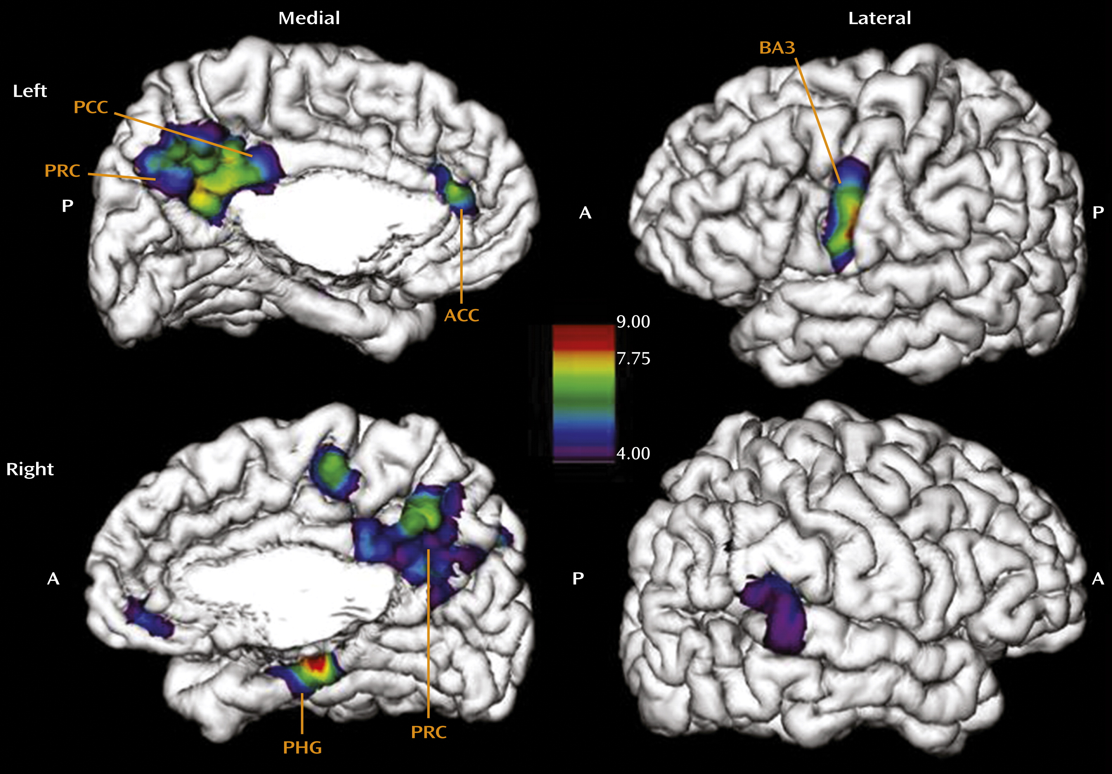
In the regression of cortical thickness against CTQ total score across all subscales, the results revealed a widespread effect of cortical thinning as a function of childhood adversity (Figure 1). The area most prominently affected in the left hemisphere was the somatosensory cortex laterally (local maxima at Talairach coordinates x=−19, y=−31, z=78; F=19.3, p<0.001; x=−62, y=−7, z=34; F=16.4, p<0.001), in locations associated with sensory representation of the female clitoris and surrounding genital area as well as the mouth area (for details regarding the precise location of the somatosensory genital field as identified using functional MRI of neural response to stimulation, see references 22 and 23). Medially, our results indicated effects of the CTQ total score on thinning of the anterior cingulate gyrus (x=−2, y=36, z=18; F=12.7, p<0.001), the precuneus (x=−3, y=−60, z=45; F=8.4, p<0.01), and the parahippocampal gyrus (x=−18, y=−20, z=−18; F=13.6, p<0.001), structures mediating emotional regulation (24), self-awareness (25, 26), and memory encoding (27, 28), respectively. In the right hemisphere, results were less pronounced, with one maxima in the right motor cortex laterally (x=14, y=−21, z=76; F=12.2, p<0.001) and in the area of index finger representation, the parahippocampal gyrus (x=17, y=−28, z=−13; F=18.8, p<0.001) and the posterior cingulate cortex medially (x=3, y=−52, z=16; F=8.9, p<0.01). The effects were maintained when we additionally controlled for PTSD. Because the CTQ total score summarizes the severity of five different types of maltreatment, these widespread effects on diverse brain regions likely reflect additive effects of several forms of abuse, which often co-occur, while specific effects of different forms of maltreatment were not considered in this analysis.
FIGURE 1. Regression of Childhood Trauma Questionnaire (CTQ) Total Score Against Cortical Thickness in Women With and Without Childhood Sexual Abusea
a Cortical thickness analysis results after regressing CTQ total score against thickness across the entire cortex. Control variables included age and depression scores. Main effects are seen in the somatosensory cortex in the female genital and mouth area on the left, the parahippocampal gyrus (PHG) bilaterally, the left anterior cingulate cortex (ACC), and the precuneus (PRC) bilaterally. For the precise location of the genital sensory field as identified using functional MRI of neural response to stimulation, see references 22 and 23. BA3=Brodmann’s area 3; PCC=posterior cingulate cortex; A=anterior; P=posterior. The color scale refers to the F values of the linear regression (significance threshold: F>4.33).
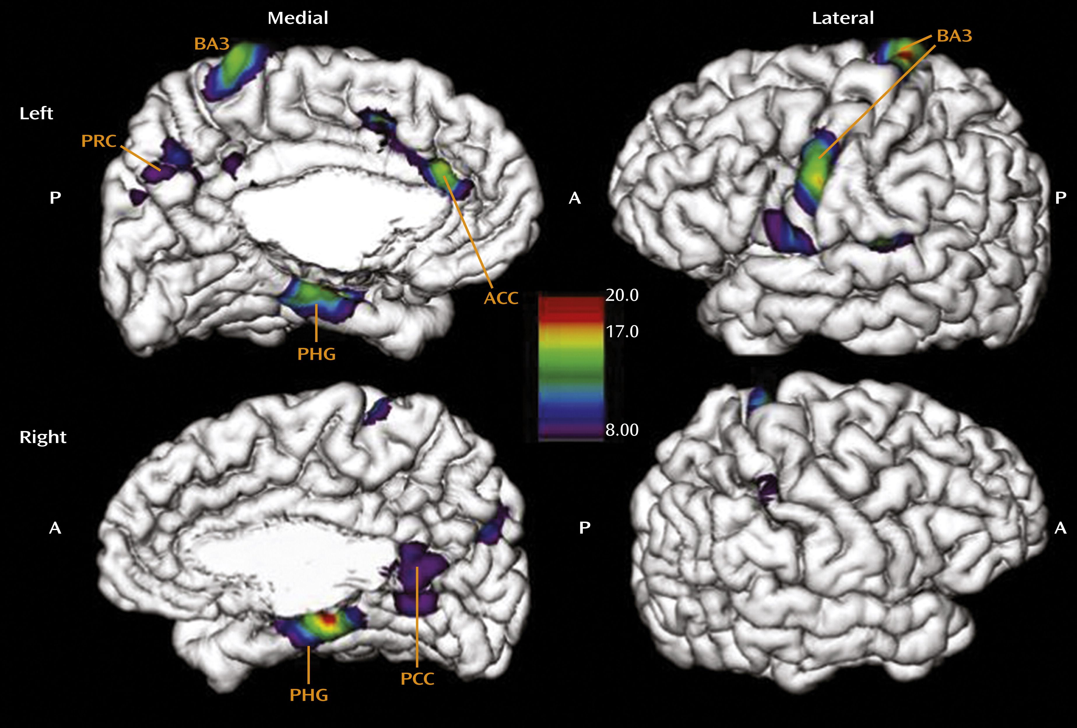
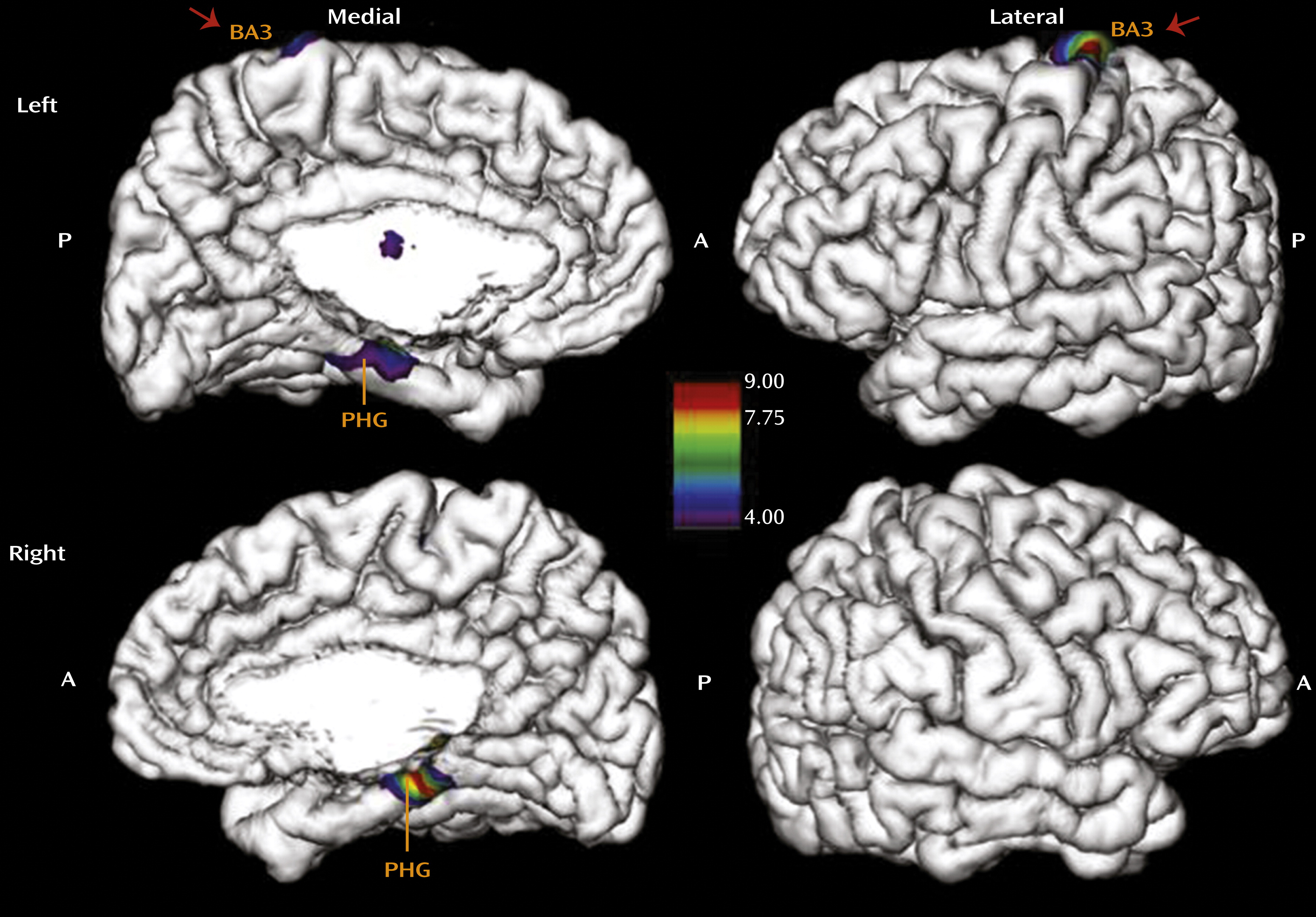
We therefore investigated whether the pronounced effect of total CTQ score on the somatosensory cortex in the genital representation field might be specific to childhood sexual abuse. To isolate the effects of sexual abuse on cortical thinning, we ran a separate analysis with the CTQ subscale score for sexual abuse as the main regressor, while controlling for age, depression, and the other four CTQ subscales entered as covariates (Figure 2). This analysis revealed a highly specific effect of sexual abuse experience on thinning of the somatosensory cortex representing the female clitoris and surrounding genital area (x=−16, y=−32, z=76; F=8.6, p<0.01) in the left hemisphere (22, 23). The parahippocampal gyrus in the left (x=−20, y=−17, z=−21; F=4.6, p<0.05) and right (x=17, y=−28, z=−14; F=8.9, p<0.01) hemispheres was also affected by sexual abuse. To further isolate the specific effects of different types of maltreatment on cortical thickness, we determined whether emotional abuse has a specific effect on cortical thickness that can be distinguished from the effect of sexual abuse (Figure 3). Indeed, emotional abuse specifically affects the areas of the left (x=−3, y=−61, z=45; F=7.8, p<0.05) and right (x=6, y=−49, z=51; F=6.2, p<0.05) precuneus and the left anterior (x=−4, y=40, z=11; F=6.9, p<0.05) and posterior cingulate cortex (x=−2, y=−47, z=28; F=8.1, p<0.01). We also observed thinning in the face region of the somatosensory cortex (x=−56, y=−12, z=45; F=22.7, p<0.001). Hence, emotional abuse, which likely represents experiences of parental rejection and is often considered most detrimental in terms of altered concept of “self,” is associated with the cortical thinning of regions implicated in mediating self-reflection, self-awareness, and first-person perspective (2426).
FIGURE 2. Regression of Childhood Trauma Questionnaire (CTQ) Sexual Abuse Score Against Cortical Thickness in Women With and Without Childhood Sexual Abusea
a Cortical thickness analysis results after regressing CTQ sexual abuse score against thickness across the entire cortex. Control variables included age, depression, and all other CTQ subscales. Main effects are seen in the somatosensory cortex in the female genital area on the left and the parahippocampal gyrus (PHG) bilaterally. For the precise location of the genital sensory field as identified using functional MRI of neural response to stimulation, see references 22 and 23. BA3=Brodmann’s area 3; A=anterior; P=posterior. The color scale refers to the F values of the linear regression (significance threshold: F>4.33).
FIGURE 3. Regression of Childhood Trauma Questionnaire (CTQ) Emotional Abuse Score Against Cortical Thickness in Women With and Without Childhood Sexual Abusea
a Cortical thickness analysis results after regressing CTQ emotional abuse score against thickness across the entire cortex. Control variables included age, depression, and all other CTQ subscales. Main effects are seen in the left and right precuneus (PRC), left anterior cingulate cortex (ACC), right parahippocampal gyrus (PHG), and left somatosensory cortex in the area of the face. BA3=Brodmann’s area 3; PCC=posterior cingulate cortex; A=anterior; P=posterior. The color scale refers to the F values of the linear regression (significance threshold: F>4.33).
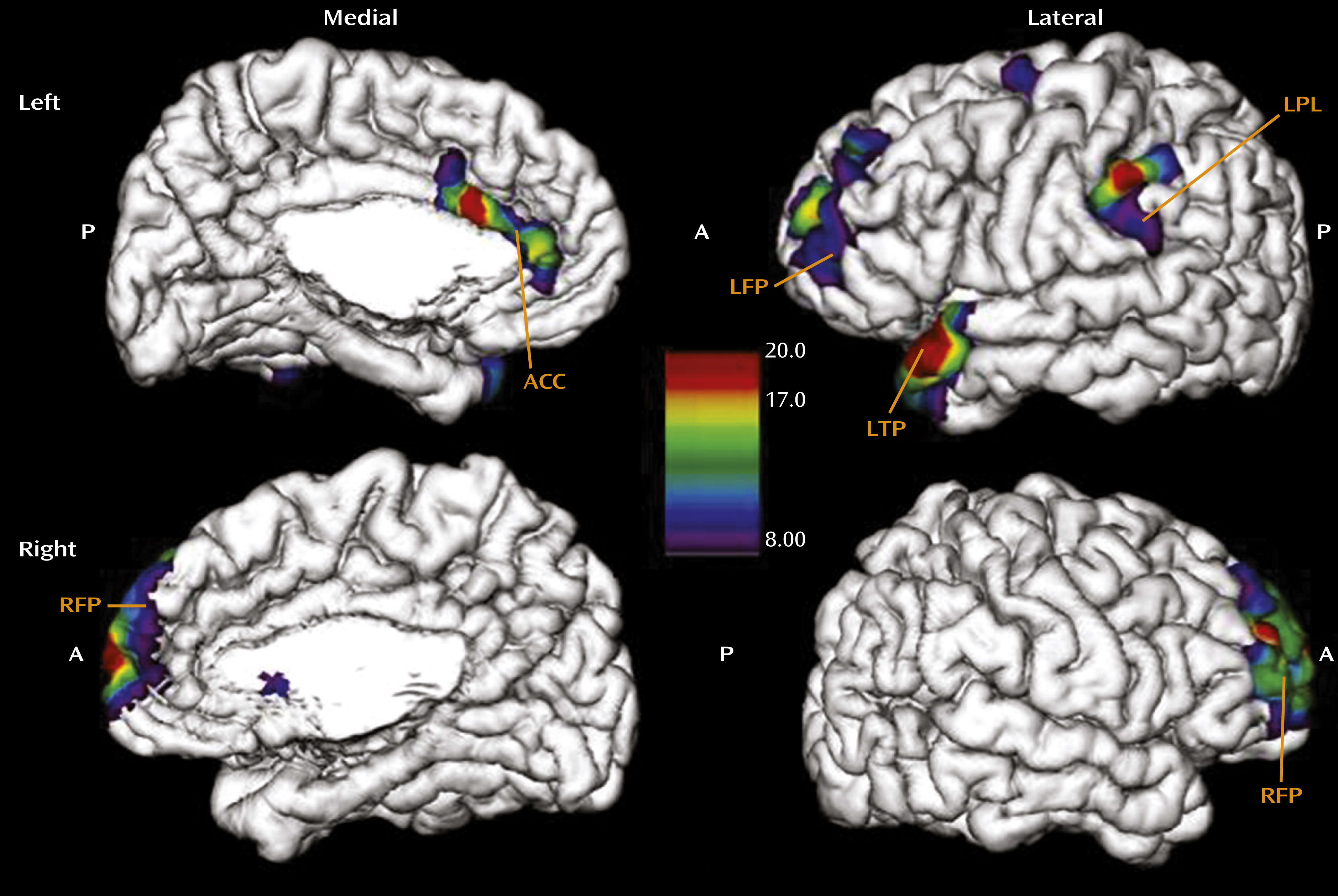
Finally, we investigated the effect of age at onset of any abuse on cortical thickness in the group of women reporting moderate to severe exposure. This analysis revealed that earlier exposure was associated with thinning of the left temporal pole (x=−49, y=22, z=−26; F=19.9, p<0.001), the left parietal lobe (x=−63, y=−32, z=45; F=18.9, p<0.001), the left frontal pole (x=−28, y=63, z=12; F=16.8, p<0.001), and the right frontal pole (x=11, y=69, z=4; F=18.6, p<0.001), areas associated with autobiographic memory, as well as thinning of the anterior cingulate cortex (x=−2, y=21, z=22; F=8.13, p<0.01) (Figure 4). This finding is concordant with reports that abuse occurring earlier in childhood is often associated with absence of memories concerning the abuse (29). We did not see an effect of duration of the abuse, which supports the assumption that the observed effects reflect developmental programming rather than consequences of cumulative exposure to maltreatment over time.
FIGURE 4. Regression of Onset Age of Maltreatment Against Cortical Thickness in Women With and Without Childhood Sexual Abusea
a Cortical thickness analysis results after regressing age at onset of abuse against thickness across the entire cortex in the group of women with moderate to severe exposure according to Childhood Trauma Questionnaire cutoff scores. Control variables included age and depression. Main effects are seen in the left frontal pole (LFP), right frontal pole (RFP), left temporal pole (LTP), left parietal lobe (LPL), and left anterior cingulate cortex (ACC). A=anterior; P=posterior. The color scale refers to the F values of the linear regression (significance threshold: F>4.33).

Discussion

Our results suggest that exposure to childhood adversity is associated with pronounced neuroplastic changes in the cortex in the form of cortical thinning that occurs in a regionally highly specific manner, determined by the type of maltreatment that was experienced. Because the study design was cross-sectional, a causal interpretation of these results is impossible. Nevertheless, our results suggest that early adverse experience is associated with neuroplastic adaptation resulting in altered cortical representation of sensory and processing areas that are directly implicated in the abusive experience. We suggest that cortical thinning in these regions may represent the most adaptive and protective response of the developing brain, potentially serving to shield the child from highly adverse environmental conditions by gating sensory experiences and processing related to the abuse. While this idea of sensory gating of abusive experiences is clearly speculative, the general principle of sensory gating is a well-known phenomenon that has been described in terms of filtering auditory input by personal relevance (30) and in terms of gating acute pain stimuli at the level of the spine (31). Whether or not abusive experiences can be gated in such ways is unknown, but this is an intriguing possibility raised by our results and should be further scrutinized.
A number of mechanisms may explain how excessively adverse sensory experiences might lead to impaired neurodevelopmental and hence structural change in adulthood. Although neocortical thickness is determined by the number of neurons as well as by the number of synapses and dendritic branches, the effects observed in this study likely reflect variations in synaptic density because the number of neurons in these cortical areas likely remains stable (32). A study of monocular visual deprivation (33) suggested that early deprivation leaves a long-term structural trace by increasing cortical dendritic spine density in the visual representation field of the opposite eye that outlasts the original experience and remains unaffected by subsequent experimental variation of visual input. Our observation of thinning in the primary somatosensory cortex after sexual abuse may be understood as a consequence of both top-down and bottom-up mechanisms.
Evidence for top-down mechanisms comes from studies investigating the role of painful stimuli on somatosensory cortex activation (34, 35). These studies demonstrate that pain perception in the somatosensory cortex is subject to cognitive modulation. By directing attention away from the painful stimulus, activation of the area of the somatosensory cortex associated with the painful stimulus can be dramatically reduced. If such top-down cognitive modulation directing perception away from the painful stimulation occurs during the critical time of synapse formation and development, as can be envisioned when children experience sexual abuse, the final formation of synaptic connections in specific parts of the neocortex would be significantly decreased.
Complementary to the cognitive modulation theory is a potential bottom-up mechanism. Decreases in blood flow have been shown to occur in the somatosensory cortex as a consequence of painful stimulation. This has been demonstrated for noxious stimuli (36) as well as for painful heat stimuli (37). Physical and sexual abuse during the period of synapse formation might lead to inhibition, and as a consequence, the number of synapses in the somatosensory cortex might be reduced. The basic cellular mechanisms implicated in such inhibition have been reported to depend on long-term depression and potentiation, in conjunction with N-methyl-d-aspartate receptors in thalamocortical circuits (38). Of note, the effect of sexual abuse on thinning of the somatosensory genital field was present only in the left hemisphere. Because the genitals are centralized in the body, this lateralization can be hypothesized to be based on central mechanisms of inhibition that may be lateralized. Future studies should scrutinize the origin of this lateralization.
Later in life, when the stimulation would be developmentally appropriate, the lack of development of the genital sensory representation field may lead to impaired sexual perception, explaining the frequent clinical reports of sexual dysfunction. However, in the absence of data linking cortical thickness with sexual behavior and the experience of sexual pleasure, we cannot further substantiate this notion. Decreased thickness of the genital sensory representation field may also contribute to genital or pelvic pain in women with a history sexual abuse, in light of studies showing that increased thickness of the somatosensory cortex is associated with increased pain threshold (39), perhaps suggesting that less differentiated sensory processing decreases the pain threshold. Assuming similar mechanisms, the most affected cortical regions implicated in the processing of emotional abuse should be brain areas associated with emotional regulation and the perception and evaluation of one’s self, as observed in the present study. If adverse stimulation during critical developmental periods of emotional regulation circuits also leads to cognitive avoidance and inhibition in association with emotional processing, then the areas of the precuneus and cingulate cortex would be primary targets of such effects, given their dominant role in these functions. Of note, decreased anterior cingulate cortex volume has been reported in studies of adults with histories of childhood trauma, although these studies did not distinguish between different types of trauma (40, 41).
An alternative explanation of our results might assume a reverse direction of effect. Individuals with sexual abuse experiences may avoid sexual contact in adulthood. Decreased frequency of sexual behavior may result in cortical thinning of the somatosensory representation field, in accordance with the general principle that connections that are less frequently used are diminished, while connections that are more often used are strengthened (5). Similarly, emotionally abused children might grow into rejection-sensitive adults who avoid situations requiring evaluation of self and therefore underuse these cortical regions. In the absence of longitudinal MRI and behavioral data, we are not able to test the competing hypotheses, which is a limitation of the study. Another limitation is reliance on retrospective self-reports of childhood experiences, which may be hampered by simple forgetting, nonawareness, nondisclosure, and reporting biases. However, a recent meta-analysis of studies using external corroboration of self-reports revealed that false negative reports are more frequent than false positive ones, leading to downward biases in estimated associations between childhood abuse and outcome variables (42).
In conclusion, our observations lend support for the hypothesis that neural plasticity occurs during development, as demonstrated by the thinning of cortical regions that mediate the sensory perception and processing of specific abusive experiences. We hope that our results will serve as an impetus for developing longitudinal and mechanistic studies designed to further scrutinize the association between childhood adversity and experience-dependent neural plasticity. Such studies have the potential to elucidate the biological basis of the detrimental behavioral effects of childhood trauma, which may lead to improved strategies for the prevention and intervention of disorders such as sexual dysfunction by targeting the substantial capacity of the human brain for neural plasticity.
Acknowledgments

The authors thank Dr. Beate Ditzen for assistance in data preparation.

References

1.
Scott KM, Smith DR, Ellis PM: Prospectively ascertained child maltreatment and its association with DSM-IV mental disorders in young adults. Arch Gen Psychiatry 2010; 67:712–719
2.
Shonkoff JP, Garner AS; Committee on Psychosocial Aspects of Child and Family Health; Committee on Early Childhood, Adoption, and Dependent Care; Section on Developmental and Behavioral Pediatrics: The lifelong effects of early childhood adversity and toxic stress. Pediatrics 2012; 129:e232–e246
3.
Paras ML, Murad MH, Chen LP, Goranson EN, Sattler AL, Colbenson KM, Elamin MB, Seime RJ, Prokop LJ, Zirakzadeh A: Sexual abuse and lifetime diagnosis of somatic disorders: a systematic review and meta-analysis. JAMA 2009; 302:550–561
4.
American College of Obstetricians and Gynecologists; Committee on Health Care for Underserved Women: Committee opinion no 498: adult manifestations of childhood sexual abuse. Obstet Gynecol 2011; 118:392–395
5.
Elbert T, Rockstroh B: Reorganization of human cerebral cortex: the range of changes following use and injury. Neuroscientist 2004; 10:129–141
6.
Maguire EA, Gadian DG, Johnsrude IS, Good CD, Ashburner J, Frackowiak RS, Frith CD: Navigation-related structural change in the hippocampi of taxi drivers. Proc Natl Acad Sci USA 2000; 97:4398–4403
7.
Flor H, Elbert T, Knecht S, Wienbruch C, Pantev C, Birbaumer N, Larbig W, Taub E: Phantom-limb pain as a perceptual correlate of cortical reorganization following arm amputation. Nature 1995; 375:482–484
8.
Elbert T, Flor H, Birbaumer N, Knecht S, Hampson S, Larbig W, Taub E: Extensive reorganization of the somatosensory cortex in adult humans after nervous system injury. Neuroreport 1994; 5:2593–2597
9.
Wiesel TN, Hubel DH: Single-cell responses in striate cortex of kittens deprived of vision in one eye. J Neurophysiol 1963; 26:1003–1017
10.
Elbert T, Pantev C, Wienbruch C, Rockstroh B, Taub E: Increased cortical representation of the fingers of the left hand in string players. Science 1995; 270:305–307
11.
Bernstein DP, Stein JA, Newcomb MD, Walker E, Pogge D, Ahluvalia T, Stokes J, Handelsman L, Medrano M, Desmond D, Zule W: Development and validation of a brief screening version of the Childhood Trauma Questionnaire. Child Abuse Negl 2003; 27:169–190
12.
Bremner JD, Vermetten E, Mazure CM: Development and preliminary psychometric properties of an instrument for the measurement of childhood trauma: the Early Trauma Inventory. Depress Anxiety 2000; 12:1–12
13.
First MB, Spitzer RL, Gibbon M, Williams JB: Structured Clinical Interview for DSM-IV-TR Axis I Disorders, Research Version. New York, Biometrics Research, New York State Psychiatric Institute, 2002
14.
Lerch JP, Evans AC: Cortical thickness analysis examined through power analysis and a population simulation. Neuroimage 2005; 24:163–173
15.
Lerch JP, Pruessner JC, Zijdenbos A, Hampel H, Teipel SJ, Evans AC: Focal decline of cortical thickness in Alzheimer’s disease identified by computational neuroanatomy. Cereb Cortex 2005; 15:995–1001
16.
Lyttelton O, Boucher M, Robbins S, Evans AC: An unbiased iterative group registration template for cortical surface analysis. Neuroimage 2007; 34:1535–1544
17.
Sled JG, Zijdenbos AP, Evans AC: A nonparametric method for automatic correction of intensity nonuniformity in MRI data. IEEE Trans Med Imaging 1998; 17:87–97
18.
Collins DL, Neelin P, Peters TM, Evans AC: Automatic 3D intersubject registration of MR volumetric data in standardized Talairach space. J Comput Assist Tomogr 1994; 18:192–205
19.
Zijdenbos AP, Forghani R, Evans AC: Automatic “pipeline” analysis of 3-D MRI data for clinical trials: application to multiple sclerosis. IEEE Trans Med Imaging 2002; 21:1280–1291
20.
MacDonald D, Kabani N, Avis D, Evans AC: Automated 3-D extraction of inner and outer surfaces of cerebral cortex from MRI. Neuroimage 2000; 12:340–356
21.
Genovese CR, Lazar NA, Nichols T: Thresholding of statistical maps in functional neuroimaging using the false discovery rate. Neuroimage 2002; 15:870–878
22.
Michels L, Mehnert U, Boy S, Schurch B, Kollias S: The somatosensory representation of the human clitoris: an fMRI study. Neuroimage 2010; 49:177–184
23.
Kell CA, von Kriegstein K, Rösler A, Kleinschmidt A, Laufs H: The sensory cortical representation of the human penis: revisiting somatotopy in the male homunculus. J Neurosci 2005; 25:5984–5987
24.
Mohanty A, Engels AS, Herrington JD, Heller W, Ho MH, Banich MT, Webb AG, Warren SL, Miller GA: Differential engagement of anterior cingulate cortex subdivisions for cognitive and emotional function. Psychophysiology 2007; 44:343–351
25.
Lou HC, Luber B, Stanford A, Lisanby SH: Self-specific processing in the default network: a single-pulse TMS study. Exp Brain Res 2010; 207:27–38
26.
Kjaer TW, Nowak M, Lou HC: Reflective self-awareness and conscious states: PET evidence for a common midline parietofrontal core. Neuroimage 2002; 17:1080–1086
27.
Guedj E, Bettus G, Barbeau EJ, Liégeois-Chauvel C, Confort-Gouny S, Bartolomei F, Chauvel P, Cozzone PJ, Ranjeva JP, Guye M: Hyperactivation of parahippocampal region and fusiform gyrus associated with successful encoding in medial temporal lobe epilepsy. Epilepsia 2011; 52:1100–1109
28.
Luck D, Danion JM, Marrer C, Pham BT, Gounot D, Foucher J: The right parahippocampal gyrus contributes to the formation and maintenance of bound information in working memory. Brain Cogn 2010; 72:255–263
29.
Chu JA, Frey LM, Ganzel BL, Matthews JA: Memories of childhood abuse: dissociation, amnesia, and corroboration. Am J Psychiatry 1999; 156:749–755
30.
Mayer AR, Hanlon FM, Franco AR, Teshiba TM, Thoma RJ, Clark VP, Canive JM: The neural networks underlying auditory sensory gating. Neuroimage 2009; 44:182–189
31.
Melzack R, Wall PD: Pain mechanisms: a new theory. Science 1965; 150:971–979
32.
Andersen SL: Trajectories of brain development: point of vulnerability or window of opportunity? Neurosci Biobehav Rev 2003; 27:3–18
33.
Hofer SB, Mrsic-Flogel TD, Bonhoeffer T, Hübener M: Experience leaves a lasting structural trace in cortical circuits. Nature 2009; 457:313–317
34.
Bushnell MC, Duncan GH, Hofbauer RK, Ha B, Chen JI, Carrier B: Pain perception: is there a role for primary somatosensory cortex? Proc Natl Acad Sci USA 1999; 96:7705–7709
35.
Jones AK, Friston K, Frackowiak RS: Localization of responses to pain in human cerebral cortex. Science 1992; 255:215–216
36.
Tommerdahl M, Delemos KA, Vierck CJ, Favorov OV, Whitsel BL: Anterior parietal cortical response to tactile and skin-heating stimuli applied to the same skin site. J Neurophysiol 1996; 75:2662–2670
37.
Apkarian AV, Stea RA, Manglos SH, Szeverenyi NM, King RB, Thomas FD: Persistent pain inhibits contralateral somatosensory cortical activity in humans. Neurosci Lett 1992; 140:141–147
38.
Feldman DE: Synaptic mechanisms for plasticity in neocortex. Annu Rev Neurosci 2009; 32:33–55
39.
Grant JA, Courtemanche J, Duerden EG, Duncan GH, Rainville P: Cortical thickness and pain sensitivity in zen meditators. Emotion 2010; 10:43–53
40.
Treadway MT, Grant MM, Ding Z, Hollon SD, Gore JC, Shelton RC: Early adverse events, HPA activity and rostral anterior cingulate volume in MDD. PLoS ONE 2009; 4:e4887
41.
Dannlowski U, Stuhrmann A, Beutelmann V, Zwanzger P, Lenzen T, Grotegerd D, Domschke K, Hohoff C, Ohrmann P, Bauer J, Lindner C, Postert C, Konrad C, Arolt V, Heindel W, Suslow T, Kugel H: Limbic scars: long-term consequences of childhood maltreatment revealed by functional and structural magnetic resonance imaging. Biol Psychiatry 2012; 71:286–293
42.
Hardt J, Rutter M: Validity of adult retrospective reports of adverse childhood experiences: review of the evidence. J Child Psychol Psychiatry 2004; 45:260–273

Information & Authors

Information

Published In

Go to American Journal of Psychiatry
American Journal of Psychiatry
Pages: 616 - 623
PubMed: 23732967

History

Received: 20 July 2012
Revision received: 10 December 2012
Accepted: 28 January 2013
Published online: 1 June 2013
Published in print: June 2013

Authors

Details

Christine M. Heim, Ph.D.
From the Institute of Medical Psychology, Charité University Medicine Berlin, Germany; the Department of Psychiatry and Behavioral Sciences, Emory University School of Medicine, Atlanta; the Department of Psychiatry and Behavioral Sciences, University of Miami Miller School of Medicine, Miami; and the McGill Center for Studies in Aging and the Departments of Psychology and of Psychiatry, Neurology, and Neurosurgery, McGill University, Montreal.
Helen S. Mayberg, M.D.
From the Institute of Medical Psychology, Charité University Medicine Berlin, Germany; the Department of Psychiatry and Behavioral Sciences, Emory University School of Medicine, Atlanta; the Department of Psychiatry and Behavioral Sciences, University of Miami Miller School of Medicine, Miami; and the McGill Center for Studies in Aging and the Departments of Psychology and of Psychiatry, Neurology, and Neurosurgery, McGill University, Montreal.
Tanja Mletzko, M.S.
From the Institute of Medical Psychology, Charité University Medicine Berlin, Germany; the Department of Psychiatry and Behavioral Sciences, Emory University School of Medicine, Atlanta; the Department of Psychiatry and Behavioral Sciences, University of Miami Miller School of Medicine, Miami; and the McGill Center for Studies in Aging and the Departments of Psychology and of Psychiatry, Neurology, and Neurosurgery, McGill University, Montreal.
Charles B. Nemeroff, M.D., Ph.D.
From the Institute of Medical Psychology, Charité University Medicine Berlin, Germany; the Department of Psychiatry and Behavioral Sciences, Emory University School of Medicine, Atlanta; the Department of Psychiatry and Behavioral Sciences, University of Miami Miller School of Medicine, Miami; and the McGill Center for Studies in Aging and the Departments of Psychology and of Psychiatry, Neurology, and Neurosurgery, McGill University, Montreal.
Jens C. Pruessner, Ph.D.
From the Institute of Medical Psychology, Charité University Medicine Berlin, Germany; the Department of Psychiatry and Behavioral Sciences, Emory University School of Medicine, Atlanta; the Department of Psychiatry and Behavioral Sciences, University of Miami Miller School of Medicine, Miami; and the McGill Center for Studies in Aging and the Departments of Psychology and of Psychiatry, Neurology, and Neurosurgery, McGill University, Montreal.

Notes

Address correspondence to Dr. Heim ([email protected]) and Dr. Pruessner ([email protected]).

Funding Information

Dr. Mayberg has received consulting fees from St. Jude Medical Neuromodulation. Dr. Nemeroff has received consulting fees from Takeda and Xhale; holds stock or other financial interests in CeNeRx BioPharma, NovaDel Pharma, PharmaNeuroBoost, Revaax Pharma, and Xhale; has patents for methods and devices for transdermal delivery of lithium and for a method of assessing antidepressant drug therapy via transport inhibition of monoamine neurotransmitters by ex vivo assay; has served on scientific advisory boards for the American Foundation for Suicide Prevention, the Anxiety Disorders Association of America, CeNeRx BioPharma, NARSAD, NovaDel Pharma, and PharmaNeuroBoost; has served on the board of directors for the American Foundation for Suicide Prevention and NovaDel Pharma; and has received research grants from the Agency for Healthcare Research and Quality and NIH. Dr. Heim, Ms. Mletzko, and Dr. Pruessner report no financial relationships with commercial interests.Supported by NIH (MH 073698 and MH 58922), NARSAD, and Charité Research Funds.

Metrics & Citations

Metrics

Citations

Export Citations

If you have the appropriate software installed, you can download article citation data to the citation manager of your choice. Simply select your manager software from the list below and click Download.

For more information or tips please see 'Downloading to a citation manager' in the Help menu.

Format
Citation style
Style
Copy to clipboard

View Options

View options

PDF/EPUB

View PDF/EPUB

Get Access

Login options

Already a subscriber? Access your subscription through your login credentials or your institution for full access to this article.

Personal login Institutional Login Open Athens login
Purchase Options

Purchase this article to access the full text.

PPV Articles - American Journal of Psychiatry

PPV Articles - American Journal of Psychiatry

Not a subscriber?

Subscribe Now / Learn More

PsychiatryOnline subscription options offer access to the DSM-5-TR® library, books, journals, CME, and patient resources. This all-in-one virtual library provides psychiatrists and mental health professionals with key resources for diagnosis, treatment, research, and professional development.

Need more help? PsychiatryOnline Customer Service may be reached by emailing [email protected] or by calling 800-368-5777 (in the U.S.) or 703-907-7322 (outside the U.S.).

Media

Figures

Other

Tables

Share

Share

Share article link

Share