Abstract

Objective:

Bipolar disorder is associated with high risk for suicidal behavior that often develops in adolescence and young adulthood. Elucidation of involved neural systems is critical for prevention. This study of adolescents and young adults with bipolar disorder with and without a history of suicide attempts combines structural, diffusion tensor, and functional MR imaging methods to investigate implicated abnormalities in the morphology and structural and functional connectivity within frontolimbic systems.

Method:

The study had 26 participants with bipolar disorder who had a prior suicide attempt (the attempter group) and 42 participants with bipolar disorder without a suicide attempt (the nonattempter group). Regional gray matter volume, white matter integrity, and functional connectivity during processing of emotional stimuli were compared between groups, and differences were explored for relationships between imaging modalities and associations with suicide-related symptoms and behaviors.

Results:

Compared with the nonattempter group, the attempter group showed significant reductions in gray matter volume in the orbitofrontal cortex, hippocampus, and cerebellum; white matter integrity in the uncinate fasciculus, ventral frontal, and right cerebellum regions; and amygdala functional connectivity to the left ventral and right rostral prefrontal cortex. In exploratory analyses, among attempters, there was a significant negative correlation between right rostral prefrontal connectivity and suicidal ideation and between left ventral prefrontal connectivity and attempt lethality.

Conclusions:

Adolescent and young adult suicide attempters with bipolar disorder demonstrate less gray matter volume and decreased structural and functional connectivity in a ventral frontolimbic neural system subserving emotion regulation. Among attempters, reductions in amygdala–prefrontal functional connectivity may be associated with severity of suicidal ideation and attempt lethality.
As many as 56% of individuals with bipolar disorder attempt suicide, and 15%−19% die by suicide (1). Suicidal behavior is a leading cause of death and often commences during adolescence or young adulthood, when frontolimbic systems implicated in suicidal behavior are maturing. Thus, elucidating the neural underpinnings of suicidal behavior during adolescence and young adulthood may improve early identification and prevention.
Convergent neuroimaging evidence in adult suicide attempters across psychiatric disorders supports involvement of frontolimbic system structural and functional abnormalities. Structural MR imaging (sMRI) and functional neuroimaging (functional MRI [fMRI] and positron emission tomography) studies in adult attempters have shown decreased gray matter volume and frontal dysfunction during emotion stimulus processing (2), particularly in the ventral prefrontal cortex. Postmortem findings in people who have died by suicide suggest decreased ventral prefrontal neuroreceptor binding and neurotrophic factors (3, 4). Also implicated are ventral prefrontal connection sites s1bserving emotion and impulse processing and regulation, including the anterior cingulate cortex, insula, thalamus, striatum, and amygdala (5).
Suicidal behavior has been linked to reduced structural integrity and functional frontolimbic connections in studies of adults, including postmortem (6), sMRI, diffusion tensor imaging (DTI), and fMRI (5) studies, although few studies were conducted in bipolar disorder specifically.
Data on neural circuitry associated with suicide attempts in adolescents and young adults are scant. Initial reports suggest consistencies with adults in reduced frontal gray matter, altered frontotemporal system responses to negative emotional stimuli, and white matter abnormalities (5). Adolescent sample sizes have been low, and most participants have had major depressive disorder.
Single modality structural and functional imaging studies in those who attempt suicide implicate structure-function relationships, suggesting a need for multimodal studies. Few studies have examined associations of imaging measures with suicide-related symptoms and behaviors, such as attempt lethality, ideation, intent, impulsivity, and hopelessness. In adults who have attempted suicide, ventral prefrontal volume decreases and dysfunction have been linked to lethality (7, 8), frontal dysfunction and white matter abnormalities to ideation (9, 10), and orbitofrontal white matter fractional anisotropy decreases to impulsivity in bipolar disorder (11). Imaging studies of suicidal ideation are rare (1); one study of ideation in pediatric epilepsy showed decreased orbitofrontal white matter (12).
This study of bipolar disorder in adolescents and young adults who have attempted suicide, compared with those who have not, combines sMRI, DTI, and fMRI connectivity methods to study gray matter volume, white matter integrity, and functional connectivity. We hypothesized that participants with a history of suicide attempt would show lower ventral frontal and mesial temporal gray matter, and lower white matter fractional anisotropy and functional connectivity in their connections. We explored relationships between imaging findings across modalities and with attempt lethality, anticipating ventral prefrontal associations, and with suicidal ideation and intent, hopelessness, and impulsivity. We explored comparisons with a healthy adolescent and young adult group.

Method

Subjects

Participants were 68 individuals with bipolar disorder (age range=14–25 years); 26 individuals had ≥1 suicide attempts (attempters), and 42 had no suicide attempt (nonattempters) (Table 1). A healthy comparison group (N=45, mean age=20.8 years, SD=3.3, age range=15–25; 58% female) had no lifetime DSM-IV axis I disorder diagnosis, history of suicide attempt, or history of first-degree relative with a major mood or psychotic disorder or suicide attempt. Family history was assessed with the Family History Research Diagnostic Criteria (13). Written informed consent was obtained from participants ≥18 years, permission was obtained from parents or guardians of minors, and assent was obtained from minors, in accordance with the Yale University institutional review board. The presence or absence of DSM-IV axis I diagnoses and mood state at scanning were confirmed with the Structured Clinical Interview for DSM-IV for participants ≥18 years, and the Schedule for Affective Disorders and Schizophrenia for School-Age Children (14) was used for participants <18 years. Adolescents were in the middle to later stages of puberty. No participant had a history of loss of consciousness ≥5 minutes or a neurological or major medical disorder, except two attempters (8% of this group) and one nonattempter (2% of this group) were treated for hypothyroidism.
TABLE 1. Demographic and Clinical Characteristics of Study Participants With Bipolar Disorder
CharacteristicBipolar Disorder Participants With a History of Suicide Attempts (N=26)Bipolar Disorder Participants Without a History of Suicide Attempts (N=42)
 MeanSDMeanSD
Age (years)a20.53.020.63.2
Beck Hopelessness Scale score7.35.65.84.4
Barratt Impulsivity Scale, Version 11, total score75.113.270.210.0
Beck Scale for Suicide Ideation score18.19.75.17.4
 N%N%
Female20772355
Rapid cycling14541331
Mood state at scan    
 Euthymic11422355
 Depressed6231024
 Elevated935921
Lifetime psychosis12461536
Unmedicated8311638
Medications    
 Lithium carbonate519921
 Anticonvulsants1038921
 Antipsychotics9352150
 Antidepressants519410
 Stimulants519819
 Benzodiazepines519614
 Methadone1412
 Levothyroxine2812
 Adrenergic agonists37
 Naltrexone14
 Nonbenzodiazepine hypnotic12
Comborbidity    
 Lifetime substance use disorders8311536
  Substance abuse415410
  Substance dependence519512
  Alcohol abuse312819
  Alcohol dependence28
  Cannabis abuse312410
  Cannabis dependence31237
  Cocaine abuse1412
  Cocaine dependence1425
  Opioid dependence2825
  Amphetamine abuse14
  Polysubstance dependence12
  Sedative-hypnotic dependence12
  Other substance dependence12
 Lifetime other psychiatric disorders    
  Panic disorder623512
  Posttraumatic stress disorder519512
  Social phobia2837
  Specific phobia2812
  Obsessive-compulsive disorder2812
  Generalized anxiety disorder1425
  Anorexia nervosa1412
  Bulimia nervosa37
  Eating disorder not otherwise specified1412
  Attention deficit hyperactivity disorderb233113
a
The age range of participants was 14–25 years.
b
Assessed only in individuals <18 years of age.
Suicide attempts, defined as self-injurious acts committed with at least some intent to die, were assessed using the Columbia Suicide History Form (15), and medical lethality was assessed with the Beck Medical Lethality Scale (16). The Beck Scale for Suicide Ideation (17) assessed suicidal ideation severity, and the Suicide Intent Scale (16) assessed past intent to die. Additional assessments used were the Beck Hopelessness Scale (18) and the Barratt Impulsivity Scale, Version 11 (19).

MRI Acquisition

The scanning session for each subject included high-resolution sMRI, DTI, and fMRI using a single 3-Tesla Siemens Trio MR scanner (Siemens, Erlangen, Germany). Sagittal sMRI images were acquired with a three-dimensional T1-weighted magnetization-prepared rapid gradient-echo sequence with the following parameters: TR=1,500 ms, TE=2.83 ms, matrix=256×256, field of view=256 mm×256 mm, 160 1-mm slices without gap and two averages. DTI acquisition was aligned with the anterior commissure–posterior commissure plane. Diffusion sensitizing gradients were applied along 32 noncollinear directions with b=1,000 seconds/mm2 and an acquisition without diffusion weighting with b=0, TR=7,400 ms, TE=115 ms, matrix=128×128, field of view=256 mm×256 mm, 40 3-mm slices without gap. The fMRI data were acquired with a single-shot echo planar imaging sequence aligned to DTI with parameters: TR=2,000 ms, TE=25 ms, matrix=64×64, field of view=240 mm×240 mm, 32 3-mm slices without gap. Participants performed an fMRI event-related emotion face processing task (20) in which they viewed faces depicting happy, neutral, or fearful expressions and, with a button press, indicated if each face was female or male.

sMRI Processing

Images were processed with SPM5 (http://www.fil.ion.ucl.ac.uk/spm), with the segmentation function implemented for bias correction, spatial normalization, and segmentation of the original structural images in the same model. SPM5 tissue probability maps (voxels 2×2×2 mm3) guided normalization and segmentation. To ensure overall tissue amount in a class was not altered, a modulation step was used during normalization. Gray matter images were smoothed with an 8-mm full width at half maximum isotropic kernel (21).

DTI Processing

Diffusion-weighted data were interpolated to 1.5-mm isotropic voxels and denoised by a three-dimensional isotropic Sigma 2-mm full width at half maximum Gaussian kernel. Diffusion eigenvectors and corresponding eigenvalues (lambda 1, lambda 2, and lambda 3) were acquired after diagonalization of DTI data. Fractional anisotropy was calculated, and whole-brain fractional anisotropy maps were normalized with SPM5 to Montreal Neurological Institute (MNI) space using a tissue probability map of white matter template, resampled to 2×2×2 mm3 during normalization and spatially smoothed at 10-mm full width at half maximum (22).

Functional Connectivity Processing

The fMRI processing was performed with SPM8 (20). The first two images of each run were discarded to account for hemodynamic delay. Remaining images were realigned, then spatially normalized to MNI space. The fMRI data were resampled to voxels 3×3×3 mm3 during normalization and smoothed at 8-mm full width at half maximum. Event-related response amplitudes were estimated using the general linear model (23) at the individual subject level for each of the three emotion event types: happy, neutral, and fearful expressions. For each subject, this approach created statistical images of the blood-oxygen-level-dependent signal change related to the three emotional event types relative to the baseline fixation crosshair control (24). A bilateral amygdala seed region of interest was defined with the Wake Forest University PickAtlas Tool (http://www.fmri.wfubmc.edu/download.htm). The amygdala was chosen because it is implicated in suicidal behavior (5), it has strong connectivity with other implicated regions showing decreased connectivity to it in adolescents and adults with bipolar disorder (20), and it may be less likely than the cortex to show age-related changes in health and bipolar disorder (25, 26). For each participant, a mean time series for the amygdala for each emotion type (happy, neutral, and fearful) was calculated by averaging the time series for all region of interest voxels. Correlational analyses were performed between the time series for the bilateral amygdala region of interest with that for each brain voxel, resulting in a correlation map including the correlation coefficient for each voxel with that of the amygdala region of interest, for each emotion type (20, 22). Correlation coefficients were transformed to z values using Fisher’s r-to-z transformation.

Statistical Analyses

Potential differences in age and gender across all three groups were assessed by one-way analysis of variance (ANOVA) and chi-square tests, respectively. Within the bipolar disorder sample, two-sample t tests and chi-square tests were used to assess potential group (attempter, nonattempter) differences in impulsivity, hopelessness, suicide ideation severity, medication status (on or off), lifetime comorbid substance abuse or dependence (yes or no), mood state (euthymic, depressed, or elevated), psychosis, and rapid cycling.

Within imaging modality analyses.

For primary hypothesis testing, two-sample t tests were conducted in SPM to assess group differences in gray matter volume and fractional anisotropy, with age as a covariate. Findings were considered significant at p<0.005 (uncorrected) if they also met a spatial extent empirically determined by Monte Carlo simulation implemented in AlphaSim at a corrected significance of p<0.05. Group differences in functional connectivity were analyzed using two-sample t tests in SPM, with age as a covariate and functional connectivity correlation coefficients (z scores) from the amygdala to brain voxels as the dependent variables for each facial condition (happy, neutral, and fearful). Findings underwent a Bonferroni correction for the three emotion conditions at p<0.0016 and a spatial extent empirically determined by Monte Carlo simulation. Results that survived to p<0.001, with and without AlphaSim spatial extent thresholding (p<0.05, corrected), are noted. Gray matter volumes, fractional anisotropy, and functional connectivity correlation coefficients (z scores) were extracted from each cluster in which group differences were detected (MarsBaR toolbox, http://marsbar.sourceforge.net).
Gender and medication effects on group differences were assessed with analysis of covariance (ANCOVA), but they were not significant and were dropped for parsimony. Exploratory analyses employed two-tailed two-sample t tests to determine whether extracted values in regions of difference differed by clinical factors (medication status, lifetime substance use disorders, psychosis, or rapid cycling) and by one-way ANOVA for mood state.

Associations between imaging modalities and with behaviors.

Correlational analyses were performed between gray matter volumes, fractional anisotropy, and functional connectivity correlation coefficients (z scores) from each cluster in which group differences were detected. Relationships of gray matter, white matter, and functional connectivity abnormalities to suicide-related symptoms or behaviors (maximum lethality of attempts, intent for most lethal attempt, most severe ideation, and current hopelessness and impulsivity) were assessed using Spearman (rs) or Pearson (r) correlations as appropriate. Correlational analyses were performed in attempters only, and for all subjects, partial correlations (radj) were estimated controlling for group (attempter, nonattempter) using ANCOVA. Given the exploratory nature of analyses, correlations were considered significant at p<0.05, uncorrected.

Healthy group comparisons.

In secondary analyses, gray matter volumes, fractional anisotropy, and functional connectivity correlation coefficients (z scores) were extracted for the healthy comparison group in each region of attempter and nonattempter group differences. Three-group (healthy comparison, attempter, and nonattempter) one-way ANOVAs were performed, followed by Dunnett’s t tests to control for multiple comparisons.

Results

Attempters and nonattempters did not differ from each other or from healthy comparison subjects in age or gender. Attempters and nonattempters differed significantly in suicidal ideation severity (t=5.95, df=60, p<0.001) but not in impulsivity, hopelessness, medication status, lifetime comorbid substance abuse or dependence, mood state, psychosis, or rapid cycling.

Group Differences Within Imaging Modalities

sMRI.

Attempters demonstrated significantly lower gray matter volume than nonattempters in the right orbitofrontal cortex and hippocampus, as well as bilaterally in the cerebellum extending into the vermis (Table 2, Figure 1). Results for all regions survived to p<0.001, and the right orbitofrontal cortex and hippocampus survived AlphaSim spatial extent thresholding (p<0.05, corrected). No areas showed significantly increased gray matter. Results were unrelated to the clinical factors explored.
TABLE 2. Areas of Significant Difference Between Individuals With Bipolar Disorder With, Relative to Those Without, a History of Suicide Attemptsa
RegionBrodmann’s AreaCluster SizeMontreal Neurological Institute Coordinatest
xyz
Gray matter volume (p<0.005)      
 Right orbitofrontal11/472151636–244.16
 Right hippocampus43328–3484.23
 Cerebellum410–4–80–183.35
Fractional anisotropy (p<0.005)      
 Right uncinate fasciculus636420–284.78
 Left uncinate/ventral prefrontal900–40–22–263.79
 Right cerebellum57928–50–424.06
Functional connectivity (p<0.0016)      
 Happy face      
  Left ventral prefrontal11/47, 10, 32109–1836–154.31
 Neutral face      
  Left ventral prefrontal11/47, 10, 24/32190–1527–94.21
  Right rostral prefrontal10711563154.22
 Fearful face      
  Left ventral prefrontal11/47, 1020–1836–184.11
a
The table includes all regions that met the AlphaSim spatial extent threshold for p<0.05, corrected, with the addition of the left ventral prefrontal region in the fearful face condition for functional connectivity that was below the AlphaSim threshold.
FIGURE 1. Decreased Gray Matter Volume in Adolescents and Young Adults With Bipolar Disorder With, Relative to Those Without, a History of Suicide Attempta
a The structural MR T1 axial-oblique and sagittal images display the right orbitofrontal cortex, right hippocampal, and cerebellum regions where gray matter volume was decreased in the group with bipolar disorder with a history of suicide attempts, compared with the group without a history of suicide attempts (p<0.005, meeting AlphaSim spatial extent threshold for p<0.05, corrected). The numbers below the images are the Montreal Neurological Institute coordinates (mm) for the corresponding plane. The color bar represents the range of t values. The right side of the axial-oblique images is on the right side of the brain.

DTI.

Attempters demonstrated significantly lower fractional anisotropy than nonattempters in a left uncinate fasciculus region extending into left ventral frontal areas, a right uncinate region, and the right cerebellum (Table 2, Figure 2). Results for all regions survived to p<0.001, and the right uncinate and right cerebellum survived AlphaSim spatial extent thresholding (p<0.05, corrected). No areas showed significantly increased fractional anisotropy. Results were unrelated to clinical factors except for fractional anisotropy in the right uncinate region, which was lower in bipolar disorder participants with rapid cycling compared with those without rapid cycling (t=2.07, df=65, p=0.042).
FIGURE 2. Decreased Fractional Anisotropy in Adolescents and Young Adults With Bipolar Disorder With, Relative to Those Without, a History of Suicide Attempta
a Axial-oblique images display areas of decreased fractional anisotropy, in regions of the bilateral uncinate fasciculus, extending on the left into the left ventral frontal cortex, and cerebellum in the group with bipolar disorder with a history of suicide attempts, compared with the group without a history of suicide attempts (p<0.005, meeting AlphaSim spatial extent threshold for p<0.05, corrected). The numbers below the images are the Montreal Neurological Institute coordinates (mm) for the z plane. The color bar represents the range of t values. The right side of the axial-oblique images is on the right side of the brain.

Functional connectivity.

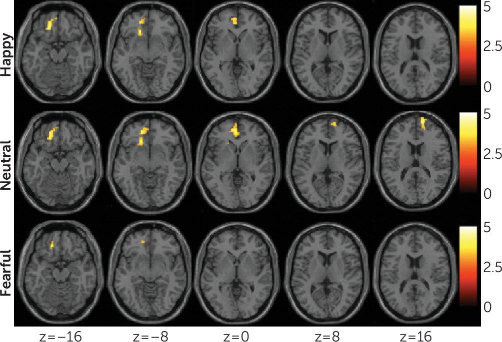
Attempters demonstrated significantly lower functional connectivity than nonattempters from the amygdala to a left ventral prefrontal region, including orbitofrontal, rostral prefrontal, and ventral anterior cingulate areas, in happy and neutral conditions (Table 2, Figure 3). A similar region showed lower connectivity in the fearful condition; however, the group difference was below the AlphaSim spatial extent threshold. There was a region of significantly lower connectivity in the right rostral prefrontal cortex in the neutral condition. Significance for all regions survived to p<0.001, and all regions but the left ventral prefrontal region in the fear condition survived AlphaSim spatial extent thresholding (p<0.05, corrected). No areas showed significantly increased functional connectivity. Results were unrelated to the clinical factors explored.
FIGURE 3. Decreased Functional Connectivity in Adolescents and Young Adults With Bipolar Disorder With, Relative to Those Without, a History of Suicide Attempta
a Axial-oblique images display the regions of decreased functional connectivity from the amygdala, in the group with bipolar disorder with a history of suicide attempts, compared with the group without a history of suicide attempts, to the left ventral prefrontal cortex during happy (top row, p<0.0016, meeting AlphaSim spatial extent threshold for p<0.05, corrected), neutral (middle row, p<0.0016, meeting AlphaSim spatial extent threshold for p<0.05, corrected), and fearful (bottom row, p<0.0016, uncorrected) facial conditions and to the right rostral prefrontal cortex in the neutral facial (second row, right two images, p<0.0016, meeting AlphaSim spatial extent threshold for p<0.05, corrected) condition. The numbers below the images are the Montreal Neurological Institute coordinates (mm) for the z plane. Functional connectivity in the right rostral prefrontal cortex was negatively associated with severity of suicidal ideation (p<0.05). Functional connectivity in the left ventral prefrontal cortex was negatively associated with lethality of most severe attempt in all three facial conditions (p<0.05). The color bar represents the range of t values. The right side of the axial-oblique images is on the right side of the brain.

Associations Between Imaging Modalities and With Behaviors

Bilateral cerebellum gray matter volume was significantly correlated with right uncinate fasciculus fractional anisotropy among attempters only (r=0.53, p=0.008) and with the right and left uncinate among all subjects (radj=0.32, p=0.009, radj=0.28, p=0.027). Among all subjects, right orbitofrontal and bilateral cerebellum gray matter volumes were significantly correlated with right cerebellum fractional anisotropy (radj=0.25, p=0.047, radj=0.29, p=0.022).
Among attempters, suicidal ideation severity was negatively correlated with functional connectivity in the right rostral prefrontal cortex (rs=−0.490, p=0.033). This correlation was not significant in the overall sample adjusted for group. Attempt lethality was negatively correlated with left ventral prefrontal functional connectivity during happy, neutral, and fearful conditions (rs=−0.47, p=0.031; rs=−0.49, p=0.025; and rs=−0.58, p=0.006, respectively).

Healthy Group Comparisons

sMRI.

Right hippocampal volume in the attempter group, but not in the nonattempter group, was significantly lower than that in the healthy comparison group (Dunnett’s p<0.05).

DTI.

Fractional anisotropy in the attempter group, but not in the nonattempter group, was significantly lower than that in the healthy comparison group in the left and right uncinate fasciculus regions (Dunnett’s p<0.05), and this measure fell short of significance (Dunnett’s p=0.06) in the right cerebellum region.

Functional connectivity.

Secondary analyses indicated that functional connectivity in the attempter group, but not in the nonattempter group, was significantly lower than that in the healthy comparison group in the left ventral prefrontal cortex region for the three facial conditions (Dunnett’s p<0.05), and this measure fell short of significance (Dunnett’s p=0.07) in the right rostral prefrontal cortex region in the neutral condition.

Discussion

This study demonstrated reductions in gray matter volume and integrity of structural and functional connections in a frontolimbic system in a sample of individuals with bipolar disorder who have attempted suicide, compared with nonattempters. Decreases were observed in gray matter volumes in the right orbitofrontal cortex and hippocampus, and bilateral cerebellum. Diminished integrity of white matter in the uncinate and ventral frontal and right cerebellum regions, and decreased functional connectivity between the amygdala and left ventral prefrontal and right rostral prefrontal regions, were also observed. Cerebellar gray matter volume was associated with uncinate and cerebellar fractional anisotropy, and orbitofrontal gray matter volume was associated with cerebellar fractional anisotropy. Among attempters, decreases in rostral and ventral prefrontal functional connectivity were associated with severity of suicidal ideation and attempt lethality, respectively.
Gray matter volume reductions associated with suicide attempts in bipolar disorder were in the orbitofrontal cortex, hippocampus, and cerebellum, regions implicated in emotion processing and regulation and in bipolar disorder (27). The orbitofrontal cortex plays a central role in regulating emotion responses, including through its connections to the amygdala, hippocampus, and cerebellum (27). Volume decreases in the orbitofrontal cortex have been reported in adult attempters with major depressive disorder (28). The hippocampus may also influence emotion reactions and regulatory processes, given its role in encoding and recall of the emotional significance of events. Moreover, impaired memory processes have been associated with suicide attempts, implicating the hippocampus (29). Cerebellar connections with the prefrontal cortex and amygdala suggest a role for the cerebellum in emotion regulation, consistent with emotion dysregulation observed with cerebellum lesions (27). Hippocampus and cerebellum volume decreases have been observed in adult attempters (30, 31) but have been studied primarily in attempters with major depression, and some findings are inconsistent (28, 32). We did not detect differences in amygdala volume between the bipolar disorder attempters and nonattempters. Varying findings may be attributed to differences in imaging methodology and subject samples.
White matter findings were in regions of the bilateral uncinate, extending into left ventral frontal regions, and in the right cerebellum. The uncinate provides major connections between the ventral prefrontal cortex and amygdala and therefore is implicated in emotion regulation. In bipolar disorder, white matter orbitofrontal decreases have been reported and were associated with impulsivity (11). The white matter decreases observed herein in attempters included more caudal frontal and temporal areas and were not associated with impulsivity. Right uncinate fractional anisotropy values were lower in association with rapid cycling, a potential risk factor for suicide in bipolar disorder (33), suggesting a link to mood dysregulation. Decreased white matter volumes have been observed in the cerebellum in attempters with major depression (31). The cerebellum finding in attempters with bipolar disorder suggests it may be a structural abnormality associated with attempts across these mood disorders.
Exploratory analyses also provided preliminary evidence for associations between some of the gray and white matter findings. These findings raise the possibility of interactions between the development of gray and white matter in suicide risk. We cannot address this possibility using this study’s data. However, reciprocal relationships between neurons and glia during development may contribute to concomitant gray and white matter developmental changes (3436). It is also possible that gray and white matter associations are the results of other biological processes, as opposed to a direct interaction between their developmental processes. For example, they may reflect responses to common genetic or environmental factors.
Functional connectivity between the amygdala and the ventral prefrontal cortex is critical in emotion regulation and showed significant decreases during the processing of happy and neutral faces, and decreases during the processing of fearful faces that did not reach significance, in attempters compared with nonattempters. Studies suggest emotion regulation deficits in adolescent suicide attempters, and adolescents may attempt suicide in an effort to dampen intolerable emotional states (37). Moreover, adolescents with higher affect dysregulation are more likely to have multiple suicide attempts (38). Previous fMRI studies have shown increased responses to negative emotional faces, specifically angry faces, in frontal circuitry in adolescents (39), similar to adults (2). Depression and suicidal ideation have been associated with altered responses to neutral face stimuli in adults, particularly with projecting a negative bias (40). Together with significant reductions in functional connectivity between the amygdala and ventral prefrontal cortex in the happy condition—and in the fearful condition, although this did not reach significance—the observation regarding the processing of neutral face stimuli suggests that more widespread affective dysregulation may be present in suicide attempters with bipolar disorder, limiting modulation of emotional responses and impairing effective management of affect.
Decreased functional connectivity from the amygdala was also noted in the right rostral prefrontal cortex in the neutral condition, suggested by exploratory analyses to be associated with severity of suicidal ideation. Less is known about the role of the rostral prefrontal cortex in suicidal behavior. The experience of mental pain in attempters during recall of suicidal episodes showed associations with decreased functioning of the rostral prefrontal cortex (41), a region implicated in self-referential and introspective mental functions (42) altered in suicidal ideation and during suicidal acts (43). Frontal dysfunction was observed in a study of adults with suicidal ideation (9). In depressed youths, maladaptive emotion regulation response tendencies increased the likelihood of suicidal ideation, planning, and action, while adaptive regulatory responses decreased suicidal behavior (44). Because the rostral prefrontal cortex is important in the development of regulation of emotions and adaptive response tendencies (45), it is possible that emotion dysregulation, resulting from disruptions in prefrontal system functional connectivity, contributes to suicidal ideation and risk for suicidal behavior.
The magnitude of functional connectivity between the amygdala and the left ventral prefrontal cortex was negatively associated with attempt lethality. Individuals who make high-lethality attempts may be most at risk for suicide death (8). Our finding comports with adult neuroimaging studies demonstrating associations between attempt lethality and ventral prefrontal hypometabolism (8). High lethality has been associated with highly planned, less impulsive attempts (8). Together with the lack of associations between impulsivity and reductions in structural and functional integrity among attempters in this study, this finding suggests that altered ventral prefrontal functioning in attempt lethality may not, at least for some individuals, be related to its role in controlling impulses but may relate to emotion dysregulation. However, impulsivity was assessed at scanning. It is possible that associations with ventral prefrontal alterations may exist at the time of attempt.
For most regions in which attempters differed from nonattempters, attempters, but not nonattempters, also differed significantly from healthy comparison subjects. Although this suggests that the findings may be related to suicidal tendencies, comparisons with individuals with other disorders are needed. It is also possible that the findings are a consequence of bipolar disorder or of suicide attempts. Future longitudinal studies are needed to elucidate the predictive value of these results. Orbitofrontal and cerebellar gray matter volume decreases in healthy comparison subjects relative to suicide attempters did not reach significance. This suggests that gray matter volumes in these regions may better differentiate attempter status within bipolar disorder. Orbitofrontal volume decreases in attempters compared with nonattempters have been reported previously in bipolar disorder (46). Orbitofrontal decreases have also been reported, relative to healthy comparison groups, in attempter groups with major depressive disorder and borderline personality disorder (5). It is possible that aspects of our sample or methods decreased the ability to detect significant differences between attempters and healthy comparison subjects. Cerebellar findings are mixed (5), and additional studies are warranted.
Study limitations include the modest sample size. While no effects in the regions of group differences were detected for gender, medication, substance comorbidity, mood state, or psychosis, the number of participants in the subgroups for the factors assessed was small, limiting power. There were not sufficient numbers of subjects to perform meaningful analyses for subtypes of medications and nonsubstance psychiatric comorbidities. Age range was large, and the sample included pubertal teenagers and young adults. While age was included as a covariate, developmental brain changes could still have affected results. Although suicide attempt and ideation history were assessed systematically, assessments were based on retrospective subject reporting, which could decrease accuracy. Results were reported with a primary threshold of p<0.005, with AlphaSim spatial extent thresholding (p<0.05, corrected), in order to balance the effort to avoid premature strict correction that may limit report of findings that could prove useful in the future to the field, especially as there are rare similar data in the relatively young field of suicide research, with potential report of spurious results. However, caution should be taken in considering the results because lower primary thresholds may lead to spurious findings when utilizing cluster-extent-based thresholds (47). We noted which results survived to a threshold of p<0.001, with and without AlphaSim spatial extent thresholding (p<0.05, corrected), to provide this further information. Correlational analyses should be considered preliminary. Exploratory correlational analyses were not controlled for multiple comparisons. Some associations across imaging modalities and with behaviors were in directions anticipated but did not reach significance, potentially owing to the sample size. Directionality of the DTI acquisition was limited. Additional analyses (e.g., analyses of group differences in activation and/or functional connectivity analyses using psychophysiological interaction analysis methods to further isolate stimulus-related connectivity) could aid in further interpretation of the data. However, as in the previous published work of our group (20, 22) and of other research groups, there can be complex interactions between mood state and the valence of emotional stimuli. Thus, the sample sizes in the present study limit the ability to fully assess further interactions with suicide history, and additional comprehensive analyses are beyond the scope of this article.
In summary, this multimodal structural and functional neuroimaging study of adolescent and young adult suicide attempters with bipolar disorder demonstrates decreases in gray matter and white matter structural and functional connectivity in a ventral frontolimbic neural system that subserves emotion regulation. This study provides preliminary evidence for gray matter and white matter relationships and associations of system features with suicidal ideation and attempt lethality. Integrated study of brain structure and function, and their associations with symptoms and behavior, in suicide research is important in elucidating risk mechanisms that may involve complex, parallel, and interacting relationships between developing circuitry and symptoms and behaviors. Longitudinal studies are needed to more fully investigate development of system features, their role in the transition from suicidal ideation to behavior and risk for completed suicide, and the role of emotion regulation in suicidal behavior in bipolar disorder and whether it generalizes to other disorders.

Acknowledgments

The authors thank Susan Quatrano, B.A., and Philip Markovich, B.A., for their assistance with research; Cheryl Lacadie, B.S., Karen A. Martin, B.A., R.T.(R.)M.R., Terry Hickey, R.T.(R., N.M., M.R.), and Hedy Sarofin, R.T.(R.)M.R., for their technical expertise; and the research subjects for their participation.

References

1.
Abreu LN, Lafer B, Baca-Garcia E, et al: Suicidal ideation and suicide attempts in bipolar disorder type I: an update for the clinician. Rev Bras Psiquiatr 2009; 31:271–280
2.
Jollant F, Lawrence NS, Giampietro V, et al: Orbitofrontal cortex response to angry faces in men with histories of suicide attempts. Am J Psychiatry 2008; 165:740–748
3.
Arango V, Underwood MD, Gubbi AV, et al: Localized alterations in pre- and postsynaptic serotonin binding sites in the ventrolateral prefrontal cortex of suicide victims. Brain Res 1995; 688:121–133
4.
Dwivedi Y: Brain-derived neurotrophic factor and suicide pathogenesis. Ann Med 2010; 42:87–96
5.
Cox Lippard ET, Johnston JAY, Blumberg HP: Neurobiological risk factors for suicide: insights from brain imaging. Am J Prev Med 2014; 47(Suppl 2):S152–S162
6.
Torres-Platas SG, Hercher C, Davoli MA, et al: Astrocytic hypertrophy in anterior cingulate white matter of depressed suicides. Neuropsychopharmacology 2011; 36:2650–2658
7.
Soloff PH, Pruitt P, Sharma M, et al: Structural brain abnormalities and suicidal behavior in borderline personality disorder. J Psychiatr Res 2012; 46:516–525
8.
Oquendo MA, Placidi GPA, Malone KM, et al: Positron emission tomography of regional brain metabolic responses to a serotonergic challenge and lethality of suicide attempts in major depression. Arch Gen Psychiatry 2003; 60:14–22
9.
Marchand WR, Lee JN, Garn C, et al: Striatal and cortical midline activation and connectivity associated with suicidal ideation and depression in bipolar II disorder. J Affect Disord 2011; 133:638–645
10.
Yurgelun-Todd DA, Bueler CE, McGlade EC, et al: Neuroimaging correlates of traumatic brain injury and suicidal behavior. J Head Trauma Rehabil 2011; 26:276–289
11.
Mahon K, Burdick KE, Wu J, et al: Relationship between suicidality and impulsivity in bipolar I disorder: a diffusion tensor imaging study. Bipolar Disord 2012; 14:80–89
12.
Caplan R, Siddarth P, Levitt J, et al: Suicidality and brain volumes in pediatric epilepsy. Epilepsy Behav 2010; 18:286–290
13.
Andreasen NC, Endicott J, Spitzer RL, et al: The family history method using diagnostic criteria: reliability and validity. Arch Gen Psychiatry 1977; 34:1229–1235
14.
Kaufman J, Birmaher B, Brent D, et al: Schedule for Affective Disorders and Schizophrenia for School-Age Children-Present and Lifetime Version (K-SADS-PL): initial reliability and validity data. J Am Acad Child Adolesc Psychiatry 1997; 36:980–988
15.
Oquendo MA, Halberstam B, Mann JJ: Risk factors for suicidal behavior: the utility and limitations of research instruments, in Standardized Evaluation in Clinical Practice. Edited by First MB. Arlington, Va, American Psychiatric Publishing, 2003, pp 103–130
16.
Beck AT, Beck R, Kovacs M: Classification of suicidal behaviors: I: quantifying intent and medical lethality. Am J Psychiatry 1975; 132:285–287
17.
Beck AT, Kovacs M, Weissman A: Assessment of suicidal intention: the Scale for Suicide Ideation. J Consult Clin Psychol 1979; 47:343–352
18.
Beck AT, Weissman A, Lester D, et al: The measurement of pessimism: the hopelessness scale. J Consult Clin Psychol 1974; 42:861–865
19.
Patton JH, Stanford MS, Barratt ES: Factor structure of the Barratt impulsiveness scale. J Clin Psychol 1995; 51:768–774
20.
Wang F, Bobrow L, Liu J, et al: Corticolimbic functional connectivity in adolescents with bipolar disorder. PLoS One 2012; 7:e50177
21.
Wang F, Kalmar JH, Womer FY, et al: Olfactocentric paralimbic cortex morphology in adolescents with bipolar disorder. Brain 2011; 134:2005–2012
22.
Wang F, Kalmar JH, He Y, et al: Functional and structural connectivity between the perigenual anterior cingulate and amygdala in bipolar disorder. Biol Psychiatry 2009; 66:516–521
23.
Friston KJ, Holmes AP, Worsley KJ, et al: Statistical parametric maps in functional imaging: a general linear approach. Hum Brain Mapp 1994; 2:189–210
24.
Shah MP, Wang F, Kalmar JH, et al: Role of variation in the serotonin transporter protein gene (SLC6A4) in trait disturbances in the ventral anterior cingulate in bipolar disorder. Neuropsychopharmacology 2009; 34:1301–1310
25.
Uematsu A, Matsui M, Tanaka C, et al: Developmental trajectories of amygdala and hippocampus from infancy to early adulthood in healthy individuals. PLoS One 2012; 7:e46970
26.
Blumberg HP, Fredericks C, Wang F, et al: Preliminary evidence for persistent abnormalities in amygdala volumes in adolescents and young adults with bipolar disorder. Bipolar Disord 2005; 7:570–576
27.
Blond BN, Fredericks CA, Blumberg HP: Functional neuroanatomy of bipolar disorder: structure, function, and connectivity in an amygdala-anterior paralimbic neural system. Bipolar Disord 2012; 14:340–355
28.
Monkul ES, Hatch JP, Nicoletti MA, et al: Fronto-limbic brain structures in suicidal and non-suicidal female patients with major depressive disorder. Mol Psychiatry 2007; 12:360–366
29.
Richard-Devantoy S, Berlim MT, Jollant F: Suicidal behaviour and memory: a systematic review and meta-analysis. World J Biol Psychiatry 2015; 16:544–566
30.
Colle R, Chupin M, Cury C, et al: Depressed suicide attempters have smaller hippocampus than depressed patients without suicide attempts. J Psychiatr Res 2015; 61:13–18
31.
Hwang JP, Lee TW, Tsai SJ, et al: Cortical and subcortical abnormalities in late-onset depression with history of suicide attempts investigated with MRI and voxel-based morphometry. J Geriatr Psychiatry Neurol 2010; 23:171–184
32.
Baldaçara L, Nery-Fernandes F, Rocha M, et al: Is cerebellar volume related to bipolar disorder? J Affect Disord 2011; 135:305–309
33.
Gao K, Tolliver BK, Kemp DE, et al: Correlates of historical suicide attempt in rapid-cycling bipolar disorder: a cross-sectional assessment. J Clin Psychiatry 2009; 70:1032–1040
34.
Giedd JN: The teen brain: insights from neuroimaging. J Adolesc Health 2008; 42:335–343
35.
Fields RD, Stevens-Graham B: New insights into neuron-glia communication. Science 2002; 298:556–562
36.
Bray S, Krongold M, Cooper C, et al: Synergistic effects of age on patterns of white and gray matter volume across childhood and adolescence. eNeuro 2015; 2:e0003-15.2015
37.
Zlotnick C, Donaldson D, Spirito A, et al: Affect regulation and suicide attempts in adolescent inpatients. J Am Acad Child Adolesc Psychiatry 1997; 36:793–798
38.
Esposito C, Spirito A, Boergers J, et al: Affective, behavioral, and cognitive functioning in adolescents with multiple suicide attempts. Suicide Life Threat Behav 2003; 33:389–399
39.
Pan LA, Hassel S, Segreti AM, et al: Differential patterns of activity and functional connectivity in emotion processing neural circuitry to angry and happy faces in adolescents with and without suicide attempt. Psychol Med 2013; 43:2129–2142
40.
Maniglio R, Gusciglio F, Lofrese V, et al: Biased processing of neutral facial expressions is associated with depressive symptoms and suicide ideation in individuals at risk for major depression due to affective temperaments. Compr Psychiatry 2014; 55:518–525
41.
Reisch T, Seifritz E, Esposito F, et al: An fMRI study on mental pain and suicidal behavior. J Affect Disord 2010; 126:321–325
42.
Gusnard DA, Akbudak E, Shulman GL, et al: Medial prefrontal cortex and self-referential mental activity: relation to a default mode of brain function. Proc Natl Acad Sci USA 2001; 98:4259–4264
43.
Marchand WR: Self-referential thinking, suicide, and function of the cortical midline structures and striatum in mood disorders: possible implications for treatment studies of mindfulness-based interventions for bipolar depression. Depress Res Treat 2012; 2012:246725
44.
Tamás Z, Kovacs M, Gentzler AL, et al: The relations of temperament and emotion self-regulation with suicidal behaviors in a clinical sample of depressed children in Hungary. J Abnorm Child Psychol 2007; 35:640–652
45.
Luna B, Padmanabhan A, O’Hearn K: What has fMRI told us about the development of cognitive control through adolescence? Brain Cogn 2010; 72:101–113
46.
Benedetti F, Radaelli D, Poletti S, et al: Opposite effects of suicidality and lithium on gray matter volumes in bipolar depression. J Affect Disord 2011; 135:139–147
47.
Woo CW, Krishnan A, Wager TD: Cluster-extent based thresholding in fMRI analyses: pitfalls and recommendations. Neuroimage 2014; 91:412–419

Information & Authors

Information

Published In

Go to American Journal of Psychiatry
American Journal of Psychiatry
Pages: 667 - 675
PubMed: 28135845

History

Received: 19 May 2015
Revision received: 18 November 2015
Revision received: 6 July 2016
Revision received: 1 September 2016
Revision received: 28 October 2016
Accepted: 17 November 2016
Published online: 31 January 2017
Published in print: July 01, 2017

Keywords

  1. Suicide
  2. Mood Disorders-Bipolar
  3. Brain Imaging Techniques
  4. Adolescents
  5. MRI
  6. fMRI
  7. DTI

Authors

Details

Jennifer A.Y. Johnston, M.A.
From the Department of Psychiatry and the Child Study Center, Yale School of Medicine, New Haven, Conn.; the Division of Psychology and Language Sciences, University College London; and the Department of Psychiatry, Columbia University Medical Center, New York.
Fei Wang, M.D., Ph.D.
From the Department of Psychiatry and the Child Study Center, Yale School of Medicine, New Haven, Conn.; the Division of Psychology and Language Sciences, University College London; and the Department of Psychiatry, Columbia University Medical Center, New York.
Jie Liu, Ph.D.
From the Department of Psychiatry and the Child Study Center, Yale School of Medicine, New Haven, Conn.; the Division of Psychology and Language Sciences, University College London; and the Department of Psychiatry, Columbia University Medical Center, New York.
Benjamin N. Blond, M.D.
From the Department of Psychiatry and the Child Study Center, Yale School of Medicine, New Haven, Conn.; the Division of Psychology and Language Sciences, University College London; and the Department of Psychiatry, Columbia University Medical Center, New York.
Amanda Wallace, M.D.
From the Department of Psychiatry and the Child Study Center, Yale School of Medicine, New Haven, Conn.; the Division of Psychology and Language Sciences, University College London; and the Department of Psychiatry, Columbia University Medical Center, New York.
Jiacheng Liu, M.D., Ph.D.
From the Department of Psychiatry and the Child Study Center, Yale School of Medicine, New Haven, Conn.; the Division of Psychology and Language Sciences, University College London; and the Department of Psychiatry, Columbia University Medical Center, New York.
Linda Spencer, B.S.
From the Department of Psychiatry and the Child Study Center, Yale School of Medicine, New Haven, Conn.; the Division of Psychology and Language Sciences, University College London; and the Department of Psychiatry, Columbia University Medical Center, New York.
Elizabeth T. Cox Lippard, Ph.D.
From the Department of Psychiatry and the Child Study Center, Yale School of Medicine, New Haven, Conn.; the Division of Psychology and Language Sciences, University College London; and the Department of Psychiatry, Columbia University Medical Center, New York.
Kirstin L. Purves, B.Sc.
From the Department of Psychiatry and the Child Study Center, Yale School of Medicine, New Haven, Conn.; the Division of Psychology and Language Sciences, University College London; and the Department of Psychiatry, Columbia University Medical Center, New York.
Angeli Landeros-Weisenberger, M.D.
From the Department of Psychiatry and the Child Study Center, Yale School of Medicine, New Haven, Conn.; the Division of Psychology and Language Sciences, University College London; and the Department of Psychiatry, Columbia University Medical Center, New York.
Eric Hermes, M.D.
From the Department of Psychiatry and the Child Study Center, Yale School of Medicine, New Haven, Conn.; the Division of Psychology and Language Sciences, University College London; and the Department of Psychiatry, Columbia University Medical Center, New York.
Brian Pittman, M.S.
From the Department of Psychiatry and the Child Study Center, Yale School of Medicine, New Haven, Conn.; the Division of Psychology and Language Sciences, University College London; and the Department of Psychiatry, Columbia University Medical Center, New York.
Sheng Zhang, Ph.D.
From the Department of Psychiatry and the Child Study Center, Yale School of Medicine, New Haven, Conn.; the Division of Psychology and Language Sciences, University College London; and the Department of Psychiatry, Columbia University Medical Center, New York.
Robert King, M.D.
From the Department of Psychiatry and the Child Study Center, Yale School of Medicine, New Haven, Conn.; the Division of Psychology and Language Sciences, University College London; and the Department of Psychiatry, Columbia University Medical Center, New York.
Andrés Martin, M.D., M.P.H.
From the Department of Psychiatry and the Child Study Center, Yale School of Medicine, New Haven, Conn.; the Division of Psychology and Language Sciences, University College London; and the Department of Psychiatry, Columbia University Medical Center, New York.
Maria A. Oquendo, M.D.
From the Department of Psychiatry and the Child Study Center, Yale School of Medicine, New Haven, Conn.; the Division of Psychology and Language Sciences, University College London; and the Department of Psychiatry, Columbia University Medical Center, New York.
Hilary P. Blumberg, M.D. [email protected]
From the Department of Psychiatry and the Child Study Center, Yale School of Medicine, New Haven, Conn.; the Division of Psychology and Language Sciences, University College London; and the Department of Psychiatry, Columbia University Medical Center, New York.

Notes

Address correspondence to Dr. Blumberg ([email protected]).

Funding Information

National Institute of Mental Health10.13039/100000025: R01MH070902, R01MH69747, RC1MH088366, T32MH014276
National Institute on Drug Abuse10.13039/100000026: K25DA040032
American Foundation for Suicide Prevention10.13039/100001455
Brain and Behavior Research Foundation: 10.13039/100000874
International Bipolar Foundation
The John and Hope Furth Endowment
Women's Health Research at Yale
Supported by NIMH grants RC1 MH088366, R01 MH69747, and R01 MH070902 to Dr. Blumberg and T32 MH014276 to Dr. Cox Lippard, and by NIDA grant K25 DA040032 to Dr. Zhang. Also supported by the American Foundation for Suicide Prevention (Dr. Blumberg), by the International Bipolar Disorder Foundation (Dr. Blumberg), by the Brain and Behavior Research Foundation (Dr. Blumberg and Dr. Wang), by Women’s Health Research at Yale (Dr. Blumberg), and by the John and Hope Furth Endowment (Dr. Blumberg).Dr. Oquendo receives royalties for the commercial use of the Columbia Suicide Severity Rating Scale and an honorarium as president of APA, and her family owns stock in Bristol-Myers Squibb. Dr. Martin receives royalties from Wolters Kluwer and an editorial stipend from the American Academy of Child and Adolescent Psychiatry. The other authors report no financial relationships with commercial interests.

Metrics & Citations

Metrics

Citations

Export Citations

If you have the appropriate software installed, you can download article citation data to the citation manager of your choice. Simply select your manager software from the list below and click Download.

For more information or tips please see 'Downloading to a citation manager' in the Help menu.

Format
Citation style
Style
Copy to clipboard

View Options

View options

PDF/EPUB

View PDF/EPUB

Login options

Already a subscriber? Access your subscription through your login credentials or your institution for full access to this article.

Personal login Institutional Login Open Athens login
Purchase Options

Purchase this article to access the full text.

PPV Articles - American Journal of Psychiatry

PPV Articles - American Journal of Psychiatry

Not a subscriber?

Subscribe Now / Learn More

PsychiatryOnline subscription options offer access to the DSM-5-TR® library, books, journals, CME, and patient resources. This all-in-one virtual library provides psychiatrists and mental health professionals with key resources for diagnosis, treatment, research, and professional development.

Need more help? PsychiatryOnline Customer Service may be reached by emailing [email protected] or by calling 800-368-5777 (in the U.S.) or 703-907-7322 (outside the U.S.).

Media

Figures

Other

Tables

Share

Share

Share article link

Share