Abstract

Objective:

New antidepressant treatments are needed that are effective, rapid acting, safe, and tolerable. Intermittent theta-burst stimulation (iTBS) is a noninvasive brain stimulation treatment that has been approved by the U.S. Food and Drug Administration for treatment-resistant depression. Recent methodological advances suggest that the current iTBS protocol might be improved through 1) treating patients with multiple sessions per day at optimally spaced intervals, 2) applying a higher overall pulse dose of stimulation, and 3) precision targeting of the left dorsolateral prefrontal cortex (DLPFC) to subgenual anterior cingulate cortex (sgACC) circuit. The authors examined the feasibility, tolerability, and preliminary efficacy of Stanford Accelerated Intelligent Neuromodulation Therapy (SAINT), an accelerated, high-dose resting-state functional connectivity MRI (fcMRI)–guided iTBS protocol for treatment-resistant depression.

Methods:

Twenty-two participants with treatment-resistant depression received open-label SAINT. fcMRI was used to individually target the region of the left DLPFC most anticorrelated with sgACC in each participant. Fifty iTBS sessions (1,800 pulses per session, 50-minute intersession interval) were delivered as 10 daily sessions over 5 consecutive days at 90% resting motor threshold (adjusted for cortical depth). Neuropsychological testing was conducted before and after SAINT.

Results:

One participant withdrew, leaving a sample size of 21. Nineteen of 21 participants (90.5%) met remission criteria (defined as a score <11 on the Montgomery-Åsberg Depression Rating Scale). In the intent-to-treat analysis, 19 of 22 participants (86.4%) met remission criteria. Neuropsychological testing demonstrated no negative cognitive side effects.

Conclusions:

SAINT, an accelerated, high-dose, iTBS protocol with fcMRI-guided targeting, was well tolerated and safe. Double-blinded sham-controlled trials are needed to confirm the remission rate observed in this initial study.
Depression is the leading cause of disability worldwide, and approximately 800,000 suicides occur each year (13). New antidepressant treatments are needed that are safe, tolerable, rapid acting, durable, and effective.
Repetitive transcranial magnetic stimulation (rTMS) delivered to the left dorsolateral prefrontal cortex (DLPFC) is a noninvasive brain stimulation technique approved by the U.S. Food and Drug Administration (FDA) for treatment-resistant depression (4). rTMS involves passing an electrical current through a magnetic coil placed superficial to the scalp, producing a high-intensity magnetic field that passes through the scalp, skull, and meninges to excite neuronal tissue (5). Repeated high-frequency excitation of the same brain region results in strengthening of synapses through a process known as long-term potentiation (6, 7), causing changes in functional connectivity (6, 8). The mechanism of rTMS on the core depressive symptoms is hypothesized to be mediated in part through indirect inhibitory functional connectivity from the left DLPFC to the subgenual anterior cingulate cortex (sgACC) (811).
A more efficient form of rTMS, known as intermittent theta-burst stimulation (iTBS), recently approved by the FDA, has significantly shortened the duration of rTMS treatment sessions from 37 minutes to 3 minutes (12) and produces equivalent antidepressant responses (13, 14). FDA-approved rTMS and iTBS courses involve daily stimulation sessions (600 iTBS pulses) for 6 weeks, and one trial has demonstrated remission in 32% of patients and response in 49% (13). Studies suggest that the efficacy of iTBS might be improved by accelerated, spaced delivery of stimulation sessions (1518), higher overall pulse doses (1921), and individualized targeting (8, 22).
We investigated the safety, tolerability, and preliminary efficacy of an accelerated high-dose iTBS protocol using functional connectivity MRI (fcMRI)–guided targeting. This protocol involved 5 consecutive days (Monday through Friday) of 10 iTBS sessions per day (1,800 pulses per session, 50-minute intersession intervals) delivered to the region of the left DLPFC that is most functionally anticorrelated with the sgACC in each participant (23). This protocol was termed Stanford Accelerated Intelligent Neuromodulation Therapy (SAINT), to distinguish it from other attempts at accelerating TMS protocols without individualized targeting, 50-minute intersession intervals, or high pulse dose (24, 25). Our initial investigation of SAINT (23) demonstrated efficacy in a small cohort of participants with severe and treatment-refractory depression (these participants are not included in the present study). The study we describe here builds on our initial report by testing SAINT in a larger and more generalizable cohort of participants with treatment-resistant depression to examine the feasibility, safety, and preliminary efficacy of this approach.

Methods

Participants

Participants were required to be currently experiencing a nonpsychotic major depressive episode as part of either major depressive disorder or bipolar II disorder as defined by DSM-5 criteria and to have not responded to at least one antidepressant medication. At the time of screening, participants were required to have a 17-item Hamilton Depression Rating Scale (HAM-D) score ≥20, a negative urine drug screen, and a negative urine pregnancy test if female. Participants were excluded if they had any contraindications to rTMS, such as a history of seizures, metallic implants in the head, cardiac pacemakers, or a neurological disorder. Participants were recruited through the Depression Research Clinic at Stanford University, study advertisements, and clinic referrals.
Twenty-three participants (ages 19–78, 13 female) were recruited for this study. One participant was screened out after enrollment for having a very high motor threshold (>90% machine output) and one participant with a history of multiple prior therapeutic intolerances (anxiety leading to early discontinuation of intravenous ketamine infusions and conventional rTMS) dropped out after the first day of stimulation because of anxiety. This resulted in a final sample of 21 participants (ages 19–78, 12 female). Nineteen participants had a diagnosis of major depressive disorder, and two had a diagnosis of bipolar II disorder currently in a depressive episode (>1 year). Table 1 summarizes participants’ demographic characteristics and treatment history. Participants were required to maintain their antidepressant regimen throughout study enrollment (for medications taken during enrollment, see Table S1 in the online supplement). Six participants were retreated when they no longer met the criterion for remission and again met the study entry criterion (HAM-D score ≥20). Mean time between treatments was 20.5 weeks (SD=6.6).
TABLE 1. Demographic information and treatment history for all participants (N=21) in a study of Stanford Accelerated Intelligent Neuromodulation Therapy for treatment-resistant depressiona
Characteristic or Measure  
 MeanSD
Age (years)44.8617.21
Age at onset of depression (years)21.9013.11
Duration of depression (years)22.9516.30
Number of adequate antidepressant trials (lifetime)b5.863.53
Number of adequate adjunctive medications (lifetime)c1.100.94
Maudsley Staging Method score10.141.96
 N%
Female1257.1
Participants who failed adequate medication trialsb  
 1–2 trials29.5
 3–4 trials733.3
 5–6 trials314.3
 7–10 trials733.3
 >10 trials29.5
Participants who attempted FDA-approved rTMS7d33.3
Participants who attempted ECT00.0
 MeanSD
Baseline clinical measures  
 MADRS34.865.29
 HAM-D, 17-item25.904.79
 HAM-D, 6-item13.902.45
 BDI-II (N=18)28.7811.68
 Suicidal ideation  
  C-SSRS, suicidal ideation subscale (N=19)1.420.96
  HAM-D, item 31.380.67
  MADRS, item 102.380.80
a
BDI-II=Beck Depression Inventory–II; C-SSRS=Columbia-Suicide Severity Rating Scale; FDA=U.S. Food and Drug Administration; HAM-D=Hamilton Depression Rating Scale; MADRS=Montgomery-Åsberg Depression Rating Scale; rTMS=repetitive transcranial magnetic stimulation.
b
Adequate antidepressant trials was defined using the Antidepressant Treatment History Form–Short Form, version 2018.1 (ATHF-SF).
c
Medications were defined as adequate augmentation strategies according to the ATHF-SF.
d
One participant remitted with conventional rTMS; all other participants who attempted conventional rTMS did not respond to it.
All research procedures were conducted in accordance with the ethical standards outlined in the Declaration of Helsinki. The study was approved by the Stanford University Institutional Review Board, and all participants provided written informed consent before taking part in any study procedures.

Functional MRI

Before the stimulation course, each participant had both structural MRI and resting-state functional MRI (fMRI) scans. All MRI scans were acquired using a 3-T GE Discovery MR750 scanner with a 32-channel imaging coil at the Center for Cognitive and Neurobiological Imaging at Stanford University, using a simultaneous multi-slice (SMS) imaging sequence with an acceleration factor of 3 (SMS=3), which collects three equally spaced axial slices simultaneously. A total of 29 sets of three slices, for a total of 87 slices, were collected within each repetition time of 2 seconds. During the 8-minute resting-state scans, participants were instructed to let their minds wander, avoid repetitive thoughts, keep their eyes open, and keep their attention focused on a central fixation point.

Stanford Accelerated Intelligent Neuromodulation Therapy

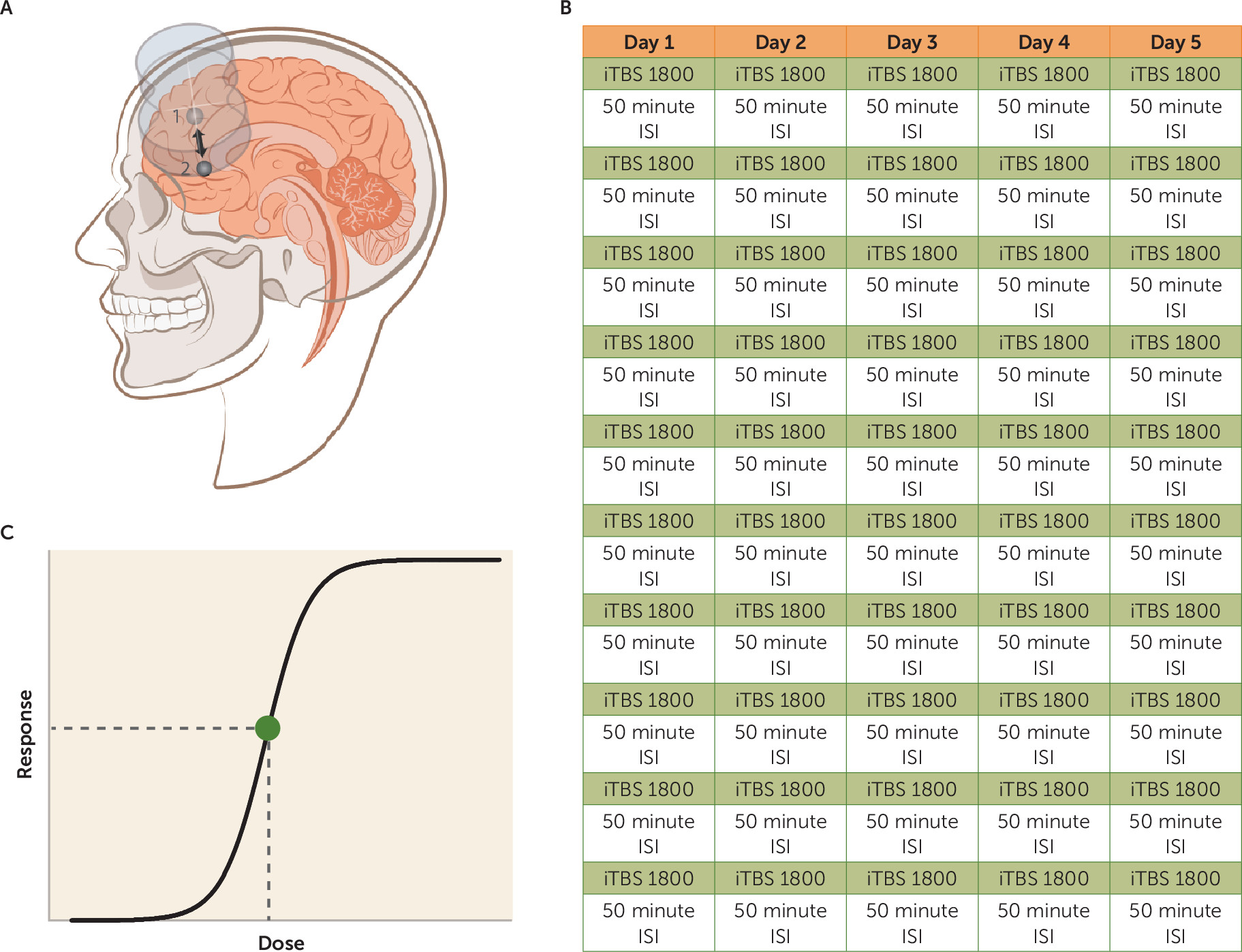
A MagVenture MagPro X100 (MagVenture A/S, Farum, Denmark) system was used to deliver sessions of iTBS; 60 cycles of 10 bursts of three pulses at 50 Hz were delivered in 2-second trains (5 Hz) with an 8-second intertrain interval. Stimulation sessions were delivered hourly (1517). Ten sessions were applied per day (18,000 pulses/day) for 5 consecutive days (90,000 pulses in total). Stimulation was delivered at 90% resting motor threshold (rMT) (13, 26, 27). A depth correction was applied (28) to consistently achieve 90% rMT at the depth of the functional target. For safety, stimulation was never delivered above 120% rMT (13). The Localite Neuronavigation System (Localite GmbH, Sankt Augustin, Germany) was used to position the TMS coil over the individualized stimulation target every session. See Figure 1 for differences between SAINT and the FDA-approved iTBS protocol. In between treatments, participants were seated in a reserved waiting area, which was not occupied with study staff or other patients. This was done both to limit the interaction time with study staff and to prevent a group effect. All iTBS sessions were delivered in the Department of Psychiatry and Behavioral Sciences at Stanford University on an outpatient basis.
FIGURE 1. The three factors that make Stanford Accelerated Intelligent Neuromodulation Therapy (SAINT) different from current FDA-approved iTBS protocolsa
a SAINT utilizes individualized targeting (panel A), accelerated, spaced delivery (panel B), and high pulse dose (both per session [panel B] and overall [panel C]). The intermittent theta-burst stimulation (iTBS) protocol approved by the U.S. Food and Drug Administration (FDA) involves one daily stimulation session of 600 pulses to the left dorsolateral prefrontal cortex (DLPFC) at an intensity of 120% resting motor threshold for 6 weeks. The left DLPFC is located using scalp measurements. In our study, we used functional MRI scans to target the region of the left DLPFC (panel A, area 1) with functional connectivity that was most anticorrelated with the subgenual anterior cingulate cortex (sgACC) (panel A, area 2). This was done because previous neuroimaging findings suggest that the higher the anticorrelation is between the stimulated region of the left DLPFC and the sgACC, the better the clinical outcome (8, 22, 75). Panel B summarizes the 10 sessions per day of 1,800 pulses per session for a total of 18,000 pulses per day (ISI=intersession interval); 1,800 pulses was chosen because it is the only pulse dose that has been explored in a blinded iTBS trial (66). Additionally, 1,800 pulses has been shown to produce long-lasting changes in cortical excitability (76) and optimally produce the intended cellular changes (77). A previous study showed that 61% of nonresponders to repetitive transcranial magnetic stimulation (rTMS) responded with further treatment (20), suggesting that FDA-approved protocols may be underdosing; in panel C, the dotted line depicts where the FDA-approved TMS dose may lie on the dose-response curve. A full 5-day course of our SAINT protocol delivered five times the pulse dose of the FDA-approved protocol. See the online supplement for more information regarding protocol development.

Clinical Assessments

Before and after SAINT, depressive symptoms and suicidal ideation were assessed using clinical and self-report assessments (HAM-D, Montgomery-Åsberg Depression Rating Scale [MADRS], Columbia-Suicide Severity Rating Scale [C-SSRS; suicidal ideation subscale], and Beck Depression Inventory–II [BDI-II]). At the end of each day’s 10 stimulation sessions, depressive symptoms were assessed using the 6-item HAM-D. The Young Mania Rating Scale was completed daily to assess for hypomania (26).
A neuropsychological test battery was administered before and after SAINT to capture any neurocognitive side effects. The Hopkins Verbal Learning Test–Revised (27), the Brief Visuospatial Memory Test–Revised (29), subtests from the Wechsler Adult Intelligence Scale, 4th ed., and several tests from the Delis Kaplan Executive Function System (30) were used. See the online supplement for detailed information about the neuropsychological test battery.

fMRI Analysis for Target Generation

Personalized left DLPFC targets were generated for each participant, using the baseline resting-state scan. All analyses were conducted in the participant’s own brain space. Resting-state scans were preprocessed according to typical methods using the Statistical Parametric Mapping program (SPM12). The resting-state scans were motion corrected and resliced. T1-weighted structural scans were then co-registered with the resting-state scans. Next, the estimation parameters to warp the T1-weighted structural image into Montreal Neurological Institute (MNI) space were calculated using SPM segmentations based on tissue probability maps. These normalization parameters were inverted and applied to MNI space regions of interest for the left DLPFC (Brodmann’s area [BA] 46) and the sgACC (BA25) to map these regions of interest onto the individual participant’s brain. The participant-space regions of interest were then resliced, smoothed, and binarized to match the dimensions of the resting-state scans. See the online supplement for more detailed information regarding fMRI processing.
The participant-space regions of interest for the left DLPFC formed the search area for TMS coil placement. Two separate algorithms were used to determine coil placement. The first algorithm sorted each of the left DLPFC and left and right sgACC voxels into functional subunits using a hierarchical agglomerative clustering algorithm. The voxel time series that most accurately reflected the median time series was then created for each functional subunit, and the correlation coefficients were calculated between all selected time series extracted from all functional subunits of the left DLPFC and sgACC. Median time series were used rather than mean time series, because median values are not susceptible to high signal outliers. The second algorithm determined the optimal left DLPFC subunit to target, based on three factors: the net correlation/anticorrelation of the left DLPFC subunit with sgACC subunits, the size of the subunit, and the spatial concentration of the subunit. See the online supplement for more details on these algorithms. Three-dimensional maps of the whole brain correlation coefficient of the selected left DLPFC subunit were then created and used to target the coil placement, using the Localite TMS Navigation software program. See Figure 2 for the individual target locations.
FIGURE 2. Individual target locations used in this study of Stanford Accelerated Intelligent Neuromodulation Therapy in comparison to the average coordinates for the F3 location in the 10-20 systema
a The average F3 location (at MNI coordinates −35.5, 49.4, 32.4) is shown in blue (78). The colors of the targets represent the percent change in Montgomery-Åsberg Depression Rating Scale score, with dark red indicating greater change. The mean distance from F3 was 25.18 mm (SD=6.15).

Clinical Outcome Analysis

All statistical analyses were conducted using SPSS, version 25 (IBM, Armonk, N.Y.). The level of statistical significance was set at p=0.05. Missing data were not imputed. Statistical analyses were planned independently by two authors (B.S.B. and B.J.) and reviewed by two authors (A.F.S. and N.R.W.).
Our primary outcome measure was change in MADRS score from baseline to immediately after SAINT, and MADRS scores were used to calculate response and remission rates. Reductions in scores on the 17-item and 6-item HAM-D and the BDI-II were used as secondary outcome measures of depression severity. Response was defined as a reduction ≥50% on these scales. Remission was defined as score <11 on the MADRS (31), a score <8 on the 17-item HAM-D (32), a score <5 on the 6-item HAM-D (33), and a score <13 on the BDI-II (34). A floor effect of SAINT treatment was observed across all scales, and initial linear mixed models produced residuals that were not normally distributed, as determined by the Shapiro-Wilk test. Thus, changes in scores on the MADRS, the 6- and 17-item HAM-D, and the BDI-II were assessed with generalized linear mixed models that used a compound symmetry covariance structure, Satterthwaite approximation of degrees of freedom, and robust estimation of coefficients to handle violations of model assumptions. Fixed effects of time, treatment course (initial versus retreatment), and a treatment history of nonresponse to conventional rTMS and their interactions were assessed. All post hoc pairwise comparisons were Bonferroni corrected.
Daily 6-item HAM-D scores were used to calculate the number of days of stimulation required to reach the response criterion (a reduction ≥50% from baseline) and the remission criterion (a score <5). Kaplan-Meier survival analysis using the Breslow test of equality of survival distributions was used to determine whether there were significant differences in the number of days to reach response and remission criteria for participants who had a history of nonresponse to conventional rTMS compared with those who did not.
Suicidality was assessed using the suicidal ideation subscale of the C-SSRS, item 3 of the 17-item HAM-D, and item 10 of the MADRS. Response was defined as a reduction ≥50% in these scores from baseline, and remission was defined as a score of 0. Response was calculated only if the baseline score was >0. Scores were ordinal, and changes in scores were assessed with generalized linear models with a multinomial link, compound symmetry covariance structure, Satterthwaite approximation of degrees of freedom, and robust estimation of coefficients to handle violations of model assumptions.
Scores on the neuropsychological tests before and after SAINT were compared using paired t tests. The data for total score on the Hopkins Verbal Learning Test–Revised, score on the delayed recall trial from the Brief Visuospatial Memory Test–Revised, and the number of rule violations on the tower test from the Delis Kaplan Executive Function System violated the assumption of normality, so Wilcoxon signed-rank tests were used to evaluate SAINT-induced changes in performance on these three measures. Neuropsychological test data were available for 17 participants.

Results

Safety

No serious adverse events occurred. As noted earlier, one participant with a history of multiple therapeutic intolerances (anxiety leading to early discontinuation of intravenous ketamine infusions and conventional rTMS) dropped out after the first day of stimulation because of anxiety. The only side effects reported by other participants were fatigue and some discomfort at both the stimulation site and in the facial muscles during stimulation. The neuropsychological test battery showed no negative cognitive side effects following SAINT. Performance significantly improved on measures of cognitive inhibition (Delis Kaplan Executive Function System color-word inhibition task, t=4.92, df=16, p<0.001; d=1.19; and color-word inhibition switching task, t=3.77, df=16, p=0.002, d=0.91). These improvements survived correction for multiple comparisons (Bonferroni-corrected significance level, p<0.004). There were no significant changes on any of the other neurocognitive tasks (see the online supplement).

Depression Symptoms

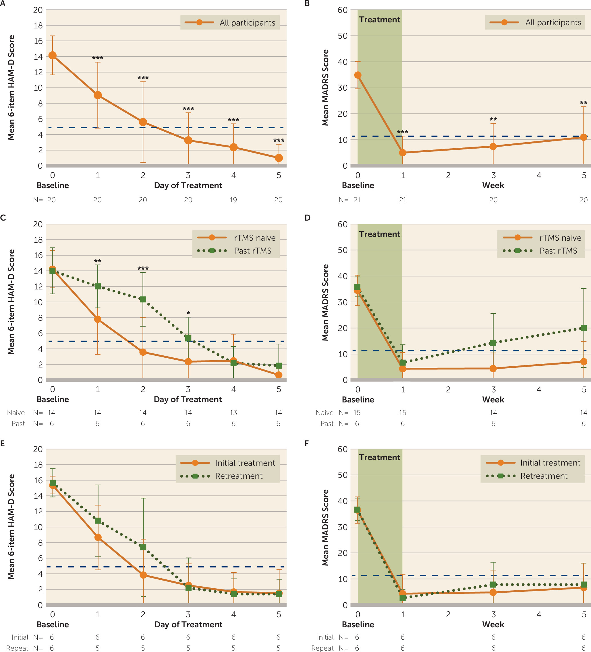
Generalized linear mixed-model analysis revealed a significant effect of day on mean 6-item HAM-D scores (F=62.70, df=5, 43, p<0.001) (Figure 3A) and a significant effect of week (F=90.42, df=3, 9, p<0.001) (Figure 3B) on mean MADRS scores, with scores at all follow-up time points being significantly lower than at baseline (Bonferroni-corrected pairwise comparisons, p<0.01). These results were recapitulated for the 17-item HAM-D (F=51.77, df=3, 12, p<0.001) and the BDI-II (F=19.04, df=3, 19, p<0.001). The response rate (a reduction ≥50% from baseline in MADRS score) was 90.48%, and all responders were in remission after SAINT (MADRS score <11). In the intent-to-treat analysis, 19 of 22 participants (86.4%) met remission criteria (see Table S2 in the online supplement).
FIGURE 3. Changes in depression score during and after Stanford Accelerated Intelligent Neuromodulation Therapy (SAINT) in participants with treatment-resistant depressiona
a Panel A shows the mean score on the 6-item Hamilton Depression Rating Scale (HAM-D) with each day of SAINT for all participants. Panel B shows the mean Montgomery-Åsberg Depression Rating Scale (MADRS) for all participants at baseline (week 0), just after SAINT (week 1), and 2 and 4 weeks after SAINT (weeks 3 and 5). Panels C and D show daily 6-item HAM-D scores and weekly MADRS scores, respectively, separated by participants with and without past treatment with repetitive transcranial magnetic stimulation (rTMS). For the six participants who were retreated with the same SAINT protocol after they relapsed to depression and again met study entry criteria, panels E and F show daily 6-item HAM-D scores and weekly MADRS scores, respectively, for the initial treatment and retreatment. During the 5 days of SAINT treatment, the daily mean HAM-D scores were equivalent between initial treatment and retreatment. The mean MADRS scores at baseline (week 0) and immediately after SAINT (week 1) were equivalent between initial treatment and retreatment. This effect remained equivalent 2 and 4 weeks after treatment (weeks 3 and 5). The dashed horizontal lines in all panels indicate remission criteria, and error bars indicate standard deviation. For panels A and B, significance is compared with baseline; for panels C–F, significance is between groups. Indicated p values are Bonferroni corrected.
*p<0.05. **<0.01. ***<0.001.
Results were similar across all clinical assessments (Table 2). One month after SAINT, 70% of participants continued to meet response criteria (see Table 2 for response and remission rates at 1 month).
TABLE 2. Clinical assessment scores for participants immediately after and 1 month after Stanford Accelerated Intelligent Neuromodulation Therapy (SAINT) for treatment-resistant depressiona
 Post-SAINTOne Month Post-SAINT
MeasureMeanSDNResponse (%)NRemission (%)NMeanSDNResponse (%)NRemission (%)N
MADRS5.006.372190.482190.482110.9511.762070.002060.0020
HAM-D, 17-item4.294.432190.482180.95218.058.312075.002065.0020
HAM-D, 6-item2.243.102185.712185.71214.404.722075.002070.0020
BDI-II4.475.7615100.001293.331512.2513.061657.141462.5016
Suicidal ideation              
 C-SSRSb0.000.0018100.0014100.00180.000.0019100.0014100.0019
 HAM-D, item 30.050.2221100.001995.24210.100.3120100.001890.0020
 MADRS, item 100.100.442195.242195.24210.350.752090.002080.0020
a
Response was defined as a reduction ≥50% in score from baseline; remission was defined as a score <8 on the 17-item HAM-D (32), a score <5 on the 6-item HAM-D (33), a score <11 on the MADRS (31), a score <13 on the BDI-II (34), and a score of zero on the C-SSRS (74). Data for the intent-to-treat sample are presented in Table S2 in the online supplement. BDI-II=Beck Depression Inventory–II; C-SSRS=Columbia-Suicide Severity Rating Scale; HAM-D=Hamilton Depression Rating Scale; MADRS=Montgomery-Åsberg Depression Rating Scale.
b
Suicidal ideation subscale.
A hypothesis of SAINT is that conventional rTMS delivers insufficient cumulative stimulation to induce response and remission from depression for some patients. We tested this in part by including participants who had a history of nonresponse to conventional rTMS (rTMS nonresponders, N=6). When comparing the nonresponder group to the rest of the participants, we found that conventional rTMS nonresponders had similar 6-item HAM-D (Figure 3C) and MADRS (Figure 3D) scores at baseline. Generalized linear mixed-model analysis revealed a main effect of group (F=7.85, df=1, 23, p=0.010) and a group-by-treatment day interaction (F=4.45, df=5, 43, p=0.002), with Bonferroni-corrected post hoc comparisons demonstrating significantly higher 6-item HAM-D scores among participants with a prior history of TMS nonresponse on treatment days 1, 2, and 3 (Figure 3C). Although TMS nonresponders had greater mean MADRS scores at every follow-up time point after SAINT, neither the main effect of group (F=5.67, df=1, 4, p=0.072) nor the group-by-time interaction reached statistical significance (F=1.08, df=3, 9, p=0.405) (Figure 3D), indicating that participants with a history of conventional rTMS nonresponse took more time to respond but ultimately had a treatment effect similar to that of the other participants.

Days of Treatment Until Response and Remission

The mean number of days of SAINT completed until participants met the response criterion (a reduction ≥50% from baseline in 6-item HAM-D score) was 2.30 (SD=1.13; ∼23 10-minute treatments; N=20; daily 6-item HAM-D scores missing for one participant), and the mean number of days to achieve remission (6-item HAM-D score <5) was 2.63 (SD=1.21; ∼26 10-minute treatments; N=19; one participant did not achieve remission by the 6-item HAM-D criterion).
Kaplan-Meier survival analysis revealed that participants who had previously not responded to a 6-week rTMS treatment course (N=6) required more days of treatment to achieve the responder criterion (χ2=4.36, p=0.037; mean=3.00 days, SD=0.63; ∼30 10-minute treatments), and this approached statistical significance for remission criterion as well (χ2= 3.56, p=0.057; mean=3.20 days, SD=0.84; ∼32 10-minute treatments; N=5; one participant did not meet criteria for remission).

Suicidality Scales

Of the 21 participants in the per-protocol analysis, 19 reported some degree of suicidality at the time of screening on the C-SSRS, 20 reported suicidality on item 3 of the 17-item HAM-D, and all 21 reported suicidality on item 10 of the MADRS. Changes in suicidality scale scores were assessed with a generalized linear model with a multinomial link. After SAINT, there were significant reductions in the C-SSRS (χ2= 16.40, df=1, p<0.001), item 3 of the 17-item HAM-D (χ2= 31.06, df=3, p<0.001), and item 10 of the MADRS (χ2=46.86, df=3, p<0.001) at all follow-up time points (chi-square tests, p values <0.001) (see Table 2). One month after SAINT, 80%−100% of participants remained in remission on these measures (see Table 2).

Retreatment Efficacy

Six participants were retreated after they no longer met the remission criterion and again met study entry criteria (the mean time between treatments was 20.5 weeks, SD=6.6). There were no significant differences in daily 6-item HAM-D scores (generalized linear mixed model, F=2.60, df=1, 18, p=0.124) (Figure 3E) or in weekly MADRS scores between treatment courses (F=0.00, df=1, 65, p=0.985) (Figure 3F). There were no treatment-by-time interactions (MADRS: F=2.25, df=3, 98, p=0.087; 6-item HAM-D: F=0.91, df=5, 59, p=0.481). Baseline and follow-up daily 6-item HAM-D and weekly MADRS scores were all similar between treatment courses (Bonferroni-corrected pairwise comparisons, p>0.05). See Figures 3E–F and Table S4 in the online supplement for MADRS scores for initial SAINT and retreatment for each participant.

Discussion

The aim of this study was to examine the safety, feasibility, and preliminary efficacy of an accelerated, high-dose, fcMRI-guided iTBS treatment protocol (SAINT) for treatment-resistant depression. We found that SAINT significantly reduced depressive symptoms and suicidal ideation in patients with treatment-resistant depression within 5 days, without negative cognitive side effects. The remission rate we observed is higher than reported open-label remission rates for standard FDA-approved rTMS protocols (37%) (13, 35, 36), ECT (∼48%) (37), and ketamine (31%) (38) for treating treatment-resistant depression (see Table S5 in the online supplement). The difference between the observed remission rate for SAINT and remission rates reported for standard rTMS protocols may be due to the spaced stimulation sessions, accelerated delivery, high pulse dose, individualized targeting, higher sham effect, or a combination of these. The individual contribution of each of these elements cannot be determined from this study, as they were not investigated separately. Double-blinded trials will be needed to determine the contribution of the sham effect.
The 50-minute spacing in our SAINT protocol complements evidence from basic neuroscience research and human physiology data, which suggest that multiple, spaced daily iTBS sessions have an enhanced effect compared with the same number of single daily sessions (1517, 3942). Multiple, spaced sessions have also been shown to produce accumulating nonlinear improvements in clinical symptoms (41, 43). The duration of intersession intervals is likely to be important, as stimulation sessions with intersession intervals of 50–90 minutes have been shown to have a cumulative effect on synaptic strengthening, whereas sessions with intersession intervals of 40 minutes or less do not show this cumulative effect (1517, 44). Similarly, some studies have shown that two theta-burst stimulation sessions delivered to the motor cortex 15 minutes apart do not increase cortical excitability compared with a single session (42, 45). Finally, left DLPFC activity has been shown to be correlated with sgACC activation 10 minutes after rTMS; this correlation was reduced 27 minutes after rTMS, and the correlation between these regions reached its nadir 45 minutes after rTMS (10), the hypothetically optimal time for stimulation. Taken together, these data could explain the limited response rate (39%) of a previously reported accelerated iTBS stimulation protocol for depression (24, 25), which used an intersession interval of 15 minutes, with 20 sessions delivered over 4 days. In comparison, a previous study utilizing intervals of 1 hour between sessions (46) obtained a similar response rate (43%) after only 15 sessions of conventional rTMS treatment over 2 days. However, the study with 15-minute intersession intervals was a randomized controlled trial, so the response rates cannot be directly compared with ours in the present study.
The functional connectivity–guided targeting method used in our study may have contributed to the 90% remission rate we observed. The left DLPFC is a large brain area that consists of several subregions, some of which are correlated and some anticorrelated with sgACC activity (47). Recent work suggests that these correlated and anticorrelated subregions are part of different affective circuits; stimulating a subregion of the left DLPFC that is anticorrelated with the sgACC reduces melancholic symptoms, resulting in lower MADRS score (48, 49). In contrast, targeting a left DLPFC subregion correlated with the sgACC reduces anxiosomatic symptoms and results in reduced anxiety ratings (48). The anxiosomatic target is within range of the 5-cm rule coil position, whereas the melancholic target is more anterior and lateral within BA46 (48). Our targeting approach was in broad agreement with the target identified to produce maximum clinical change in previous studies (22), which has been demonstrated to be centered in BA46 (8). However, we further extend this strategy by parsing the BA46 region into functional subunits based on the degree of correlation/anticorrelation with the functional subunits of the sgACC (23).
The individualization of our targeting approach may also be important; a trial in healthy individuals showed that stimulating the left DLPFC using personalized functional connectivity–guided targeting induced the desired change in functional connectivity between the left DLPFC and the sgACC (10). A recent interleaved TMS-fMRI study showed that when using individualized functional connectivity–guided targeting, stimulation propagated from the left DLPFC to the sgACC in all participants (50). In comparison, a separate study defined the left DLPFC anatomically (border of BA9/BA46) and stimulation propagated to the sgACC in only 44% of participants (51). By stimulating the subregion of the left DLPFC that is most anticorrelated with the sgACC in each individual, we may have reduced variability in signal propagation to the intended brain target as well as improved treatment efficacy of the core depressive symptoms (8, 48). Additional studies are needed to determine how important individualized fcMRI-guided targeting methods are relative to fcMRI-guided methods based on group-average fMRI data. Clinical studies are needed to directly compare remission rates following iTBS/rTMS protocols with and without individualized targeting.
Our SAINT protocol administered five times the overall pulse dose of the FDA-approved iTBS protocol, as well as a higher density of stimulation (90,000 pulses in 5 days, compared with the standard 18,000 iTBS pulses in 6 weeks [13]). Previous studies found that 61% of individuals who do not respond to an rTMS treatment course responded with additional rTMS treatment sessions (20) and that higher overall pulse doses are associated with higher efficacy (52, 53). A recent report demonstrated nonasymptotic negative linear relationships between the number of rTMS treatments and depression symptom scores (54). This suggests that higher overall pulse doses might further reduce depression symptoms. The apparent need for higher pulse dose is consistent with deep brain stimulation in other neuropsychiatric disorders, where ∼500,000 pulses of stimulation are delivered each day (55). Our SAINT protocol applies an amount of stimulation equivalent to a 6-week standard iTBS treatment protocol (18,000 pulses) each day of stimulation (13). Thirty percent of participants in our study met response criteria after the first day of stimulation (N=6/20; daily 6-item HAM-D scores missing for one participant), which is equivalent to response rates for iTBS/rTMS for this treatment resistance level (5660). None of the nonresponders to prior rTMS in our study responded after a single day of SAINT (see Figure 3), whereas 83% of these prior rTMS nonresponders did respond by the end of the 5-day protocol. Our study administered the highest number of TMS pulses per day and the highest overall TMS pulse dose of any study we are aware of. It is possible that standard FDA-approved rTMS protocols may benefit from higher overall pulse doses.
Prior rTMS nonresponders in our study required more stimulation sessions to induce a clinically significant response. It is possible that depressed individuals with a higher degree of treatment resistance display neuroplasticity impairments (61). Thus, highly treatment-resistant individuals may require a higher pulse dose to induce an antidepressant response, and individuals with the highest degree of treatment resistance may require maintenance iTBS therapy (62) or even an implanted cortical stimulator (63, 64) to induce and sustain antidepressant response (20).
Our study has several limitations, including a small sample size and an open-label design. The small sample size in our study means that the treatment effect may have been influenced by unknown factors related to sampling biases, which emphasizes the need for larger double-blinded sham-controlled trials. Moreover, without a sham-control group we cannot rule out the possibility that our results are primarily due to sham effect; a multicenter rTMS trial in veterans with treatment-resistant depression found equivalent remission rates in the active (40.7%) and sham (37.4%) groups (65). The sham effect for SAINT may be particularly high because of the frequency of the treatment sessions (10 per day) and the perceived novelty of the method. However, a previous study found that individuals with high treatment refractoriness, like many of the participants in this study, showed no sham response to iTBS sessions of 1,800 pulses (13, 66), and an observational study monitoring 124 individuals with treatment-resistant depression receiving treatment as usual (medications, psychotherapy, and ECT) showed a 3.6% remission rate after 1 year of treatment (67), demonstrating the low incidence of spontaneous remission in this population. The remission rate observed in this study is higher than rates reported in previous open-label interventions for treatment-resistant depression (see Table S5 in the online supplement). Double-blinded sham-controlled trials are needed to determine the contribution of the sham effect to the high remission rate observed for SAINT.
Further methodological uncertainties include stimulation of a single brain region (68), fixed stimulation frequencies (54, 69), fixed intersession intervals (69, 70), and the lack of state-dependent stimulation (71). Individualized stimulation frequencies may result in quicker and more durable responses (69, 72), and different cortical excitability profiles may require different intersession intervals (70, 73). Finally, recent studies have shown that applying stimulation in particular brain states using real-time EEG-triggered TMS can increase cortical responses to stimulation (71).
In conclusion, SAINT, our high-dose, accelerated, fcMRI-guided iTBS protocol, is preliminarily safe, feasible, and associated with a high rate of remission from depression. The potential efficacy of SAINT in treating suicidal ideation and the short duration of the protocol suggest that SAINT could provide a means of rapidly ensuring the safety of suicidal patients. However, larger, double-blinded, sham-controlled trials are required to confirm the results from this initial study. We remain cautious in our enthusiasm about the present results, as this is an open-label study with all the shortcomings associated with uncontrolled studies.
Footnotes

Dr. Cole, Ms. Stimpson, and Dr. Bentzley share first authorship. Dr. Sudheimer and Dr. Williams share senior authorship.
ClinicalTrials.gov identifier: NCT03240692.

Supplementary Material

File (appi.ajp.2019.19070720.ds001.pdf)

References

1.
Reddy MS: Depression: the disorder and the burden. Indian J Psychol Med 2010; 32:1–2
3.
Friedrich MJ: Depression is the leading cause of disability around the world. JAMA 2017; 317:1517
4.
George MS, Taylor JJ, Short EB: The expanding evidence base for rTMS treatment of depression. Curr Opin Psychiatry 2013; 26:13–18
5.
Hallett M: Transcranial magnetic stimulation and the human brain. Nature 2000; 406:147–150
6.
Huerta PT, Volpe BT: Transcranial magnetic stimulation, synaptic plasticity, and network oscillations. J Neuroeng Rehabil 2009; 6:7
7.
Ogiue-Ikeda M, Kawato S, Ueno S: The effect of repetitive transcranial magnetic stimulation on long-term potentiation in rat hippocampus depends on stimulus intensity. Brain Res 2003; 993:222–226
8.
Fox MD, Buckner RL, White MP, et al: Efficacy of transcranial magnetic stimulation targets for depression is related to intrinsic functional connectivity with the subgenual cingulate. Biol Psychiatry 2012; 72:595–603
9.
Kito S, Hasegawa T, Takamiya A, et al: Transcranial magnetic stimulation modulates resting EEG functional connectivity between the left dorsolateral prefrontal cortex and limbic regions in medicated patients with treatment-resistant depression. J Neuropsychiatry Clin Neurosci 2017; 29:155–159
10.
Singh A, Erwin-Grabner T, Sutcliffe G, et al: Intermittent theta burst stimulation at personalized targets reduces the functional connectivity of the default mode network in healthy subjects. bioRxiv, May 24, 2019 (https://doi.org/10.1101/646265)
11.
Padmanabhan JL, Cooke D, Joutsa J, et al: A human depression circuit derived from focal brain lesions. Biol Psychiatry 2019; 86:749–758
12.
Huang YZ, Rothwell JC: The effect of short-duration bursts of high-frequency, low-intensity transcranial magnetic stimulation on the human motor cortex. Clin Neurophysiol 2004; 115:1069–1075
13.
Blumberger DM, Vila-Rodriguez F, Thorpe KE, et al: Effectiveness of theta burst versus high-frequency repetitive transcranial magnetic stimulation in patients with depression (THREE-D): a randomised non-inferiority trial. Lancet 2018; 391:1683–1692
14.
Blumberger DM, Vila-Rodriguez F, Dunlop K, et al: Intermittent theta-burst versus 10 Hz left dorsolateral prefrontal rTMS for treatment resistant depression: preliminary results from a two-site, randomized, single blind non-inferiority trial (abstract). Brain Stimul 2015; 8:329
15.
Smolen P, Zhang Y, Byrne JH: The right time to learn: mechanisms and optimization of spaced learning. Nat Rev Neurosci 2016; 17:77–88
16.
Kramár EA, Babayan AH, Gavin CF, et al: Synaptic evidence for the efficacy of spaced learning. Proc Natl Acad Sci USA 2012; 109:5121–5126
17.
Lynch G, Kramár EA, Babayan AH, et al: Differences between synaptic plasticity thresholds result in new timing rules for maximizing long-term potentiation. Neuropharmacology 2013; 64:27–36
18.
Thomson AC, de Graaf TA, Kenis G, et al: No additive meta plasticity effects of accelerated iTBS with short inter-session intervals. Brain Stimul 2019; 12:1301–1303
19.
George MS, Wassermann EM, Kimbrell TA, et al: Mood improvement following daily left prefrontal repetitive transcranial magnetic stimulation in patients with depression: a placebo-controlled crossover trial. Am J Psychiatry 1997; 154:1752–1756
20.
Yip AG, George MS, Tendler A, et al: 61% of unmedicated treatment resistant depression patients who did not respond to acute TMS treatment responded after four weeks of twice weekly deep TMS in the Brainsway pivotal trial. Brain Stimul 2017; 10:847–849
21.
Perera T, George MS, Grammer G, et al: The Clinical TMS Society consensus review and treatment recommendations for TMS therapy for major depressive disorder. Brain Stimul 2016; 9:336–346
22.
Weigand A, Horn A, Caballero R, et al: Prospective validation that subgenual connectivity predicts antidepressant efficacy of transcranial magnetic stimulation sites. Biol Psychiatry 2018; 84:28–37
23.
Williams NR, Sudheimer KD, Bentzley BS, et al: High-dose spaced theta-burst TMS as a rapid-acting antidepressant in highly refractory depression. Brain 2018; 141:e18
24.
Desmyter S, Duprat R, Baeken C, et al: Accelerated intermittent theta burst stimulation for suicide risk in therapy-resistant depressed patients: a randomized, sham-controlled trial. Front Hum Neurosci 2016; 10:480
25.
Duprat R, Desmyter S, Rudi R, et al: Accelerated intermittent theta burst stimulation treatment in medication-resistant major depression: a fast road to remission? J Affect Disord 2016; 200:6–14
26.
Ozten E, Sayar GH, Karamustafalioglu O: Hypomanic shift observed during rTMS treatment of patients with unipolar depressive disorder: four case reports. Ann Gen Psychiatry 2013; 12:12
27.
O’Neil-Pirozzi TM, Goldstein R, Strangman GE, et al: Test-re-test reliability of the Hopkins Verbal Learning Test–Revised in individuals with traumatic brain injury. Brain Inj 2012; 26:1425–1430
28.
Stokes MG, Chambers CD, Gould IC, et al: Simple metric for scaling motor threshold based on scalp-cortex distance: application to studies using transcranial magnetic stimulation. J Neurophysiol 2005; 94:4520–4527
29.
Benedict RHB, Schretlen D, Groninger L, et al: Revision of the Brief Visuospatial Memory Test: studies of normal performance, reliability, and validity. Psychol Assess 1996; 8:145–153
30.
Delis DC, Kramer JH, Kaplan E, et al: Reliability and validity of the Delis-Kaplan Executive Function System: an update. J Int Neuropsychol Soc 2004; 10:301–303
31.
Zimmerman M, Posternak MA, Chelminski I: Derivation of a definition of remission on the Montgomery-Asberg depression rating scale corresponding to the definition of remission on the Hamilton rating scale for depression. J Psychiatr Res 2004; 38:577–582
32.
Leucht S, Fennema H, Engel R, et al: What does the HAMD mean? J Affect Disord 2013; 148:243–248
33.
Timmerby N, Andersen JH, Søndergaard S, et al: A systematic review of the clinimetric properties of the 6-item version of the Hamilton Depression Rating Scale (HAM-D6). Psychother Psychosom 2017; 86:141–149
34.
Schulte-van Maaren YWM, Carlier IV, Zitman FG, et al: Reference values for major depression questionnaires: the Leiden Routine Outcome Monitoring Study. J Affect Disord 2013; 149:342–349
35.
Carpenter LL, Janicak PG, Aaronson ST, et al: Transcranial magnetic stimulation (TMS) for major depression: a multisite, naturalistic, observational study of acute treatment outcomes in clinical practice. Depress Anxiety 2012; 29:587–596
36.
Janicak PG, Nahas Z, Lisanby SH, et al: Durability of clinical benefit with transcranial magnetic stimulation (TMS) in the treatment of pharmacoresistant major depression: assessment of relapse during a 6-month, multisite, open-label study. Brain Stimul 2010; 3:187–199
37.
Heijnen WT, Birkenhäger TK, Wierdsma AI, et al: Antidepressant pharmacotherapy failure and response to subsequent electroconvulsive therapy: a meta-analysis. J Clin Psychopharmacol 2010; 30:616–619
38.
Lener MS, Kadriu B, Zarate CA Jr: Ketamine and beyond: investigations into the potential of glutamatergic agents to treat depression. Drugs 2017; 77:381–401
39.
Harris KM, Kater SB: Dendritic spines: cellular specializations imparting both stability and flexibility to synaptic function. Annu Rev Neurosci 1994; 17:341–371
40.
Tse NY, Goldsworthy MR, Ridding MC, et al: The effect of stimulation interval on plasticity following repeated blocks of intermittent theta burst stimulation. Sci Rep 2018; 8:8526
41.
Goldsworthy MR, Pitcher JB, Ridding MC: The application of spaced theta burst protocols induces long-lasting neuroplastic changes in the human motor cortex. Eur J Neurosci 2012; 35:125–134
42.
Nettekoven C, Volz LJ, Kutscha M, et al: Dose-dependent effects of theta burst rTMS on cortical excitability and resting-state connectivity of the human motor system. J Neurosci 2014; 34:6849–6859
43.
Nyffeler T, Cazzoli D, Hess CW, et al: One session of repeated parietal theta burst stimulation trains induces long-lasting improvement of visual neglect. Stroke 2009; 40:2791–2796
44.
Rogasch NC, Daskalakis ZJ, Fitzgerald PB: Mechanisms underlying long-interval cortical inhibition in the human motor cortex: a TMS-EEG study. J Neurophysiol 2013; 109:89–98
45.
Chung SW, Rogasch NC, Hoy KE, et al: The effect of single and repeated prefrontal intermittent theta burst stimulation on cortical reactivity and working memory. Brain Stimul 2018; 11:566–574
46.
Holtzheimer PE 3rd, McDonald WM, Mufti M, et al: Accelerated repetitive transcranial magnetic stimulation for treatment-resistant depression. Depress Anxiety 2010; 27:960–963
47.
Hoshi E: Functional specialization within the dorsolateral prefrontal cortex: a review of anatomical and physiological studies of non-human primates. Neurosci Res 2006; 54:73–84
48.
Siddiqi S, Taylor S, Cooke D, et al: Distinct symptom-specific treatment targets for antidepressant neuromodulation (abstract). Brain Stimul 2019; 12:537
49.
Siddiqi SH, Taylor SF, Cooke D, et al: Distinct symptom-specific treatment targets for circuit-based neuromodulation. Am J Psychiatry (Epub ahead of print, March 12, 2020)
50.
Oathes DJ, Zimmerman J, Duprat R, et al: Individualized non-invasive brain stimulation engages the subgenual anterior cingulate and amygdala. bioRxiv, December 21, 2018 (https://doi.org/10.1101/503441)
51.
Vink JJT, Mandija S, Petrov PI, et al: A novel concurrent TMS-fMRI method to reveal propagation patterns of prefrontal magnetic brain stimulation. Hum Brain Mapp 2018; 39:4580–4592
52.
Kaster TS, Daskalakis ZJ, Noda Y, et al: Efficacy, tolerability, and cognitive effects of deep transcranial magnetic stimulation for late-life depression: a prospective randomized controlled trial. Neuropsychopharmacology 2018; 43:2231–2238
53.
Jorge RE, Robinson RG, O’Brien J: Top cited papers in international psychogeriatrics, 5: a controlled study of repetitive transcranial magnetic stimulation as a treatment of depression in the elderly. Int Psychogeriatr 2009; 21:855–860
54.
Kaster TS, Downar J, Vila-Rodriguez F, et al: Trajectories of response to dorsolateral prefrontal rTMS in major depression: a THREE-D study. Am J Psychiatry 2019; 176:367–375
55.
Williams NR, Okun MS: Deep brain stimulation (DBS) at the interface of neurology and psychiatry. J Clin Invest 2013; 123:4546–4556
56.
Fregni F, Marcolin MA, Myczkowski M, et al: Predictors of antidepressant response in clinical trials of transcranial magnetic stimulation. Int J Neuropsychopharmacol 2006; 9:641–654
57.
George MS, Lisanby SH, Avery D, et al: Daily left prefrontal transcranial magnetic stimulation therapy for major depressive disorder: a sham-controlled randomized trial. Arch Gen Psychiatry 2010; 67:507–516
58.
Levkovitz Y, Isserles M, Padberg F, et al: Efficacy and safety of deep transcranial magnetic stimulation for major depression: a prospective multicenter randomized controlled trial. World Psychiatry 2015; 14:64–73
59.
O’Reardon JP, Solvason HB, Janicak PG, et al: Efficacy and safety of transcranial magnetic stimulation in the acute treatment of major depression: a multisite randomized controlled trial. Biol Psychiatry 2007; 62:1208–1216
60.
Hsu JH, Downar J, Vila-Rodriguez F, et al: Impact of prior treatment on remission with intermittent theta burst versus high-frequency repetitive transcranial magnetic stimulation in treatment resistant depression. Brain Stimul 2019; 12:1553–1555
61.
Pittenger C, Duman RS: Stress, depression, and neuroplasticity: a convergence of mechanisms. Neuropsychopharmacology 2008; 33:88–109
62.
Pridmore S, Erger S, Rybak M, et al: Early relapse (ER) transcranial magnetic stimulation (TMS) in treatment resistant major depression. Brain Stimul 2018; 11:1098–1102
63.
Williams NR, Short EB, Hopkins T, et al: Five-year follow-up of bilateral epidural prefrontal cortical stimulation for treatment-resistant depression. Brain Stimul 2016; 9:897–904
64.
Williams NR, Bentzley BS, Hopkins T, et al: Optimization of epidural cortical stimulation for treatment-resistant depression. Brain Stimul 2018; 11:239–240
65.
Yesavage JA, Fairchild JK, Mi Z, et al: Effect of repetitive transcranial magnetic stimulation on treatment-resistant major depression in US veterans: a randomized clinical trial. JAMA Psychiatry 2018; 75:884–893
66.
Li CT, Chen MH, Juan CH, et al: Efficacy of prefrontal theta-burst stimulation in refractory depression: a randomized sham-controlled study. Brain 2014; 137:2088–2098
67.
Dunner DL, Rush AJ, Russell JM, et al: Prospective, long-term, multicenter study of the naturalistic outcomes of patients with treatment-resistant depression. J Clin Psychiatry 2006; 67:688–695
68.
Feffer K, Fettes P, Giacobbe P, et al: 1Hz rTMS of the right orbitofrontal cortex for major depression: safety, tolerability, and clinical outcomes. Eur Neuropsychopharmacol 2018; 28:109–117
69.
Maeda F, Keenan JP, Tormos JM, et al: Interindividual variability of the modulatory effects of repetitive transcranial magnetic stimulation on cortical excitability. Exp Brain Res 2000; 133:425–430
70.
Du X, Summerfelt A, Chiappelli J, et al: Individualized brain inhibition and excitation profile in response to paired-pulse TMS. J Mot Behav 2014; 46:39–48
71.
Schaworonkow N, Triesch J, Ziemann U, et al: EEG-triggered TMS reveals stronger brain state-dependent modulation of motor evoked potentials at weaker stimulation intensities. Brain Stimul 2019; 12:110–118
72.
Chung SW, Sullivan CM, Rogasch NC, et al: The effects of individualised intermittent theta burst stimulation in the prefrontal cortex: a TMS-EEG study. Hum Brain Mapp 2019; 40:608–627
73.
Oberman L, Eldaief M, Fecteau S, et al: Abnormal modulation of corticospinal excitability in adults with Asperger’s syndrome. Eur J Neurosci 2012; 36:2782–2788
74.
Posner K, Brown GK, Stanley B, et al: The Columbia-Suicide Severity Rating Scale: initial validity and internal consistency findings from three multisite studies with adolescents and adults. Am J Psychiatry 2011; 168:1266–1277
75.
Cash RFH, Zalesky A, Thomson RH, et al: Subgenual functional connectivity predicts antidepressant treatment response to transcranial magnetic stimulation: independent validation and evaluation of personalization. Biol Psychiatry 2019; 86:e5–e7
76.
Peinemann A, Reimer B, Löer C, et al: Long-lasting increase in corticospinal excitability after 1800 pulses of subthreshold 5 Hz repetitive TMS to the primary motor cortex. Clin Neurophysiol 2004; 115:1519–1526
77.
Volz LJ, Benali A, Mix A, et al: Dose-dependence of changes in cortical protein expression induced with repeated transcranial magnetic theta-burst stimulation in the rat. Brain Stimul 2013; 6:598–606
78.
Okamoto M, Dan H, Sakamoto K, et al: Three-dimensional probabilistic anatomical cranio-cerebral correlation via the international 10-20 system oriented for transcranial functional brain mapping. Neuroimage 2004; 21:99–111

Information & Authors

Information

Published In

Go to American Journal of Psychiatry
American Journal of Psychiatry
Pages: 716 - 726
PubMed: 32252538

History

Received: 23 July 2019
Revision received: 25 November 2019
Accepted: 2 January 2020
Published online: 7 April 2020
Published in print: August 01, 2020

Keywords

  1. Repetitive Transcranial Magnetic Stimulation
  2. Intermittent Theta-Burst Stimulation
  3. Depression-Treatment Resistant
  4. Functional Connectivity
  5. Precision Medicine
  6. Targeted Brain Stimulation

Authors

Details

Eleanor J. Cole, Ph.D.
Department of Psychiatry and Behavioral Sciences, Stanford University, Palo Alto, Calif. (all authors), and Department of Psychology (Stimpson, Cherian, Choi, Aaron, Guerra, Phillips), Palo Alto University, Palo Alto, Calif.
Katy H. Stimpson, B.S.
Department of Psychiatry and Behavioral Sciences, Stanford University, Palo Alto, Calif. (all authors), and Department of Psychology (Stimpson, Cherian, Choi, Aaron, Guerra, Phillips), Palo Alto University, Palo Alto, Calif.
Brandon S. Bentzley, M.D., Ph.D.
Department of Psychiatry and Behavioral Sciences, Stanford University, Palo Alto, Calif. (all authors), and Department of Psychology (Stimpson, Cherian, Choi, Aaron, Guerra, Phillips), Palo Alto University, Palo Alto, Calif.
Merve Gulser, B.S.
Department of Psychiatry and Behavioral Sciences, Stanford University, Palo Alto, Calif. (all authors), and Department of Psychology (Stimpson, Cherian, Choi, Aaron, Guerra, Phillips), Palo Alto University, Palo Alto, Calif.
Kirsten Cherian, Ph.D.
Department of Psychiatry and Behavioral Sciences, Stanford University, Palo Alto, Calif. (all authors), and Department of Psychology (Stimpson, Cherian, Choi, Aaron, Guerra, Phillips), Palo Alto University, Palo Alto, Calif.
Claudia Tischler, B.S.
Department of Psychiatry and Behavioral Sciences, Stanford University, Palo Alto, Calif. (all authors), and Department of Psychology (Stimpson, Cherian, Choi, Aaron, Guerra, Phillips), Palo Alto University, Palo Alto, Calif.
Romina Nejad, M.S.
Department of Psychiatry and Behavioral Sciences, Stanford University, Palo Alto, Calif. (all authors), and Department of Psychology (Stimpson, Cherian, Choi, Aaron, Guerra, Phillips), Palo Alto University, Palo Alto, Calif.
Heather Pankow, B.S.
Department of Psychiatry and Behavioral Sciences, Stanford University, Palo Alto, Calif. (all authors), and Department of Psychology (Stimpson, Cherian, Choi, Aaron, Guerra, Phillips), Palo Alto University, Palo Alto, Calif.
Elizabeth Choi, B.S.
Department of Psychiatry and Behavioral Sciences, Stanford University, Palo Alto, Calif. (all authors), and Department of Psychology (Stimpson, Cherian, Choi, Aaron, Guerra, Phillips), Palo Alto University, Palo Alto, Calif.
Haley Aaron, B.S.
Department of Psychiatry and Behavioral Sciences, Stanford University, Palo Alto, Calif. (all authors), and Department of Psychology (Stimpson, Cherian, Choi, Aaron, Guerra, Phillips), Palo Alto University, Palo Alto, Calif.
Flint M. Espil, Ph.D.
Department of Psychiatry and Behavioral Sciences, Stanford University, Palo Alto, Calif. (all authors), and Department of Psychology (Stimpson, Cherian, Choi, Aaron, Guerra, Phillips), Palo Alto University, Palo Alto, Calif.
Jaspreet Pannu, B.S.
Department of Psychiatry and Behavioral Sciences, Stanford University, Palo Alto, Calif. (all authors), and Department of Psychology (Stimpson, Cherian, Choi, Aaron, Guerra, Phillips), Palo Alto University, Palo Alto, Calif.
Xiaoqian Xiao, Ph.D.
Department of Psychiatry and Behavioral Sciences, Stanford University, Palo Alto, Calif. (all authors), and Department of Psychology (Stimpson, Cherian, Choi, Aaron, Guerra, Phillips), Palo Alto University, Palo Alto, Calif.
Dalton Duvio, B.S.
Department of Psychiatry and Behavioral Sciences, Stanford University, Palo Alto, Calif. (all authors), and Department of Psychology (Stimpson, Cherian, Choi, Aaron, Guerra, Phillips), Palo Alto University, Palo Alto, Calif.
Hugh B. Solvason, M.D.
Department of Psychiatry and Behavioral Sciences, Stanford University, Palo Alto, Calif. (all authors), and Department of Psychology (Stimpson, Cherian, Choi, Aaron, Guerra, Phillips), Palo Alto University, Palo Alto, Calif.
Jessica Hawkins, B.A.
Department of Psychiatry and Behavioral Sciences, Stanford University, Palo Alto, Calif. (all authors), and Department of Psychology (Stimpson, Cherian, Choi, Aaron, Guerra, Phillips), Palo Alto University, Palo Alto, Calif.
Austin Guerra, B.A.
Department of Psychiatry and Behavioral Sciences, Stanford University, Palo Alto, Calif. (all authors), and Department of Psychology (Stimpson, Cherian, Choi, Aaron, Guerra, Phillips), Palo Alto University, Palo Alto, Calif.
Booil Jo, Ph.D.
Department of Psychiatry and Behavioral Sciences, Stanford University, Palo Alto, Calif. (all authors), and Department of Psychology (Stimpson, Cherian, Choi, Aaron, Guerra, Phillips), Palo Alto University, Palo Alto, Calif.
Kristin S. Raj, M.D.
Department of Psychiatry and Behavioral Sciences, Stanford University, Palo Alto, Calif. (all authors), and Department of Psychology (Stimpson, Cherian, Choi, Aaron, Guerra, Phillips), Palo Alto University, Palo Alto, Calif.
Angela L. Phillips, Ph.D.
Department of Psychiatry and Behavioral Sciences, Stanford University, Palo Alto, Calif. (all authors), and Department of Psychology (Stimpson, Cherian, Choi, Aaron, Guerra, Phillips), Palo Alto University, Palo Alto, Calif.
Fahim Barmak, M.D.
Department of Psychiatry and Behavioral Sciences, Stanford University, Palo Alto, Calif. (all authors), and Department of Psychology (Stimpson, Cherian, Choi, Aaron, Guerra, Phillips), Palo Alto University, Palo Alto, Calif.
James H. Bishop, Ph.D.
Department of Psychiatry and Behavioral Sciences, Stanford University, Palo Alto, Calif. (all authors), and Department of Psychology (Stimpson, Cherian, Choi, Aaron, Guerra, Phillips), Palo Alto University, Palo Alto, Calif.
John P. Coetzee, Ph.D.
Department of Psychiatry and Behavioral Sciences, Stanford University, Palo Alto, Calif. (all authors), and Department of Psychology (Stimpson, Cherian, Choi, Aaron, Guerra, Phillips), Palo Alto University, Palo Alto, Calif.
Charles DeBattista, M.D.
Department of Psychiatry and Behavioral Sciences, Stanford University, Palo Alto, Calif. (all authors), and Department of Psychology (Stimpson, Cherian, Choi, Aaron, Guerra, Phillips), Palo Alto University, Palo Alto, Calif.
Jennifer Keller, Ph.D.
Department of Psychiatry and Behavioral Sciences, Stanford University, Palo Alto, Calif. (all authors), and Department of Psychology (Stimpson, Cherian, Choi, Aaron, Guerra, Phillips), Palo Alto University, Palo Alto, Calif.
Alan F. Schatzberg, M.D.
Department of Psychiatry and Behavioral Sciences, Stanford University, Palo Alto, Calif. (all authors), and Department of Psychology (Stimpson, Cherian, Choi, Aaron, Guerra, Phillips), Palo Alto University, Palo Alto, Calif.
Keith D. Sudheimer, Ph.D. [email protected]
Department of Psychiatry and Behavioral Sciences, Stanford University, Palo Alto, Calif. (all authors), and Department of Psychology (Stimpson, Cherian, Choi, Aaron, Guerra, Phillips), Palo Alto University, Palo Alto, Calif.
Nolan R. Williams, M.D. [email protected]
Department of Psychiatry and Behavioral Sciences, Stanford University, Palo Alto, Calif. (all authors), and Department of Psychology (Stimpson, Cherian, Choi, Aaron, Guerra, Phillips), Palo Alto University, Palo Alto, Calif.

Notes

Send correspondence to Dr. Williams ([email protected]) and Dr. Sudheimer ([email protected]).

Competing Interests

Dr. Bentzley has served as a consultant for Owl Insights. Dr. DeBattista has received research support from Abbott, Biolite, Compass, Janssen, and Myriad and has served as a consultant or on advisory boards for Alkermes, Corcept, and Sage. Dr. Schatzberg has received research support from Janssen and has served as a consultant for Alkermes, Bracket, Epiodyne, Janssen, Jazz, Lundbeck/Takeda, McKinsey, Myriad, Neuronetics, and Sunovion; he holds equity in Corcept, Delpor, Dermira, Epiodyne, Gilead, Incyte Genetics, Intersect ENT, Madrigal, Merck, Owl Analytics, Seattle Genetics, Titan, and Xhale; and he is named as an inventor on Stanford University patents. Drs. Sudheimer and Williams have patents on the methodology discussed in this report. Dr. Williams has served on a scientific advisory board for Halo Neuroscience. The other authors report no financial relationships with commercial interests.

Funding Information

The Marshall and Dee Ann Payne Fund:
Neuromodulation Research Fund:
NIH: T32 035165, UL1 TR001085
Stanford CNI Innovation Award:
Stanford Psychiatry Chairman’s Small Grant:
Stanford Medical Scholars Research Scholarship:
Brain and Behavior Research Foundationhttp://dx.doi.org/10.13039/100000874: NARSAD Young Investigator Award
Department of Psychiatry and Behavioral Sciences at Stanford University:
Lehman Family:
Still Charitable Fund:
Avy L. and Robert L. Miller Foundation:
Gordie Brookstone Fund:
Charles R. Schwab:
Supported by Charles R. Schwab, the Gordie Brookstone Fund, the Marshall and Dee Ann Payne Fund, the Lehman Family, the Neuromodulation Research Fund, the Still Charitable Fund, the Avy L. and Robert L. Miller Foundation, a Stanford Psychiatry Chairman’s Small Grant, a Stanford CNI Innovation Award, NIH grants T32 035165 and UL1 TR001085, a Stanford Medical Scholars Research Scholarship, a NARSAD Young Investigator Award, and the Department of Psychiatry and Behavioral Sciences at Stanford University.

Metrics & Citations

Metrics

Citations

Export Citations

If you have the appropriate software installed, you can download article citation data to the citation manager of your choice. Simply select your manager software from the list below and click Download.

For more information or tips please see 'Downloading to a citation manager' in the Help menu.

Format
Citation style
Style
Copy to clipboard

View Options

View options

PDF/EPUB

View PDF/EPUB

Login options

Already a subscriber? Access your subscription through your login credentials or your institution for full access to this article.

Personal login Institutional Login Open Athens login
Purchase Options

Purchase this article to access the full text.

PPV Articles - American Journal of Psychiatry

PPV Articles - American Journal of Psychiatry

Not a subscriber?

Subscribe Now / Learn More

PsychiatryOnline subscription options offer access to the DSM-5-TR® library, books, journals, CME, and patient resources. This all-in-one virtual library provides psychiatrists and mental health professionals with key resources for diagnosis, treatment, research, and professional development.

Need more help? PsychiatryOnline Customer Service may be reached by emailing [email protected] or by calling 800-368-5777 (in the U.S.) or 703-907-7322 (outside the U.S.).

Media

Figures

Other

Tables

Share

Share

Share article link

Share