Full access
Published Online: 3 December 2021

Chapter 1. The Reproductive Life Cycle

Publication: Textbook of Women’s Reproductive Mental Health
Most of the chapters in this textbook are focused on the bidirectional relationship between mental health and reproductive hormone transitions. Readers already familiar with such transitions can proceed directly to those chapters. For readers who are not familiar, this chapter offers an overview of the physiology of the reproductive life cycle, an understanding of which is critical to the overall understanding of women’s mental health. This chapter focuses on the basic physiology of reproductive transitions, including descriptions of the menstrual cycle, the stages of pregnancy, labor and delivery, the postpartum period, lactation, and menopause. Each subsection covers the biological changes that define these physiological stages and their clinical impact.

Menstrual Cycle

Menarche

Menarche, defined as the first occurrence of menstruation, plays an integral role in women’s health. Menarche is achieved through regulated coordination between the hypothalamus, pituitary systems, and female reproductive structures.

Hypothalamic-Pituitary-Gonadal Axis

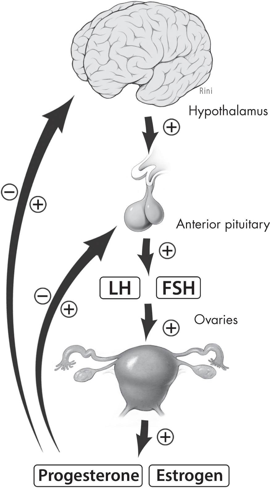
The hypothalamus plays a critical role in the production and regulation of reproductive hormones. Gonadotropin-releasing hormone (GnRH), produced and released in a pulsatile fashion by the hypothalamus, stimulates the anterior pituitary to release the gonadotropins—luteinizing hormone (LH) and follicle-stimulating hormone (FSH)—that are instrumental in the production of two ovarian hormones, estrogen and progesterone (Figure 1–1).
FSH = follicle-stimulating hormone; LH = luteinizing hormone.
Source. Image by David Rini.
Figure 1–1. The hypothalamic-pituitary-gonadal axis.
With the exception of brief periods in early embryonic and postnatal stages of life, during which it is thought to be active, the hypothalamic-pituitary-gonadal (HPG) axis is quiescent throughout childhood, likely through a primarily inhibitory mechanism (Abreu and Kaiser 2016). Pubertal maturation is initiated first by adrenarche, which is the reemergence of adrenal production, and subsequently by the reactivation of the HPG axis through a sustained increase in the pulsatile release of GnRH (Hoyt and Falconi 2015). Although the mechanism underlying the reactivation of the HPG axis is still under investigation, the inhibitory neurotransmitter GABA is thought to play a role, in conjunction with stimulatory and nutrition-dependent factors such as neuropeptide Y, kisspeptin, and glutamate (Emans and Laufer 2011).
GnRH, also known as the pulse generator, is formed in the preoptic area of the hypothalamus and is secreted from the axon terminal in the median eminence in the aforementioned pulsatile fashion required for pituitary-gonadal activation (Abreu and Kaiser 2016). LH and FSH are subsequently released in similar fashion. The significance of the pulsatile nature of GnRH release is evident because gonadotropin secretion is restored to persons lacking endogenous GnRH only when it is administered in a pulsatile fashion (Ferin 2008). GnRH administered continuously, without pulsatile release, results in decreased levels of LH and FSH (Ferin 2008).
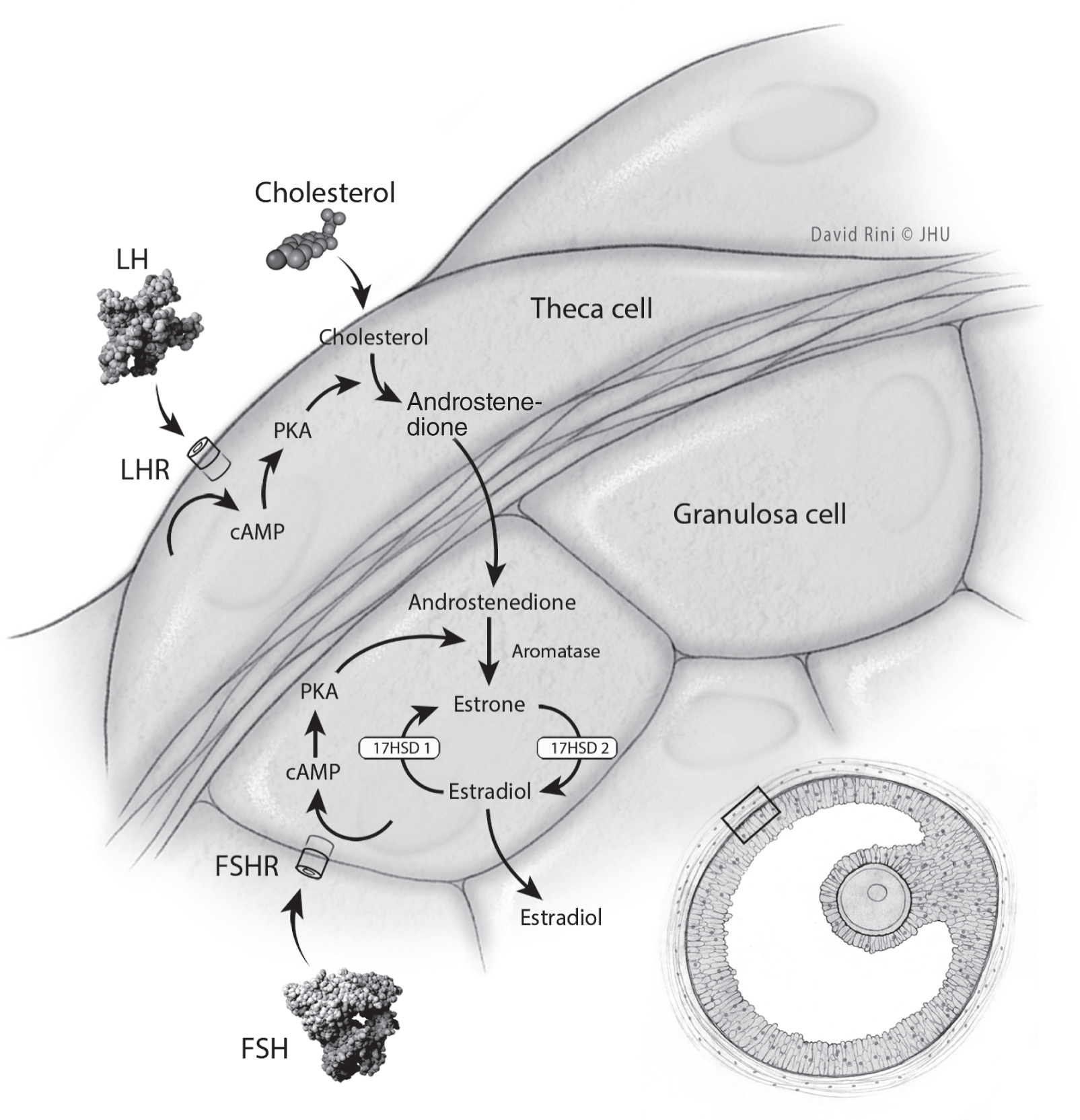
The primary effect of FSH and LH in the menstrual cycle is the development of follicles in the ovaries (Figure 1–2) (Gabbe et al. 2017). Theca cells in the ovary produce androgen precursors when stimulated by LH, whereas granulosa cells in the ovary stimulated by FSH increase the production of the enzyme aromatase, which converts androgen precursors into estrogen. The ovaries additionally produce peptides that, in addition to the androgens, trigger a feedback loop that targets both the hypothalamus and the pituitary, affecting the frequency and amplitude of GnRH release and the amount of gonadotropins released (Emans and Laufer 2011).
cAMP = cyclic adenosine monophosphate; FSHR = follicle-stimulating hormone receptor; LHR = luteinizing hormone receptor; PKA = protein kinase A.
Source. Image by David Rini.
Figure 1–2. Actions provoked by luteinizing hormone (LH) and follicle stimulating hormone (FSH) during the follicular phase.

Phases of the Menstrual Cycle

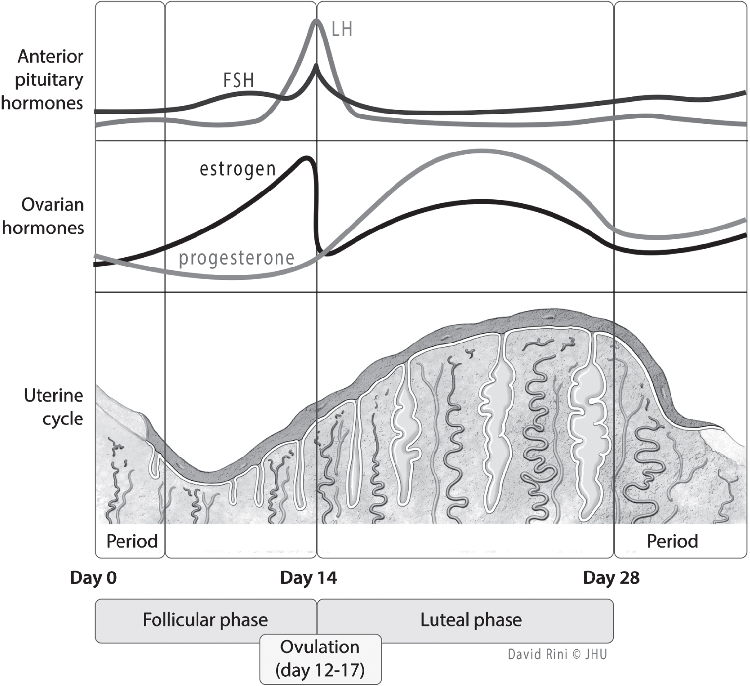
The ovarian cycle occurs, on average, every 28 days, and sequentially follows the patterns of follicular growth (the follicular phase), ovulation, and the successive formation and degeneration of the corpus luteum (the luteal phase) (Goodman 2003) (Figure 1–3).
FSH = follicle-stimulating hormone; LH = luteinizing hormone.
Source. Image by David Rini.
Figure 1–3. The menstrual and uterine cycles.

Follicular Phase

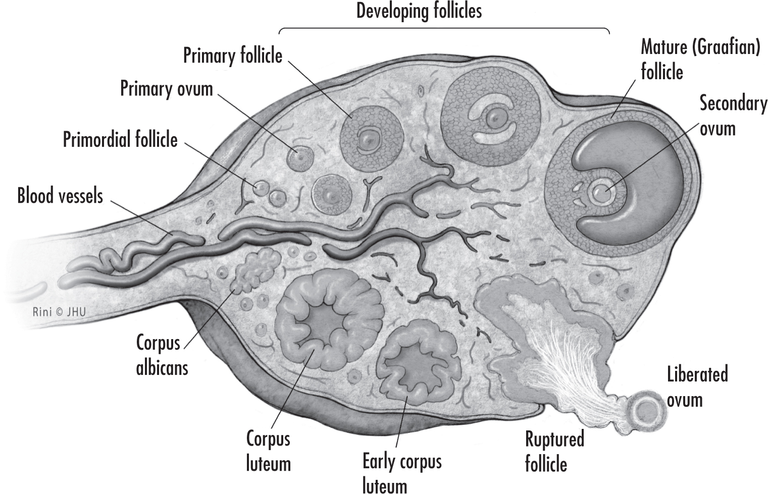
The process of follicular growth in the ovary begins before birth; by midgestation, the female fetus already has close to 7 million germ cells, which regress to 1–2 million primordial follicles by the time of birth. More than half of these will degenerate during the quiescent phase prior to puberty. After puberty, follicles gradually develop into primary, secondary, and tertiary (early antral) follicles, each over the course of about 1 year (Cox and Takov 2020). During the follicular phase, a subset of tertiary follicles during each menstrual cycle is recruited to evade the atretic process, further develop, and potentially ovulate (Ferin 2008) (Figure 1–4).
Source. Image by David Rini.
Figure 1–4. Follicle maturation.
FSH levels begin to rise toward the end of the previous cycle and are at their highest during the first week of the follicular phase (which begins on the first day of menses). Estrogen and progesterone are at low levels during the early follicular phase; this means a lack of negative feedback to the hypothalamus, which thus increases pulsatile release of GnRH, resulting in increasing levels of FSH (Chou et al. 2021). With the presence of FSH, granulosa cells hypertrophy and divide, increasing estrogen; FSH also induces the aromatase enzyme to convert androgens to estradiol (Goodman 2003). Estradiol further amplifies the effects of FSH by increasing the number of FSH receptors; meanwhile, LH promotes androgen synthesis through induction of the theca cells.
By days 5–7 of the menstrual cycle, a dominant follicle emerges and exerts negative feedback on the other developing follicles (Emans and Laufer 2011). Meanwhile, elevated estrogen levels stimulate the growth of the endometrium in the uterus by increased production of glandular and stroma cells. This process is known as the proliferative phase of the uterine cycle and coincides with the follicular phase of the ovarian cycle. Additional changes to the reproductive tract triggered by elevated estrogen levels include increased cilia formation and muscular wall contractility in the oviducts; increased growth of the myometrium and watery secretions from the cervical glands; and increased epithelial proliferation and glycogen deposition of the vagina (Goodman 2003).
By mid–follicular phase, FSH is beginning to decline, partially due to negative feedback by estradiol and inhibin on the hypothalamus and pituitary (Chou et al. 2021). By the late follicular phase, estradiol makes a sudden switch from negative to positive feedback, promoting increased release of FSH and LH by the anterior pituitary (Chou et al. 2021). LH rises 10-fold during the “LH surge” that triggers ovulation (Gabbe et al. 2017).

Ovulation

The mechanism underlying ovulation is thought to be related to the activation of proteolytic enzymes and prostaglandins that digest the collagen within the follicular wall, leading to follicular rupture and expulsion of the ovum (Reed and Carr 2018). This process is initiated by the downstream cascade of the intracellular second messenger cyclic adenosine monophosphate in the theca and granulosa cells as a result of elevated LH (Goodman 2003).

Luteal Phase

The luteal phase immediately follows ovulation and is characterized by the formation of the corpus luteum from the dominant follicle after it releases the ovum. The dominant follicle is transformed into the corpus luteum, whose purpose is to prepare the uterine lining for pregnancy and relax uterine smooth muscle. The primary product of the corpus luteum in the luteal phase is progesterone. The corpus luteum typically achieves maturity by 8–9 days after ovulation (day 22–23 of the ovarian cycle) (Ferin 2008). Progesterone, and estrogen to a lesser degree, produced by the corpus luteum promotes vascularization and thickening of the endometrial lining (called the secretory phase of the uterine cycle) in preparation for pregnancy.
When fertilization and implantation do not occur in the luteal phase, progesterone and inhibin reach their peak and inhibit further release of FSH and LH from the anterior pituitary. In the absence of FSH and LH, the corpus luteum begins to atrophy, and progesterone levels fall off. This triggers shedding of the endometrial lining, represented by day 1 of the next cycle (Gabbe et al. 2017). The endometrium releases prostaglandins that induce uterine smooth muscle contraction and sloughing of the endometrial tissue. Falling levels of progesterone and estrogen lead to a rise in FSH during the last few days of the menstrual cycle in preparation for follicle recruitment in the next cycle (Reed and Carr 2018).

Pregnancy

The three trimesters of pregnancy are delineated as the first trimester (0–12 weeks), second trimester (13–27 weeks), and third trimester (28–40 weeks). These 40 weeks are counted from the first day of the last menstrual period; thus, on the day that a woman’s next period is due, she is considered 4 weeks pregnant even though she is only 2 weeks past ovulation (and thus 2 weeks past the likely date of conception) (Chou et al. 2021).
During the three trimesters of pregnancy, a number of physiological changes occur that have profound effects on the woman, including on her mental health. Some of these physical changes can induce or mimic psychiatric symptoms (described later). Importantly, many of these changes also affect the pharmacokinetics of psychiatric medications. Considerable evidence of such effects is available for some drug classes, but for other classes almost no information is available. Moreover, little is known about whether such changes impact drug efficacy or side effects, and no guidelines exist for altering psychiatric drug dosages in pregnancy (Pariente et al. 2016). (Further details about known drug effects can be found in the specific sections that follow, as well as in Chapter 8, “A Clinical Approach to Psychiatric Diagnosis and Treatment During Pregnancy,” and the individual disorder chapters.)

Systemic Physiological Changes

Weight Gain

The mean gestational weight gain for a woman of normal weight carrying a full-term infant is typically 22–36 lb. Although recommendations have fluctuated over the past several decades, current recommendations are based on BMI. For instance, women considered underweight prior to pregnancy (BMI < 18.5 kg/m2) are recommended to gain between 28 and 40 lb, whereas women considered obese prior to pregnancy (BMI > 30 kg/m2) are recommended to gain between 11 and 20 lb (Gabbe et al. 2017).
High pre-pregnancy BMI and too much or too little weight gain during pregnancy have been correlated with preterm birth (Mitanchez and Chavatte-Palmer 2018). The correlation between obesity and preterm birth may differ according to race or ethnicity, and such differences may explain some of the racial disparity in preterm birth and maternal morbidity rates (Liu et al. 2019). For some women, changes in weight and shape across pregnancy are associated with feelings of low self-worth and low mood.

Sleep

Most pregnant women (66%–94%; Gabbe et al. 2017) report experiencing alterations in sleep patterns that lead to a subjective perception of poor sleep quality. Sleep-disordered breathing, insomnia, and sleep apnea all occur at elevated rates in pregnant women, and these may contribute to pregnancy complications such as hypertensive disorders and fetal growth restriction (Gabbe et al. 2017). Pregnant women experience varying changes in sleep patterns across the three trimesters of pregnancy. In the first trimester, women tend to experience an increase in total sleep time, including the time spent taking naps. Women can also expect an increase in daytime sleepiness and increased nocturnal insomnia during this trimester. In the second trimester, although total sleep time normalizes overall, women may experience increased awakenings. Finally, in the third trimester, total sleep time declines; women experience increases in insomnia, nocturnal awakenings, and daytime sleepiness (Gabbe et al. 2017; Santiago et al. 2001).
Sleep difficulties during pregnancy often extend through the first 2 months postpartum (Cunningham et al. 2014), when they are further complicated by a newborn’s erratic sleep patterns. Various studies have provided data on racial disparities in sleep in the general population. Black adults in the United States show significantly shorter duration and poorer-quality sleep than other racial or ethnic groups, and literature links sleep disruption to the disparity in cardiometabolic disease (Curtis et al. 2017). In pregnancy, Black women have been found to have higher short sleep prevalence compared with white women (Feinstein et al. 2020). Black women may also have different patterns of inflammation related to poor sleep than white women (Carroll et al. 2020), and poor sleep and associated inflammation may mediate the relationship between race and preterm birth (Blair et al. 2015). Significant evidence is available about the relationship between disrupted sleep and psychiatric symptoms in the perinatal period. For details, see Chapter 9, “A Clinical Approach to Psychiatric Diagnosis and Treatment in the Postpartum Period,” and Chapter 19, “Anxiety Disorders and Insomnia in the Perinatal Period.”

Physiological Changes by Organ System

Endocrine System

Reproductive hormones.
Human chorionic gonadotropin (hCG) levels remain high throughout pregnancy, returning to nonpregnant values at about 2–4 weeks postpartum, although they can take longer (Betz and Fane 2020; Reyes et al. 1985). Estrogen (in the form of estradiol) is produced first by the corpus luteum and subsequently by the placenta, and the level overall rises throughout pregnancy. Consequences of increased estrogen include suppression of the hypothalamic-pituitary-adrenal (HPA) axis and of menstruation. Concurrently, progesterone promotes pregnancy in numerous ways, including preparing the endometrial lining, modifying the maternal immune system, decreasing contractility of the smooth muscle, and inhibiting lactation (Gabbe et al. 2017). The neuroactive metabolites of progesterone, 5α-dihydroprogesterone and allopregnanolone, act centrally on GABAA receptors (McEvoy et al. 2018). Clinically, by amplifying the actions of GABA, the neuroactive metabolites play a role in suppressing oxytocin, a hormone involved in lactation and parturition (Russell and Brunton 2009). Levels of estrogen and progesterone decrease dramatically immediately after delivery but may not return completely to normal levels until about 6 months after delivery.
The high levels of estrogen and progesterone keep LH, FSH, and gonadotropins low throughout pregnancy, and they remain low for the first few weeks postpartum. The return of ovulation generally occurs 4–6 weeks after delivery, but in breastfeeding women, ovulation is inhibited by elevated prolactin stimulated by the newborn’s suckling mechanism. Increased levels of prolactin suppress the pulsatile release of GnRH, which in turn suppresses LH and contributes to inhibition of ovulation and amenorrhea (Cunningham et al. 2014; Gabbe et al. 2017). Estrogen levels are also decreased for a longer period of time postpartum in lactating women (Gabbe et al. 2017).
The extent to which GnRH is suppressed, delaying the resumption of both ovulation and menstruation, is affected by the intensity of breastfeeding and the maternal metabolic or nutritional integrity (World Health Organization Task Force on Methods for the Natural Regulation of Fertility 1998). When the frequency of breastfeeding is maintained at more than eight episodes per day, along with suckling episodes lasting longer than 7 minutes, prolactin levels are likely to remain elevated, and ovarian function continues to be inactive. For breastfeeding women with poor nutritional status, lactational energy demands may not be met, which can prolong GnRH suppression (World Health Organization Task Force on Methods for the Natural Regulation of Fertility 1998).
The mean reinstitution of ovulation for breastfeeding women is approximately 6 months, but menstruation can be delayed as long as 36 months (Gabbe et al. 2017). Maternal biological variations can also contribute to the onset of ovulation and menstruation postpartum; therefore, it is important to emphasize the variability of both ovulation and postpartum bleeding (bleeding can occur without ovulation, and ovulation can occur without menstruation). These fluctuations in reproductive hormones during the postpartum period may contribute to affective dysregulation, most likely associated with the period immediately following delivery or with weaning (Burke et al. 2019; Schiller et al. 2015).
Thyroid gland.
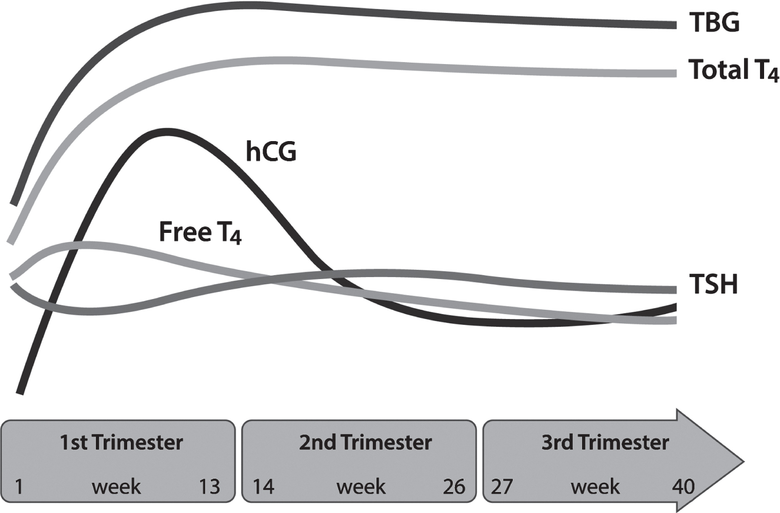
Pregnancy induces significant changes in the thyroid gland and its function (Gabbe et al. 2017). Normal pregnancy-related changes include an increase in renal iodine excretion (resulting in recommendations for increased iodine supplementation in developed countries), an increase in thyroid-binding globulin, an increase in the production of the thyroid hormones triiodothyronine (T3) and thyroxine (T4), and suppression of thyroid-stimulating hormone (TSH) (Alexander et al. 2017; Glinoer et al. 1990). Such physiological changes ultimately result in increased levels of total T4 and total T3 during pregnancy, whereas levels of free T4 and free T3 remain relatively normal (Glinoer et al. 1990). Remarkably, these myriad physiological changes occur effortlessly in healthy pregnant women (Figure 1–5); however, thyroid dysfunction can occur during pregnancy, and its evaluation is complicated by challenges in the accurate assessment of laboratory testing in the pregnant patient (Alexander et al. 2017; Gabbe et al. 2017).
hCG = human chorionic gonadotropin; T4 = thyroxine; TBG =  thyroid-binding globulin; TSH = thyroid-stimulating hormone.
Figure 1–5. Changes in the thyroid system across pregnancy.
Although routine screening for thyroid dysfunction has been debated in the literature, it is not currently endorsed by the American College of Obstetricians and Gynecologists, the American Thyroid Association, or the Endocrine Society (Committee on Patient Safety and Quality Improvement and Committee on Professional Liability 2007; Stagnaro-Green et al. 2011; Surks et al. 2004). Presently, the recommended approach is a targeted screening process that considers risk factors for thyroid dysfunction, including family or personal history of thyroid disease, morbid obesity, use of lithium, age > 30 years, and prior head or neck radiation (Melmed et al. 2019).
Psychiatrists may want to measure thyroid function to rule out thyroid dysfunction in patients with depressive or anxiety symptoms or previously known thyroid disease. It is important to use population-based, trimester-specific reference ranges for TSH and free or total T4 (Alexander et al. 2017). The first choice is a laboratory-specific pregnancy range that accounts for the characteristics of the population (Korevaar 2018). If that is not available, the upper and lower reference limits for both TSH and free T4 can be estimated using the American Thyroid Association’s clinical guidelines, which provide reference ranges for different populations found in the literature (Alexander et al. 2017).
Thyroid function generally returns to prepartum levels by about 4 weeks postpartum; however, thyroid dysfunction can occur in the postpartum period and may be an important contributor to postpartum mental illness (Bergink et al. 2011, 2018). Postpartum autoimmune thyroiditis is an important diagnosis to rule out in evaluating postpartum depression, anxiety, and other psychological disorders.
Adrenal gland.
Pregnancy is associated with marked changes in adrenocortical function, characterized by increased serum levels of aldosterone, deoxycorticosterone, corticosteroid-binding globulin, adrenocorticotropic hormone, cortisol, and free cortisol, ultimately leading to a state of physiological hypercortisolism. By the end of pregnancy, the levels of total cortisol are nearly three times higher than nonpregnant values, reaching levels equivalent to those seen in Cushing’s syndrome. These increased cortisol levels are thought to play some role in the weight gain, stretch marks, insulin resistance, and fatigue experienced in pregnancy (Gabbe et al. 2017). The degree to which pregnancy-related physiological hypercortisolemia causes clinically significant psychiatric symptoms is unknown. However, there is a theoretical correlation with depression, mood dysregulation, sleep disturbance, and cognitive dysfunction (Tang et al. 2013).
Pituitary gland.
Maternal levels of FSH and LH are decreased to undetectable amounts as a result of feedback inhibition from elevated levels of estrogen, progesterone, and inhibin during pregnancy. Serum prolactin levels begin to rise at approximately 5–8 weeks’ gestation and by full term are elevated 10-fold, which allows the body to prepare for lactation (Gabbe et al. 2017). It is not certain to what degree prolactin in pregnancy may cause psychiatric symptoms, but animal studies suggest that prolactin is an important modulator in both lactation and maternal behavior (Babb et al. 2014; Larsen and Grattan 2012).
Insulin and glucose metabolism.
During pregnancy, an increase in available glucose is required to promote fetal development while simultaneously maintaining adequate maternal nutrition. Such an increase in available glucose is associated with at least a mild peripheral insulin resistance, which develops later in pregnancy (Gabbe et al. 2017; Soma-Pillay et al. 2016). In early pregnancy, the increases in insulin secretion by insulin-secreting pancreatic β cells are met with increased insulin sensitivity. Progesterone and estrogen, in conjunction with other diabetogenic hormones (e.g., human placental lactogen, growth hormone, cortisol), are thought to play a role in mediating the insulin resistance that peaks in the third trimester. In general, standard pregnancy glucose metabolism results in slight fasting hypoglycemia, postprandial hyperglycemia, and hyperinsulinemia. Insulin resistance and relative hypoglycemia—in conjunction with increased concentrations of free fatty acids, triglycerides, and cholesterol—promote lipolysis, which allows pregnant women to preferentially metabolize fat for fuel, preserving glucose metabolism for the fetus (Cunningham et al. 2014; Soma-Pillay et al. 2016). In about 6%–9% of women, these changes in glucose and insulin will result in gestational diabetes (Centers for Disease Control and Prevention 2018). Data have shown an increased risk for gestational diabetes in Asian American and Hispanic women as compared with non-Hispanic white women, and Hispanic women have a greatly increased risk of progression to type 2 diabetes within 5 years of having had gestational diabetes (Chou et al. 2021; Pu et al. 2015).

Central Nervous System

The CNS undergoes profound changes during pregnancy and the postpartum period, utilizing significant plasticity to adapt to motherhood (Hillerer et al. 2014). For example, a functional imaging study noted that pregnancy was associated with profound and specific changes in the structure of the brain, especially in areas specific to social cognition (Hoekzema et al. 2017). During pregnancy, there is a reduction in total brain volume that reverses within 6 months of parturition (Oatridge et al. 2002); postpartum regional reductions are also seen, some of which are positively related to levels of maternal attachment (Hoekzema et al. 2017). Specific postpartum anatomical changes also occur, such as gray matter changes in the hypothalamus, amygdala, and prefrontal cortex (Kim et al. 2010).

Cardiovascular System

Profound changes occur in the cardiovascular system during pregnancy. Cardiac output increases throughout pregnancy and peaks at 30%–50% above preconception values (Chou et al. 2021). This is secondary to an increase in both heart rate and stroke volume (Pacheco et al. 2013). The increase in cardiac output is disproportionally directed to the uterus, placenta, and breasts and diminishes over time in the postpartum period, remaining above pre-pregnancy levels up to 1 year later. Maternal blood volume also increases by the second half of pregnancy and is mostly attributed to an increase in plasma volume (Gabbe et al. 2017). The compensatory experience of increased heart rate (approaching 90 bpm in the third trimester) may sometimes induce anxiety or panic attacks in women with underlying anxiety disorders.
Maternal blood pressure falls somewhat in the first and second trimesters because increased progesterone causes a decrease in systemic vascular resistance (Chou et al. 2021). As pregnancy advances, the heart is physically displaced upward and toward the left. Eccentric cardiac hypertrophy, which is a physiological phenomenon, is common in pregnancy and thought to result from expanded blood volume. This is likely an adaptive process, as seen in athletes, that enables the pregnant woman’s heart to work more efficiently. As a result of these and other physiological changes of pregnancy (e.g., drop in albumin concentration), plasma levels of medications may be diluted, creating the possibility for a decrease in efficacy (Gabbe et al. 2017).
Cardiovascular complications are the most common cause of pregnancy-related mortality (Creanga et al. 2017) and predict cardiovascular disease later in life (Margerison et al. 2019). Black women have higher rates of cardiovascular diseases such as preeclampsia in pregnancy, as well as higher associated fatality rates (Bibbins-Domingo et al. 2017). This increased risk is likely to have both physiological and socioeconomic determinants.

Hematological System

Pregnancy is considered a hypercoagulable state, in preparation for hemostasis following delivery. There are overall increases in most proclotting factors and decreases in fibrinolysis and inhibitors of coagulation. These adaptations are thought to infer protection against excessive blood loss during delivery, but they also incur risk. The risk of thromboembolic disease is increased five- to sixfold, and pulmonary embolus is a leading cause of maternal mortality in the developed world (Petersen et al. 2019). The duration of this elevated risk lasts until at least 12 weeks postpartum (Soma-Pillay et al. 2016).
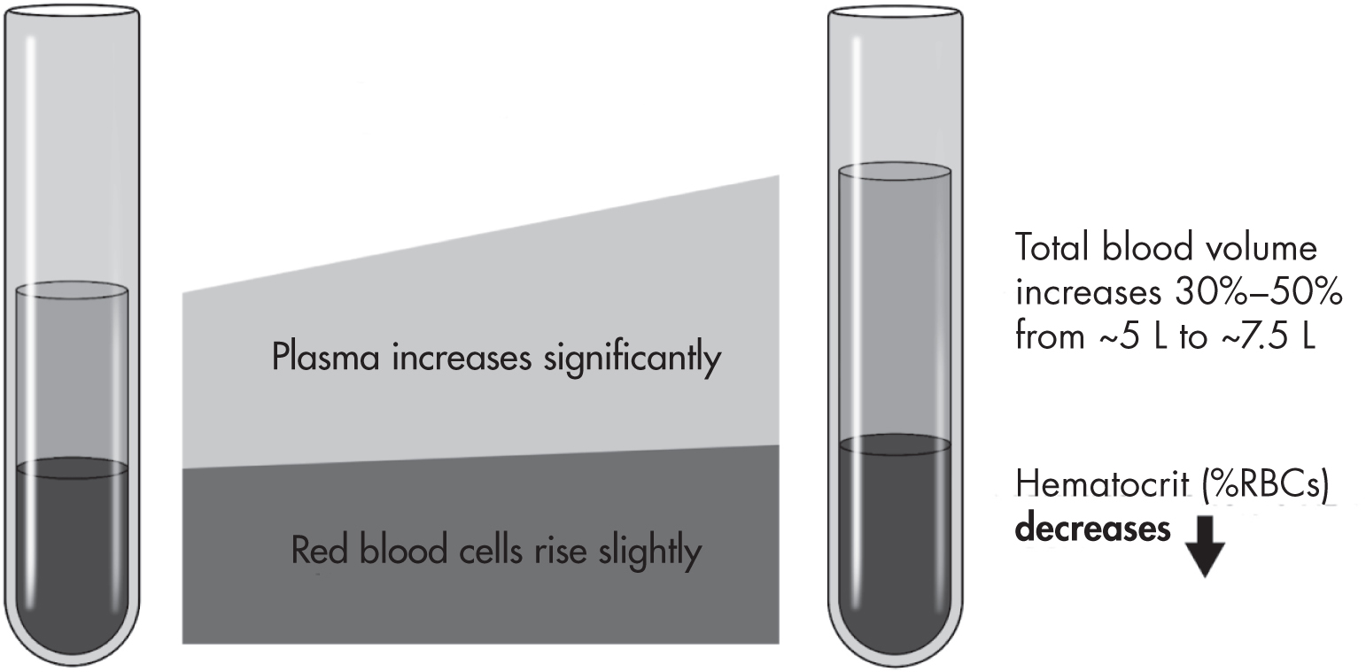
Additionally, the significant increases in plasma volume with only a slight increase in red blood cell mass result in a decrease in hemoglobin concentration, hematocrit, and red blood cell count (Figure 1–6) (Soma-Pillay et al. 2016), with Black women generally experiencing lower hematocrit and hemoglobin concentration than white women (Chou et al. 2021). This phenomenon, sometimes termed “physiological anemia of pregnancy,” may contribute to fatigue, shortness of breath, and exercise intolerance in pregnant women (Pacheco et al. 2013). It also results in decreased concentration of drug-binding proteins, altering the apparent volume of distribution and, in some cases, drug clearance (Pariente et al. 2016). Hemoglobin and hematocrit fluctuate in the first few days postpartum and return to pre-pregnancy levels by 8 weeks postpartum.
Figure 1–6. Hematological changes in pregnancy.
Finally, iron requirements during pregnancy increase by two- to threefold, attributable not only to hemoglobin synthesis but also to fetal needs, including enzyme production. There is also a 10- to 20-fold increase in the requirements for folate and a twofold increase in vitamin B12 requirements (Soma-Pillay et al. 2016).

Pulmonary System

During pregnancy, the nasopharynx mucosa becomes edematous due to increased estrogen, leading to marked nasal stuffiness and decreased nasal patency. Regarding lung volume and pulmonary function, alterations in the diaphragm and chest wall configurations lead to reduced total lung capacity and functional residual capacity. Finally, increasing progesterone levels lead to chronic hyperventilation, low maternal PaCO2, and chronic respiratory alkalosis (Gabbe et al. 2017). From a psychiatric perspective, decreased lung capacity and changes in PaCO2 may lead to increased anxiety and risk of panic attacks, particularly in patients with a history of panic disorder (Wilhelm et al. 2001).

Gastrointestinal System

Nausea and vomiting, also known as “morning sickness,” complicate up to 80% of pregnancies and can occur at any time of day. For most women, symptoms resolve by about 18 weeks of gestation, but 15%–20% of women will continue to have symptoms in the third trimester. The most severe form of nausea and vomiting is hyperemesis gravidarum, which affects 0.5%–3% of women and often leads to weight loss, dehydration, and electrolyte imbalance (Chou et al. 2021; Soma-Pillay et al. 2016).
During pregnancy, elevated levels of progesterone affect several organs, including the stomach and gallbladder. The smooth muscle tone of the stomach and the tone of the lower esophageal sphincter are both decreased, leading to increased rates of gastroesophageal reflux disease, affecting 30%–50% of pregnant women (Chou et al. 2021). The rate at which the gallbladder empties is slowed due to higher progesterone levels, increasing the risk of gallstones. In the small intestines and colon, perturbations in motility are common in pregnancy and result in an increased incidence of constipation in some women and diarrhea in others.
In the liver, total body protein increases, but serum albumin and total protein levels fall during gestation as a result of hemodilution (Gabbe et al. 2017). Pregnancy also leads to changes in both cytochrome P450 enzyme activity and glucuronidation, with corresponding implications for medication metabolism (Dallmann et al. 2018). I particular, the activity of the following is affected: the uridine 5´-diphospho-glucuronosyltransferase (UGT) isoenzymes UGT1A4 and UGT2B7 and the CYP450 isoenzymes CYP3A4, CYP2D6, CYP2C9, CYP1A2, and CYP2C19 (Pariente et al. 2016).

Genitourinary System

Physiological changes in the vagina interact with the vaginal microbiome to protect against infection and promote pregnancy maintenance. The kidneys enlarge during pregnancy, with dilation of the ureters and renal pelvis. These anatomical changes are mediated by progesterone (Soma-Pillay et al. 2016). Renal plasma flow rises 50%–80% in pregnancy, with a resulting increase in glomerular filtration rate, which reaches 40%–50% above pre-pregnancy levels by the end of the first trimester (Chou et al. 2021). This hyperfiltration leads to urinary frequency and renal excretion of various nutrients, vitamins, and renally excreted medications (e.g., lithium, risperidone, gabapentin, topiramate) (Cunningham et al. 2014). Glomerular filtration rate returns to pre-pregnancy levels by 8 weeks’ gestation, which is important to consider if medication changes were made due to physiological changes of pregnancy (Gabbe et al. 2017).

Immune System

Pregnancy is a time of dramatic changes in the immune system. It was once thought that pregnancy must be a time of total immune suppression to prevent fetal rejection. Later, evidence arose for a shift from proinflammatory to anti-inflammatory forces across pregnancy. The reality is much more complex, because the body works both to keep out foreign pathogens and to dampen cytotoxic activity (Mor and Cardenas 2010; Osborne and Monk 2013). The result is increased susceptibility to some viral and bacterial pathogens (e.g., influenza) and decreased activity for some autoimmune diseases (e.g., systemic lupus erythematosus). Some of these immune changes are still evident at 6 months postpartum and perhaps longer (Osborne et al. 2019b). In the postpartum period, an increased risk of “flare-ups” of autoimmune diseases and latent infections is associated with the return of normal immune function. In addition, increasing evidence indicates that mood and anxiety disorders experienced during the peripartum period may be related to these changes in immune functioning (Osborne et al. 2019a, 2020).

Parturition and the Postpartum Period

Phases of Parturition

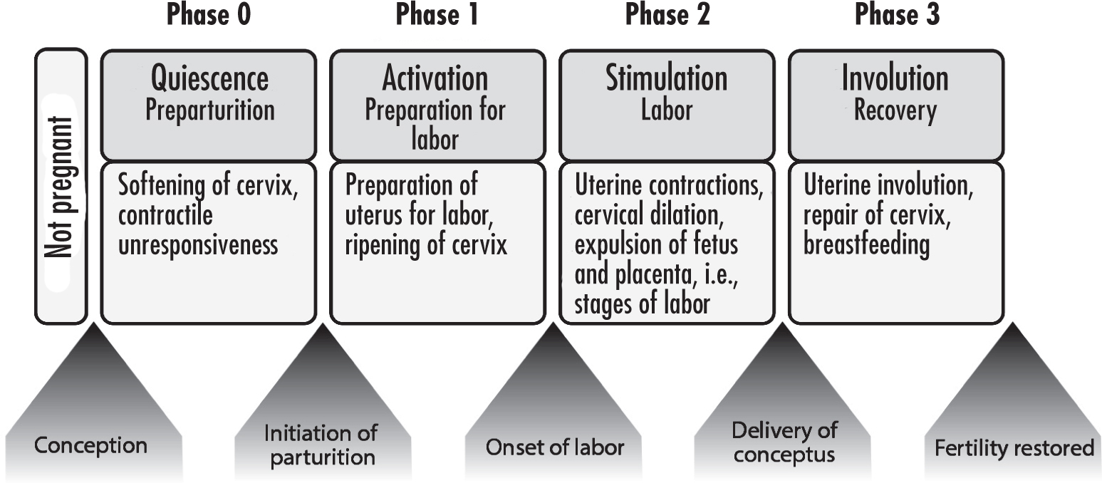
The four phases of parturition, more commonly known as labor and delivery, are described in the following subsections. Figure 1–7 provides a brief synopsis.
Figure 1–7. The phases of parturition.

Phase 0: Quiescence

Throughout most of pregnancy, the uterus is maintained in a state of quiescence, distinguished by uterine smooth muscle tranquility and cervical structural preservation (Behrman and Butler 2007). This process is mediated by inhibitory hormones, including (but not limited to) progesterone, nitric oxide, relaxin, prostacyclin, and parathyroid-related hormone. During quiescence, the uterus undergoes significant vascular and dimensional changes in preparation for contractions. Although this phase precedes uterine activation, women may nevertheless experience low-intensity myometrial (Braxton Hicks) contractions; these are not accompanied by cervical change and wane over time. During most of the quiescent phase, cervical structural integrity remains intact to support progression of the pregnancy to full term. Toward the end of this phase, the cervix undergoes a progressive remodeling, known as cervical softening, in preparation for labor (Cunningham et al. 2014).

Phase 1: Activation

The onset of the uterine activation phase typically occurs during the last 6–8 weeks of pregnancy and is defined by a series of uterine changes driven by estrogen. These include increased expression of contraction-associated proteins, including myometrial receptors for oxytocin and prostaglandin. There is also activation of certain ion channels and amplification of key proteins within gap junctions (e.g., connexin-43), which promote electrical synchrony between adjacent myometrial cells and allow for efficient uterine contractions (Challis and Gibb 1996; MacDonald 1993).
Concurrently during this phase, the cervix undergoes continued remodeling and transitions from cervical softening to cervical ripening. Cervical ripening occurs days or weeks before the onset of contractions and is characterized by degradation of cervical collagen in preparation for cervical dilation. Prostaglandins likely play a role in this process (Cunningham et al. 2014; Keirse 1979).

Phase 2: Stimulation

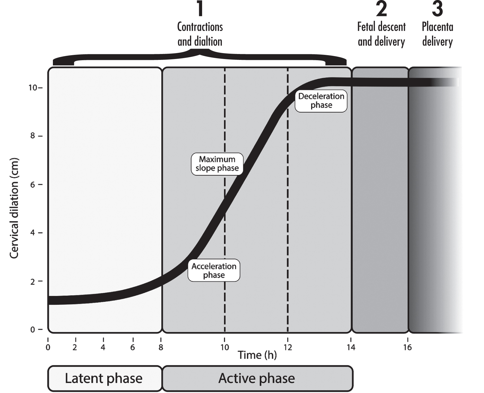
Following the activation phase, the uterus is prepared for stimulation by uterotonic agents, primarily prostaglandins and oxytocin, which promote uterine contractions. This process of regular uterine contractions occurs over days to weeks (Gabbe et al. 2017). The stimulation phase of parturition is typically recognized as active labor. Labor is divided into three sequential stages: cervical effacement and dilation, fetal descent and delivery, and placental separation and delivery (Figure 1–8) (Cunningham et al. 2014).
Figure 1–8. The stages of labor (Cunningham et al. 2014).
Stage 1: Cervical effacement and dilation.
The first stage begins at the onset of labor and continues until cervical dilation is achieved. Onset of labor is typically characterized by regular painful contractions. Consistent contractions, with sufficient intensity and duration, lead to cervical thinning and shortening, also known as cervical effacement. This stage of labor is further divided into the latent and active phases. The latent phase is characterized by a slow rate of cervical change. This is followed by rapid acceleration of cervical dilation, known as the active phase. Although variable in the literature, historically, active labor requires 80% effacement and dilation of at least 4 cm, but it has recently been recognized that it may not begin until dilation of 6 cm (Chou et al. 2021). The cervix is considered fully dilated when it reaches approximately 10 cm (Cunningham et al. 2014).
Historically (based on the “Friedman criteria”), the mean time (95th percentile) for cervical dilation from 4 cm to 10 cm was 4.6 hours (11.7) in nulliparous women and 2.4 hours (5.2) in multiparous women (Friedman 1978). More recently, Zhang et al. (2010) observed the median times (95th percentile) to be 5.3 hours (16.4) in nulliparous women and 3.8 hours (15.7) in multiparous women (Zhang et al. 2010). In general, increased maternal BMI, older maternal age, and nonoptimal fetal position correlate with longer labor times (Gabbe et al. 2017).
Stage 2: Fetal descent and delivery.
The second stage of labor begins once cervical dilation is complete and continues until the fetus is delivered. Although engagement with the fetal head (most neonates are delivered head first) can occur before labor begins, active fetal descent typically occurs after the progression of cervical dilation. As such, increased rates of fetal descent correlate with the maximal phase of cervical dilation. The increased rates are maintained until the observed fetal head reaches the perineal floor (Cunningham et al. 2014). According to Zhang et al. (2010), for nulliparous and parous women with epidural anesthesia, the median duration (95th percentile) of the second stage was 1.1 hours (3.6) and 0.4 hours (2.0), respectively. For nulliparous and parous women without epidural anesthesia, the median duration (95th percentile) was 0.6 hours (2.8) and 0.2 hours (1.3), respectively.
Stage 3: Placental separation and delivery.
The third stage of labor is defined as the time following fetal delivery until placental expulsion. Placental separation occurs as a result of the inevitably reduced size of the uterine cavity following fetal delivery creating a tension with the unchanged placental size. Once separation is achieved, the placenta is expelled through increased abdominal pressure. This is typically accomplished by compressing the fundus of the uterus while placing minimal traction on the umbilical cord (Cunningham et al. 2014).

Phase 3: Involution

Involution of the uterus takes place after fetal and placental delivery and is mediated primarily by oxytocin. Immediately following delivery, the uterus undergoes persistent contraction and retraction. Such rigidity allows compression of large uterine vessels to the point of thrombosis, with the goal of preventing hemorrhage. Uterine involution and cervical repair are components of the postdelivery remodeling processes, with the goal of restoring female organs to their nonpregnant state. These processes help protect the female reproductive tract from infection and restore endometrial responses to hormonal activity (Asgari Safdar et al. 2013; Behrman and Butler 2007).

Postpartum Period

The postpartum period, also called the puerperium, is the period following delivery of the placenta and lasts for the first several weeks after delivery. During this period, maternal anatomy and physiology return to nonpregnant states. Classically, this period is thought to last 4–6 weeks, but some body systems may not return to the nonpregnant state for up to 12 months (Porter 2013). Timing of the return to the nonpregnant state for most body systems was discussed earlier (see “Pregnancy” section); here we cover changes to the pelvic anatomy and body shape. Breast changes are discussed in greater detail later.

Pelvic Anatomy

Immediately following delivery, the fundus of the uterus is typically firm and nontender, and it lies slightly below the umbilicus. The uterus and endometrium return to nonpregnant size by approximately 8 weeks postpartum (Chou et al. 2021); however, this baseline can be affected by several factors, including uterine overdistention predelivery, multiparity, cesarean section delivery, and breastfeeding (Negishi et al. 1999). The cervix after delivery may have small lacerations. For the first few days postpartum, the cervix remains slightly dilated (width ~ 2–4 cm), but by about 1 week postpartum, the opening narrows and the cervix thickens. The external os, the opening of the uterine cervix into the vagina, never resumes its original shape. The vagina slowly contracts postpartum but does not return to its nulligravid size (Cunningham et al. 2014).

Weight and Shape

The expulsion of the fetus, placenta, and amniotic fluid combined represents a mean weight loss of approximately 10–13 lb; however, this weight loss is not immediately observed due to fluid retention postpartum, and this delay can be anxiety provoking for some women (Gabbe et al. 2017). During the first 6 weeks postdelivery, approximately one-half of the gestational weight gained during pregnancy is lost, and only approximately one-fourth of women return to their pre-pregnancy weight by 6 weeks postpartum. A slower rate of weight loss occurs during the subsequent months, for up to 6 months postpartum (Gunderson et al. 2001). If the amount of weight gained during pregnancy exceeds recommended amounts (e.g., > 35 lb in a woman with normal BMI), women are likely to have an approximate net weight gain of 11 lb. Breastfeeding does not have a significant impact on postpartum weight loss (Gabbe et al. 2017). Notable racial disparities have been shown in postpartum weight loss, with Black women demonstrating significantly greater postpartum weight retention 1 year after delivery compared with white women (Headen et al. 2012).

Breast Development and Lactation

Anatomy

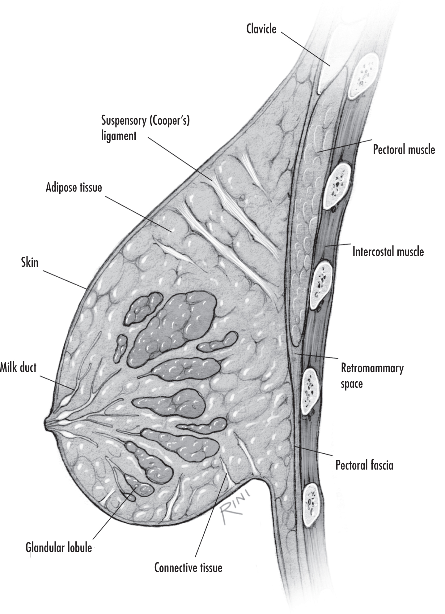
The adult female breast is made up of specialized glandular tissue and adipose tissue. The glandular tissue is organized into 15–20 lobes, which are further divided into small lobules in which breastmilk is produced (the lobules themselves are clusters of secretory epithelial cells called acini). Breastmilk is secreted through a network of ducts that arise from smaller lobular units, which then converge and eventually exit the skin via duct orifices in the nipple (Sriraman 2017). Figure 1–9 depicts the anatomical structure of a breast.
Source. Image by David Rini.
Figure 1–9. Anatomy of the breast.

Breast Development

The mammary gland first develops during embryogenesis. A mammary bud forms in the developing epidermis and extends into the subepidermal mesenchyme, while an adipose pad precursor develops underneath. A rudimentary mammary duct system then develops, canalizing and spreading through the adipose pad precursor. After birth until puberty, the mammary gland remains small, with existing ducts growing in parallel with child growth (Macias and Hinck 2012; Sriraman 2017).
Mammogenesis refers to the growth and development of the mammary glands in preparation for milk production. The process begins in puberty, with estrogen and growth factors stimulating the proliferation and branching of ducts into a treelike pattern throughout the developing adipose tissue. Initial lobular and alveolar development also occurs, which is further spurred by changes in estrogen and progesterone levels during menstruation. By the end of puberty, various lobules with a complex duct system have formed. No further changes occur until pregnancy, when further alveolar development and epithelial maturation occur in response to higher progesterone levels, leading to secretory tissue proliferation and breast enlargement. These changes are accompanied by a marked increase in vascular supply to the breast. In early pregnancy, larger, complex lobules form (Alex et al. 2020; Sriraman 2017).

Lactation

The term lactogenesis refers to the physiological changes that allow a woman to produce and secrete breastmilk. These changes begin during pregnancy and direct the alveolar epithelium to differentiate. Lactogenesis occurs in three stages: secretory initiation, secretory activation, and ongoing milk production (Pillay and Davis 2020).

Stage I: Secretory Initiation

Secretory initiation begins during the second half of pregnancy, at which point the mammary glands have become sufficiently differentiated to produce small quantities of milk components such as casein and lactose. High levels of plasma progesterone (secreted by the placenta) inhibit further secretion and gland differentiation.

Stage II: Secretory Activation

Secretory activation occurs after delivery. With the removal of the placenta, the rapid drop in progesterone and the elevated levels of prolactin, cortisol, and insulin stimulate milk secretion. Usually, at day 2 or 3 postpartum, women experience swelling of the breast along with copious milk production. In primiparous women, the secretory activation stage is slightly delayed, and early milk volume is lower. Lower milk volume is also observed in women who had cesarean births compared with those who delivered vaginally. Late onset of milk production has also been seen in women who have had retained placental fragments, diabetes, and stressful vaginal deliveries. With retained placental fragments, stage II could be inhibited by the continued secretion of progesterone and would continue to be inhibited until the remaining placental fragments are removed (Alex et al. 2020; Wagner et al. 2018).

Stage III: Galactopoiesis

The term galactopoiesis refers to ongoing milk production, which is maintained by regular removal of milk and stimulation of the nipple, which trigger prolactin release from the anterior pituitary gland and oxytocin from the posterior pituitary gland. For the ongoing synthesis and secretion of milk, the mammary gland must receive hormonal signals from both prolactin and oxytocin. Prolactin stimulates mammary gland ductal growth and epithelial cell proliferation and induces milk protein synthesis through binding to receptors on the alveolar epithelium of the mammary gland. Nipple stimulation leads to sensory transmission through the spinothalamic tracks; the hypothalamus is then stimulated, leading to decreased tonic prolactin inhibition and resulting in prolactin surges within 15 minutes of nursing (Sriraman 2017).
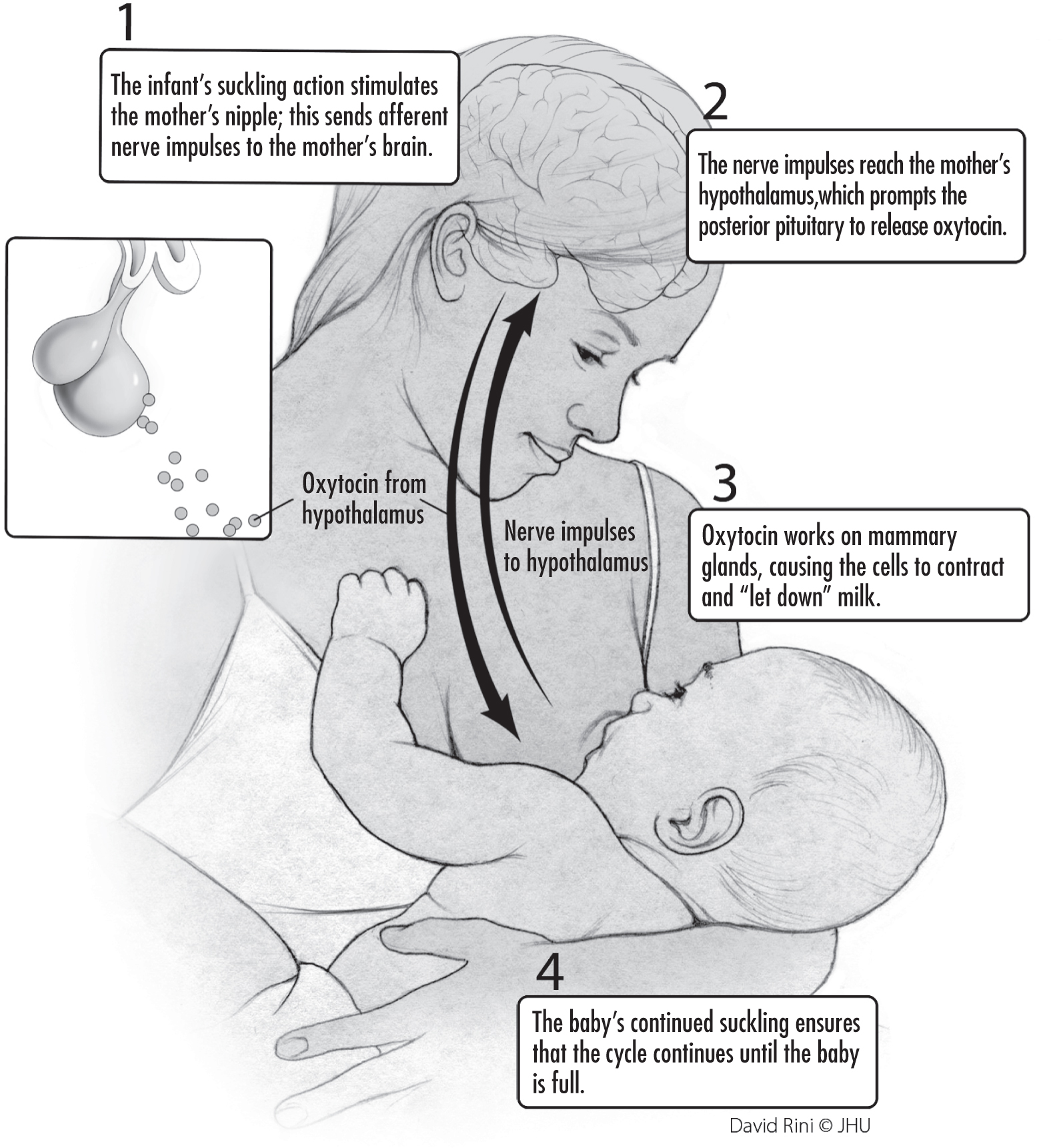
Oxytocin governs the milk ejection, or let-down, reflex (Svennersten-Sjaunja and Olsson 2005). The tactile stimulation of the nipple-areolar complex by suckling sends afferent signals to the hypothalamus, triggering release of oxytocin from the posterior pituitary. Oxytocin travels to the mammary glands and binds to G-protein-coupled oxytocin receptors located on the myoepithelial cells, causing the cells to contract. The myoepithelial cell contraction releases milk into the ducts from the alveolar lumens and through the nipple. The release of oxytocin becomes a conditioned response in the lactating woman, requiring only visual stimulation or conscious thought (Leng et al. 2005; Sriraman 2017). Figure 1–10 summarizes the let-down reflex.
Source. Image by David Rini.
Figure 1–10. The milk let-down reflex.
Once lactation is established and maintained, both physical and biochemical factors regulate milk production. If milk is not removed, intramammary pressure increases and leads to vascular stasis and accumulation of a lactation feedback inhibitor (thought to be whey protein in breastmilk); milk production consequently stops and mammary involution is initiated. Conversely, breastmilk removal decreases pressure, improves blood flow to the mammary glands, and removes the inhibitor, leading to resumed production (Institute of Medicine Committee on Nutritional Status During Pregnancy and Lactation 1991; Macias and Hinck 2012; Sriraman 2017).

Menopause

Menopause is defined as the absence of menstrual periods for a full year, and it is caused by reduced secretion of ovarian hormones, including estrogen and progesterone (Bacon 2017). An estimated 1 billion women worldwide have experienced menopause (Hoga et al. 2015). The average age of menopause in the United States is 51.5 years (Hoffman et al. 2020).
Menopause is a normal event for women and generally signals the end of a woman’s reproductive capacity. It may be experienced as a liberating end to anxieties about childbirth and to discomfort related to one’s reproductive life, or it may be viewed negatively—particularly in Western cultures—through its association with aging (Minkin 2019). (Some women might experience a combination or neither of these reactions.) Individual experiences of menopause vary, and at times women will seek medical or mental health attention for the management of symptoms (Nelson 2008). In this section, we review the physiology of the menopausal transition. See Chapter 7, “Perimenopause,” for a review of the relationship between the menopausal transition and mental health.
Menopause can be a naturally occurring event, or it can be brought about either medically or surgically as a result of—or for the prevention of—disease. Medical menopause is beyond the scope of this chapter, but it is worth noting that women with medical or surgical menopause, particularly prior to age 45 years, may have more severe and prolonged menopausal symptoms (Rodriguez and Shoupe 2015). In addition, women experiencing medical menopause have an increased risk of depression, heart disease, osteopenia or osteoporosis, sexual dysfunction, and cognitive decline compared with women in the general population (Rodriguez and Shoupe 2015).
The menopausal transition has several phases: perimenopause, menopause, and postmenopause. Perimenopause, characterized by estrogen decline and irregular menstrual cycle length, is the interval of time preceding the onset of menopause (Dutton and Rymer 2015). On average, perimenopause begins 4 years prior to the final menstrual cycle—on average, age 47—and lasts for several years (Hoffman et al. 2020). Menopause reflects the process of ovarian follicular depletion. Ovarian senescence begins in utero with programmed oocyte atresia and then continues after birth by way of follicular maturation and regression (Hoffman et al. 2020). Beginning in a woman’s later 30s to early 40s, ovarian follicles are depleted at a more rapid rate until the eventual loss of ovarian activity by the time menopause is reached; loss of ovarian function before age 40 is termed premature ovarian insufficiency (Hoffman et al. 2020).
In the early perimenopausal transition stage, the length of time between menstrual cycles increases from the typical 25–35 days during reproductive years to approximately 40–50 days (Dutton and Rymer 2015). During this time, FSH levels vary, but generally this hormone is on the rise. Estradiol levels are initially maintained steadily or, like FSH levels, are increased (Dutton and Rymer 2015). Such changes reflect diminished ovarian reserve: fewer follicles are present to secrete inhibin, a hormone that exerts negative feedback on FSH secretion (Dutton and Rymer 2015; Hoffman et al. 2020). Initially, increased FSH levels result in greater follicle recruitment and therefore an increase in estradiol; this transition period correlates with a variability in menstrual cycle length, increased amenorrhea, and more frequent anovulatory cycles (Dutton and Rymer 2015). Eventually, cycle lengths shorten and anovulatory cycles become more frequent, although unpredictably so (Hoffman et al. 2020). In addition to changes in estrogen levels, progesterone levels start to decline (Hoffman et al. 2020). As the perimenopausal period progresses and menopause is reached, FSH continues to rise and estradiol levels fall, whereas in the postmenopausal period, FSH and estradiol levels initially stabilize and eventually both decline (Dutton and Rymer 2015).
The most common physical symptom of menopause is the development of hot flashes, reported by up to 50% of menopausal women (Hoffman et al. 2020). Classically, hot flashes last 2–4 minutes and are described as a sudden sensation of heat beginning on the chest and face and then progressively spreading throughout the body. They can occur several times in a day and can be associated with perspiration, palpitations, chills, anxiety, and irritability (Hoffman et al. 2020). The proposed physiology surrounding both hot flashes and night sweats (vasomotor symptoms) reported by menopausal women is related to changes in central thermoregulatory functioning due to decreased estradiol (Dutton and Rymer 2015). Skin temperatures can increase, particularly in the fingers and toes, due to peripheral vasodilation, whereas core temperatures may slightly decrease (Hoffman et al. 2020). Recent studies estimate that hot flashes last for 7 years on average (Hoffman et al. 2020). Significant differences in the prevalence of vasomotor symptoms in women of different racial backgrounds have been reported, with Black women experiencing the highest prevalence and longest duration of all groups studied (El Khoudary et al. 2019).
Additional symptoms noted during menopause include vaginal dryness and an increase in urinary tract infections, which are functions of decreased estradiol and progesterone in the urogenital tract (Dutton and Rymer 2015). Specifically, depleted estrogen levels lead to the thinning of the epithelial lining of the vagina and subsequently, in the long term, to vaginal atrophy (the vagina shortens, narrows, and becomes less flexible) (Hoffman et al. 2020). Women may first notice a decline in vaginal lubrication during sexual activity; however, as the decreased estrogen state progresses, discomfort may be noticed during daily activities. Vaginal dryness and associated discomfort are likely among several factors in the decreased sexual function associated with menopause. Studies have shown racial differences in postmenopausal sexual functioning; compared with white women, Asian women report lower sexual desire and lower importance of sex, and Black women report lower levels of arousal and greater importance of sex (El Khoudary et al. 2019).
Estrogen depletion can also affect other organ systems and can be associated with cardiovascular disease, metabolic syndromes, and osteopenia or osteoporosis. Estrogen deficiency is proposed to play a role in the increased risk of cardiovascular disease after menopause and may be impacted in part by shifts in lipid profiles during perimenopause (Derby et al. 2009). There are also proposed shifts in body composition in the postmenopausal period, including increased fat content and decreased lean muscle. In terms of bone health, it is postulated that decreased estrogen levels lead to excessive bone resorption (Hoffman et al. 2020). Ultimately, this results in osteopenia (a condition that occurs when the body does not make new bone as quickly as it reabsorbs old bone), which is the precursor to osteoporosis, a progressive reduction in bone mass and strength that contributes to fractures (Hoffman et al. 2020). Approximately 40%–50% of postmenopausal women will experience a fracture related to osteoporosis in their lifetime (Hoffman et al. 2020). Racial differences in postmenopausal fractures have been described—rates among Black and Asian women are approximately half of those in white women—however, the physiological differences that underlie this pattern are not well understood (El Khoudary et al. 2019). For a detailed discussion of these clinical features and their relationship to mental health, see Chapter 7, “Perimenopause.”
Conclusion

Clinicians treating women for mental health conditions at times of reproductive transition must have a firm understanding of the hormonal and other physiological changes that accompany such transitions. This chapter offered an overview of the physical changes that accompany the menstrual cycle, pregnancy, parturition and the postpartum period, lactation, and menopause. The mental health sequelae of these changes are the subject of the remaining chapters of this book.

Key Points

Women’s reproductive years begin with menarche, when reproductive hormones initiate the menstrual cycle and the development of primary and secondary sexual characteristics.
The first half of the menstrual cycle is the follicular phase (days 1–14), when follicle-stimulating hormone (FSH) and luteinizing hormone (LH) spur the development of a dominant follicle, and estrogen and progesterone levels rise gradually.
Ovulation occurs at approximately day 14 and is followed by the luteal phase, when the uterine lining is prepared for implantation and the dominant follicle develops into the progesterone-producing corpus luteum.
When pregnancy does not occur, the uterine lining is shed, and the next cycle begins with menses.
The enormous changes of pregnancy are not confined to the reproductive system; they include changes in other endocrine systems as well as the central nervous, cardiovascular, hematological, pulmonary, gastrointestinal, genitourinary, and immune systems.
There are four phases to parturition: quiescence (most of pregnancy), activation (beginning of contractions), stimulation (active labor), and involution (return of the uterus to pre-pregnancy size).
The three stages of active labor are cervical dilation, delivery of the fetus, and delivery of the placenta.
Changes in breast anatomy and physiology in pregnancy prepare for lactation, which is regulated by prolactin and oxytocin.
Menopause, which represents the end of a woman’s reproductive life, is induced by changes in reproductive hormones, with a decrease in estrogen and progesterone and an increase in LH and FSH.

References

Abreu AP, Kaiser UB: Pubertal development and regulation. Lancet Diabetes Endocrinol 4(3):254–264, 2016 26852256
Alex A, Bhandary E, McGuire KP: Anatomy and physiology of the breast during pregnancy and lactation. Adv Exp Med Biol 1252:3–7, 2020 32816256
Alexander EK, Pearce EN, Brent GA, et al: 2017 guidelines of the American Thyroid Association for the diagnosis and management of thyroid disease during pregnancy and the postpartum. Thyroid 27(3):315–389, 2017 28056690
Asgari Safdar AH, Daghigh Kia H, Farhadi R: Physiology of parturition. International Journal of Advanced Biological and Biomedical Research 1(3):214–221, 2013
Babb JA, Carini LM, Spears SL, Nephew BC: Transgenerational effects of social stress on social behavior, corticosterone, oxytocin, and prolactin in rats. Horm Behav 65(4):386–393, 2014 24657520
Bacon JL: The menopausal transition. Obstet Gynecol Clin North Am 44(2):285–296, 2017 28499537
Behrman RE, Butler AS (eds): Preterm Birth: Causes, Consequences, and Prevention. Washington, DC, National Academies Press, 2007
Bergink V, Kushner SA, Pop V, et al: Prevalence of autoimmune thyroid dysfunction in postpartum psychosis. Br J Psychiatry 198(4):264–268, 2011 21343331
Bergink V, Pop VJM, Nielsen PR, et al: Comorbidity of autoimmune thyroid disorders and psychiatric disorders during the postpartum period: a Danish nationwide register-based cohort study. Psychol Med 48(8):1291–1298, 2018 28929982
Betz D, Fane K: Human Chorionic Gonadotropin (hCG). Treasure Island, FL, StatPearls Publishing, 2020
Bibbins-Domingo K, Grossman DC, Curry SJ, et al: Screening for preeclampsia: U.S. Preventive Services Task Force Recommendation Statement. JAMA 317(16):1661–1667, 2017 28444286
Blair LM, Porter K, Leblebicioglu B, et al: Poor sleep quality and associated inflammation predict preterm birth: heightened risk among African Americans. Sleep (Basel) 38(8):1259–1267, 2015 25845693
Burke CS, Susser LC, Hermann AD: GABAA dysregulation as an explanatory model for late-onset postpartum depression associated with weaning and resumption of menstruation. Arch Women Ment Health 22(1):55–63, 2019 29968131
Carroll JE, Rentscher KE, Cole SW, et al: Sleep disturbances and inflammatory gene expression among pregnant women: differential responses by race. Brain Behav Immun 88:654–660, 2020 32360438
Centers for Disease Control and Prevention: Diabetes during pregnancy. CDC.gov, June 12, 2018. Available at: https://ww.cdc.gov/reproductivehealth/maternalinfanthealth/diabetes-during-pregnancy.htm. Accessed December 30, 2020.
Challis JRG, Gibb W: Control of parturition. Prenat Neonatal Med 1:283, 1996
Chou B, Bienstock JL, Satin AJ (eds): Johns Hopkins Manual of Gynecology and Obstetrics, 6th Edition. Baltimore, MD, Wolters Kluwer, 2021
Committee on Patient Safety and Quality Improvement, Committee on Professional Liability: ACOG Committee Opinion No. 381: subclinical hypothyroidism in pregnancy. Obstet Gynecol 110(4):959–960, 2007 17906045
Cox E, Takov V: Embryology, Ovarian Follicle Development. Treasure Island, FL, StatPearls Publishing, 2020
Creanga AA, Syverson C, Seed K, et al: Pregnancy-related mortality in the United States, 2011–2013. Obstet Gynecol 130(2):366–373, 2017 28697109
Cunningham FH, Leveno KJH, Bloom SL, et al: Williams Obstetrics, 24th Edition. New York, McGraw-Hill Education, 2014
Curtis DS, Fuller-Rowell TE, El-Sheikh M, et al: Habitual sleep as a contributor to racial differences in cardiometabolic risk. Proc Natl Acad Sci USA 114(33):8889–8894, 2017 28760970
Dallmann A, Ince I, Coboeken K, et al: A physiologically based pharmacokinetic model for pregnant women to predict the pharmacokinetics of drugs metabolized via several enzymatic pathways. Clin Pharmacokinet 57(6):749–768, 2018 28924743
Derby CA, Crawford SL, Pasternak RC, et al: Lipid changes during the menopause transition in relation to age and weight: the Study of Women’s Health Across the Nation. Am J Epidemiol 169(11):1352–1361, 2009 19357323
Dutton PJ, Rymer JM: Physiology of the menstrual cycle and changes in perimenopause, in Managing the Menopause: 21st Century Solution. Cambridge, UK, Cambridge University Press, 2015, pp 1–10
El Khoudary SR, Greendale G, Crawford SL, et al: The menopause transition and women’s health at midlife: a progress report from the Study of Women’s Health Across the Nation (SWAN). Menopause 26(10):1213–1227, 2019 31568098
Emans S, Laufer M: Emans, Laufer, Goldstein’s Pediatric and Adolescent Gynecology. Philadelphia, PA, Wolters Kluwer Health/Lippincott Williams & Wilkins Health, 2011
Feinstein L, McWhorter KL, Gaston SA, et al: Racial/ethnic disparities in sleep duration and sleep disturbances among pregnant and non-pregnant women in the United States. J Sleep Res 29(5):e13000, 2020 32112620
Ferin M: The hypothalamic-hypophyseal-ovarian axis and the menstrual cycle, in Global Library of Women’s Medicine. London, The Foundation for the Global Library of Women’s Medicine, 2008. Available at: www.glowm.com/section_view/heading/the-hypothalamic-hypophyseal-ovarian-axis-and-the-menstrual-cycle/item/282. Accessed December 30, 2020.
Friedman EA: Labor: Clinical Evaluation and Management, 2nd Edition. New York, Appleton-Century-Crofts, 1978
Gabbe S, Niebyl J, Simpson J: Obstetrics: Normal and Problem Pregnancies, 7th Edition. Philadelphia, PA, Elsevier, 2017
Glinoer D, de Nayer P, Bourdoux P, et al: Regulation of maternal thyroid during pregnancy. J Clin Endocrinol Metab 71(2):276–287, 1990 2116437
Goodman HM: Basic Medical Endocrinology, 3rd Edition. New York, Academic Press, 2003
Gunderson EP, Abrams B, Selvin S: Does the pattern of postpartum weight change differ according to pregravid body size? Int J Obes Relat Metab Disord 25(6):853–862, 2001 11439300
Headen IE, Davis EM, Mujahid MS, et al: Racial-ethnic differences in pregnancy-related weight. Adv Nutr 3(1):83–94, 2012 22332106
Hillerer KM, Jacobs VR, Fischer T, Aigner L: The maternal brain: an organ with peripartal plasticity. Neural Plast 574159:2014, 2014 24883213
Hoekzema E, Barba-Müller E, Pozzobon C, et al: Pregnancy leads to long-lasting changes in human brain structure. Nat Neurosci 20(2):287–296, 2017 27991897
Hoffman B, Schorge J, Halvorson L, et al: Williams Gynecology, 4th Edition. New York, McGraw-Hill, 2020
Hoga L, Rodolpho J, Gonçalves B, et al: Women’s experience of menopause: a systematic review of qualitative evidence. JBI Database Syst Rev Implement Reports 13(8):250–337, 2015 26455946
Hoyt LT, Falconi AM: Puberty and perimenopause: reproductive transitions and their implications for women’s health. Soc Sci Med 132:103–112, 2015 25797100
Institute of Medicine Committee on Nutritional Status During Pregnancy and Lactation: Nutrition During Lactation. Washington, DC, National Academies Press, 1991
Keirse MJNC: Endogenous prostaglandins in human parturition, in Human Parturition. Edited by Keirse MJNC, Anderson ABM, Bennebroek-Gravenhorstm J. Leiden, The Netherlands, Leiden University Press, 1979, pp 101–142
Kim P, Leckman JF, Mayes LC, et al: The plasticity of human maternal brain: longitudinal changes in brain anatomy during the early postpartum period. Behav Neurosci 124(5):695–700, 2010 20939669
Korevaar TIM: The upper limit for TSH during pregnancy: why we should stop using fixed limits of 2.5 or 3.0 mU/L. Thyroid Res 11:5, 2018 29942352
Larsen CM, Grattan DR: Prolactin, neurogenesis, and maternal behaviors. Brain Behav Immun 26(2):201–209, 2012 21820505
Leng G, Caquineau C, Sabatier N: Regulation of oxytocin secretion. Vitam Horm 71:27–58, 2005 16112264
Liu B, Xu G, Sun Y, et al: Association between maternal pre-pregnancy obesity and preterm birth according to maternal age and race or ethnicity: a population-based study. Lancet Diabetes Endocrinol 7(9):707–714, 2019 31395506
MacDonald PC: Parturition: Biomolecular and physiologic process, in Williams Obstetrics, 19th Edition. Edited by Cunningham FG, MacDonald PC, Gant NF, et al. Norwalk, CT, Appleton & Lange, 1993, p 297–361
Macias H, Hinck L: Mammary gland development. Wiley Interdiscip Rev Dev Biol 1(4):533–557, 2012 22844349
Margerison CE, Catov J, Holzman C: Pregnancy as a window to racial disparities in hypertension. J Womens Health (Larchmt) 28(2):152–161, 2019 29927696
McEvoy K, Payne JL, Osborne LM: Neuroactive steroids and perinatal depression: a review of recent literature. Curr Psychiatry Rep 20(9):78, 2018 30094520
Melmed S, Koenig R, Rosen C, et al: Williams Textbook of Endocrinology, 14th Edition. New York, Elsevier, 2019
Minkin MJ: Menopause: hormones, lifestyle, and optimizing aging. Obstet Gynecol Clin North Am 46(3):501–514, 2019 31378291
Mitanchez D, Chavatte-Palmer P: Review shows that maternal obesity induces serious adverse neonatal effects and is associated with childhood obesity in their offspring. Acta Paediatr 107(7):1156–1165, 2018 29421859
Mor G, Cardenas I: The immune system in pregnancy: a unique complexity. Am J Reprod Immunol 63(6):425–433, 2010 20367629
Negishi H, Kishida T, Yamada H, et al: Changes in uterine size after vaginal delivery and cesarean section determined by vaginal sonography in the puerperium. Arch Gynecol Obstet 263(1–2):13–16, 1999 10728621
Nelson HD: Menopause. Lancet 371(9614):760–770, 2008 18313505
Oatridge A, Holdcroft A, Saeed N, et al: Change in brain size during and after pregnancy: study in healthy women and women with preeclampsia. AJNR Am J Neuroradiol 23(1):19–26, 2002 11827871
Osborne LM, Monk C: Perinatal depression: the fourth inflammatory morbidity of pregnancy? Theory and literature review. Psychoneuroendocrinology 38(10):1929–1952, 2013 23608136
Osborne LM, Brar A, Klein SL: The role of Th17 cells in the pathophysiology of pregnancy and perinatal mood and anxiety disorders. Brain Behav Immun 76:7–16, 2019a 30465878
Osborne LM, Yenokyan G, Fei K, et al: Innate immune activation and depressive and anxious symptoms across the peripartum: an exploratory study. Psychoneuroendocrinology 99:80–86, 2019b 30195110
Osborne LM, Gilden J, Kamperman AM, et al: T-cell defects and postpartum depression. Brain Behav Immun 87:397–403, 2020 31926288
Pacheco LD, Costantine MM, Hankins GD: Physiologic changes during pregnancy. Obstet Gynecol 1:5–16, 2013
Pariente G, Leibson T, Carls A, et al: Pregnancy-associated changes in pharmacokinetics: a systematic review. PLoS Med 13(11):e1002160, 2016 27802281
Petersen EE, Davis NL, Goodman D, et al: Vital signs: pregnancy-related deaths, United States, 2011–2015, and strategies for prevention, 13 states, 2013–2017. MMWR Morb Mortal Wkly Rep 68(18):423–429, 2019 31071074
Pillay J, Davis TJ: Physiology, Lactation. Treasure Island, FL, StatPearls Publishing, 2020
Porter SB (ed): Tidy’s Physiotherapy, 15th Edition. New York, Churchill Livingstone Elsevier, 2013
Pu J, Zhao B, Wang EJ, et al: Racial/Ethnic differences in gestational diabetes prevalence and contribution of common risk factors. Paediatr Perinat Epidemiol 29(5):436–443, 2015 26201385
Reed BG, Carr BR: The Normal Menstrual Cycle and the Control of Ovulation. Edited by Feingold KR, Anawalt B, Boyce A. South Dartmouth, MA, Endotext, MDText.com, 2018
Reyes FI, Winter JS, Faiman C: Postpartum disappearance of chorionic gonadotropin from the maternal and neonatal circulations. Am J Obstet Gynecol 153(5):486–489, 1985 4061509
Rodriguez M, Shoupe D: Surgical Menopause. Endocrinol Metab Clin North Am 44(3):531–542, 2015 26316241
Russell JA, Brunton PJ: Oxytocin (peripheral/central actions and their regulation), in Encyclopedia of Neuroscience. New York, Elsevier, 2009
Santiago JR, Nolledo MS, Kinzler W, et al: Sleep and sleep disorders in pregnancy. Ann Intern Med 134(5):396–408, 2001 11242500
Schiller CE, Meltzer-Brody S, Rubinow DR: The role of reproductive hormones in postpartum depression. CNS Spectr 20(1):48–59, 2015 25263255
Soma-Pillay P, Nelson-Piercy C, Tolppanen H, get al: Physiological changes in pregnancy. Cardiovasc J Afr 27(2):89–94, 2016 27213856
Sriraman NK: The nuts and bolts of breastfeeding: anatomy and physiology of lactation. Curr Probl Pediatr Adolesc Health Care 47(12):305–310, 2017 29246381
Stagnaro-Green A, Abalovich M, Alexander E, et al: Guidelines of the American Thyroid Association for the diagnosis and management of thyroid disease during pregnancy and postpartum. Thyroid 21(10):1081–1125, 2011 21787128
Surks MI, Ortiz E, Daniels GH, et al: Subclinical thyroid disease: scientific review and guidelines for diagnosis and management. JAMA 291(2):228–238, 2004 14722150
Svennersten-Sjaunja K, Olsson K: Endocrinology of milk production. Domest Anim Endocrinol 29(2):241–258, 2005 15876512
Tang A, O’Sullivan AJ, Diamond T, et al: Psychiatric symptoms as a clinical presentation of Cushing’s syndrome. Ann Gen Psychiatry 12(1):23, 2013 23866099
Wagner CL, Baatz JE, Newton D, et al: Analytical considerations and general diagnostic and therapeutic ramifications of milk hormones during lactation. Best Pract Res Clin Endocrinol Metab 32(1):5–16, 2018 29549960
Wilhelm FH, Gevirtz R, Roth WT: Respiratory dysregulation in anxiety, functional cardiac, and pain disorders: assessment, phenomenology, and treatment. Behav Modif 25(4):513–545, 2001 11530714
World Health Organization Task Force on Methods for the Natural Regulation of Fertility: The World Health Organization Multinational Study of Breast-feeding and Lactational Amenorrhea. II. Factors associated with the length of amenorrhea. Fertil Steril 70(3):461–471, 1998 9757874
Zhang J, Landy HJ, Branch DW, et al: Contemporary patterns of spontaneous labor with normal neonatal outcomes. Obstet Gynecol 116(6):1281–1287, 2010 21099592

Information & Authors

Information

Published In

Go to Textbook of Women’s Reproductive Mental Health
Textbook of Women’s Reproductive Mental Health
Pages: 1 - 30

History

Published in print: 3 December 2021
Published online: 5 December 2024
© American Psychiatric Association Publishing

Authors

Details

Alexis Lighten Wesley, M.D.
Lisa A. Catapano, M.D., Ph.D.

Metrics & Citations

Metrics

Citations

If you have the appropriate software installed, you can download article citation data to the citation manager of your choice. Simply select your manager software from the list below and click Download.

For more information or tips please see 'Downloading to a citation manager' in the Help menu.

Format
Citation style
Style
Copy to clipboard

View Options

View options

PDF/EPUB

View PDF/EPUB

Login options

Already a subscriber? Access your subscription through your login credentials or your institution for full access to this article.

Personal login Institutional Login Open Athens login

Not a subscriber?

Subscribe Now / Learn More

PsychiatryOnline subscription options offer access to the DSM-5-TR® library, books, journals, CME, and patient resources. This all-in-one virtual library provides psychiatrists and mental health professionals with key resources for diagnosis, treatment, research, and professional development.

Need more help? PsychiatryOnline Customer Service may be reached by emailing [email protected] or by calling 800-368-5777 (in the U.S.) or 703-907-7322 (outside the U.S.).

Media

Figures

Other

Tables

Share

Share

Share article link

Share