Abstract

This review synthesizes information about application of four noninvasive neuromodulation therapies (e.g., electroconvulsive therapy, magnetic seizure therapy) in treating major depressive disorder. The focus is on antidepressant effects, neurocognitive effects, and possible mechanisms of action.

Abstract

There is a plethora of current and emerging antidepressant therapies in the psychiatric armamentarium for the treatment of major depressive disorder. Noninvasive neuromodulation therapies are one such therapeutic category; they typically involve the transcranial application of electrical or magnetic stimulation to modulate cortical and subcortical brain activity. Although electroconvulsive therapy (ECT) has been used since the 1930s, with the prevalence of major depressive disorder and treatment-resistant depression (TRD), the past three decades have seen a proliferation of noninvasive neuromodulation antidepressant therapeutic development. The purpose of this critical review was to synthesize information regarding the clinical effects, neurocognitive effects, and possible mechanisms of action of noninvasive neuromodulation therapies, including ECT, transcranial magnetic stimulation, magnetic seizure therapy, and transcranial direct current stimulation. Considerable research has provided substantial information regarding their antidepressant and neurocognitive effects, but their mechanisms of action remain unknown. Although the four therapies vary in how they modulate neurocircuitry and their resultant antidepressant and neurocognitive effects, they are nonetheless useful for patients with acute and chronic major depressive disorder and TRD. Continued research is warranted to inform dosimetry, algorithm for administration, and integration among the noninvasive neuromodulation therapies and with other antidepressant strategies to continue to maximize their safety and antidepressant benefit.
There exist a variety of current and emerging antidepressant therapies in the psychiatric armamentarium for the treatment of major depressive disorder. Noninvasive neuromodulation therapies are one such therapeutic category; they involve the transcranial application of electrical or magnetic stimulation to modulate cortical and subcortical brain activity (13). One of the earliest noninvasive neuromodulation strategies to be applied in clinical practice, in the late 1930s, was electroconvulsive therapy (ECT) (4). Subsequently, from the 1940s to the 1980s, psychiatric research centered around developing pharmacotherapeutic and psychotherapeutic antidepressant strategies (5, 6).
Unfortunately, many patients with major depressive disorder find no benefit from or may be unable to tolerate pharmacotherapeutic and psychotherapeutic strategies (7). That type of major depressive disorder is referred to as treatment-resistant depression (TRD), which is defined as a lack of improvement with adequate antidepressant treatment. This diagnosis takes into account depression severity and duration (8) and may require other antidepressant strategies (911).
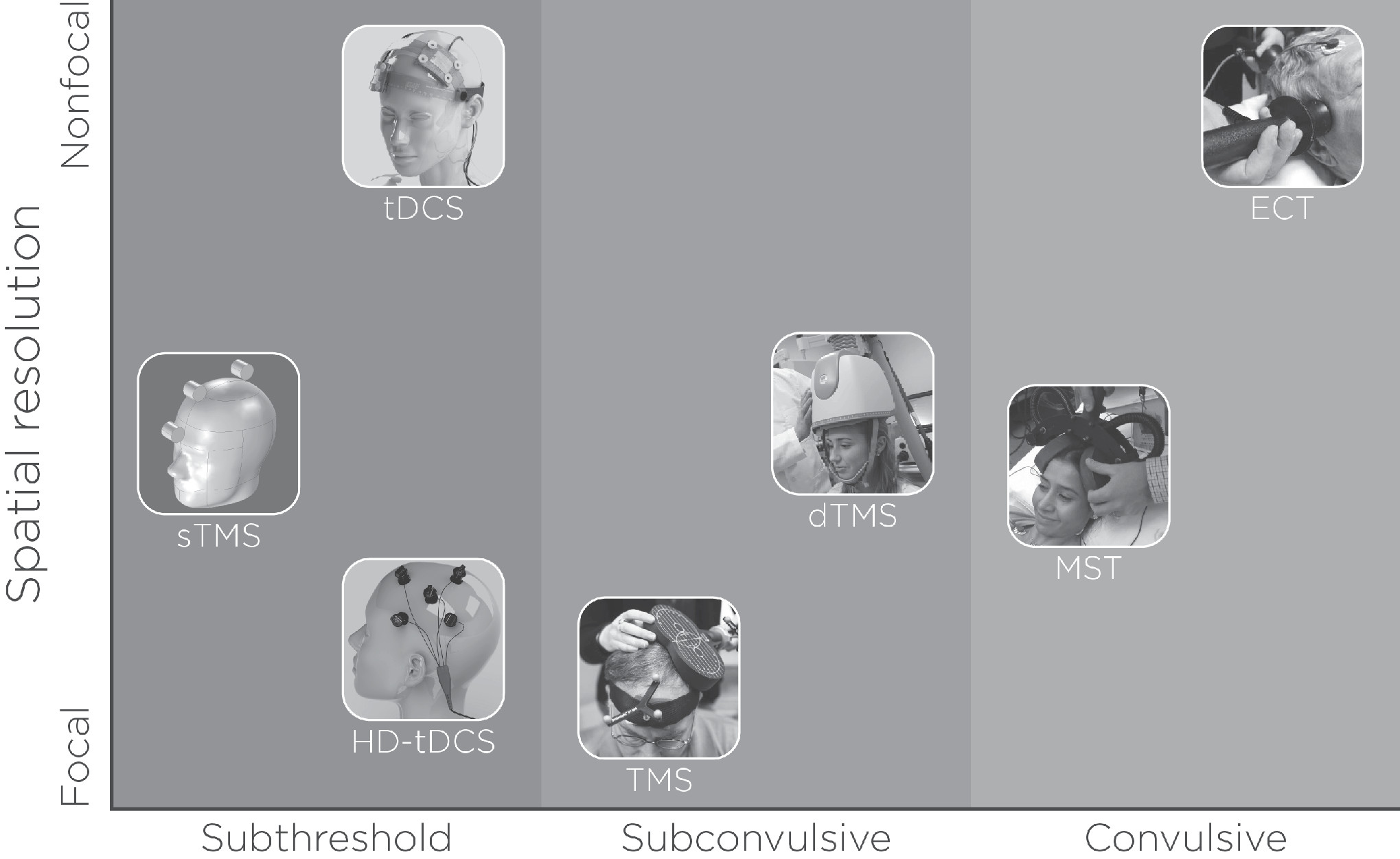
With the prevalence of major depressive disorder and TRD, as well as the associated increased functional impairment, morbidity, and mortality (12), the past three decades have seen a proliferation of noninvasive neuromodulation therapies (see Figure 1). In addition to ECT, such therapies include transcranial magnetic stimulation (TMS), magnetic seizure therapy (MST), and transcranial direct current stimulation (tDCS). To date, of these four strategies, only ECT and TMS are cleared for the treatment of depression in the United States by the U.S. Food and Drug Administration (FDA) (13, 14). MST and tDCS are considered investigational.
FIGURE 1. Noninvasive neuromodulation therapiesa
aNoninvasive neuromodulation therapies vary in their spatial resolution (focal and nonfocal) and intensity (subthreshold, subconvulsive, and convulsive). Transcranial direct current stimulation (tDCS) is subthreshold and can be focal or nonfocal, depending on the electrode configuration (HD-tDCS, high-definition transcranial direct current stimulation). Transcranial magnetic stimulation (TMS) is subconvulsive and is relatively focal, depending on the coil type (dTMS, deep transcranial magnetic stimulation; sTMS, synchronized transcranial magnetic stimulation). Electroconvulsive therapy (ECT) and magnetic seizure therapy (MST) are both convulsive; the former is nonfocal, and the latter is relatively focal.
Although all four approaches are considered noninvasive neuromodulation antidepressant therapies, they are unique in their application method, behavioral (antidepressant and neurocognitive) effects, and possible mechanisms of action. Considerable research has found consistent, although at times mixed, findings for the four therapies. Indeed, because each therapy can be applied in a multitude of ways to modulate cortical and subcortical brain regions and brain connectivity, there can be an infinite number of brain and behavior relationships (15).
The purpose of this critical review article is to synthesize comprehensive information regarding the application of four noninvasive neuromodulation therapies (ECT, TMS, MST, and tDCS) in the treatment of MDD and TRD. Specific information focuses on antidepressant effects, neurocognitive effects, and possible mechanisms of action.

ECT

ECT is a noninvasive neuromodulation therapy that uses electrical stimuli to induce a therapeutic tonic-clonic convulsive seizure. Diagnostic indications for ECT include major depressive disorder or TRD, bipolar disorder, schizophrenia, and schizoaffective disorders (16). ECT is a first-line treatment when a rapid and definitive response is needed urgently, as in the case of catatonia, risk of harm to self or others, or major depressive disorder with psychotic features. ECT can also be a second-line treatment when medication trials and augmentation strategies have failed to treat an acute mood or psychotic episode.
ECT can be divided into acute, continuation, and maintenance phases of treatment. The acute (also known as series or index) phase of ECT consists of approximately 12 sessions administered either twice or three times weekly. Treatment includes individualized end points of remission, clinical plateau, or nonresponse, usually defined by a standardized depression symptom severity measure. Continuation ECT includes the first six months after an ECT acute course and involves either a scheduled taper of treatments from weekly to monthly or symptom-triggered treatments to prolong remission (17, 18). Maintenance ECT includes regularly scheduled treatments at fixed intervals six months after the ECT acute course to prevent the recurrence of the psychiatric episode (e.g., major depressive disorder) (19).
ECT has very large effect sizes relative to sham ECT (Cohen’s d=0.91) and pharmacotherapy (Cohen’s d=0.80) (20), but the ability to predict an individual patient’s response with clinical and demographic factors is modest (21, 22). Treatment resistance to pharmacotherapy and longer duration of a major depressive episode (longer than two years) are the primary factors predictive of ECT nonresponse. Other factors, such as age, sex, diagnosis (bipolar versus unipolar), and depressive subtype (atypical, melancholic), have not been found to be predictive of clinical outcomes (21, 22). Although such techniques are not ready for clinical use, recent research has focused on neuroimaging (i.e., magnetic resonance imaging [MRI]) data-driven methods to improve prediction of ECT response (2326) and relapse (27).
Both the stimulus delivery and the seizure induction seem to work in synergy with respect to clinical response. ECT electrode configuration (d’Elia right unilateral, bifrontal, or bitemporal) dictates the geometry of the induced electric field in the brain (28). ECT clinicians often start with a focal (right unilateral) and switch to a less focal (bitemporal, bifrontal) electrode configuration in the context of modest improvement midway through the acute ECT course (29, 30). The ECT pulse train includes pulse width, amplitude, frequency, and train duration.
Although research on quantitative electroencephalography (EEG) analysis has yet to identify the characteristics of the seizure associated with efficacy (31), most ECT clinicians focus on seizure duration (minimum of 15–20 seconds), with the rationale that shorter seizure duration is reflective of inadequate electric stimulation (16). Adjustments to total charge to ensure adequate seizure duration include sequential increases in pulse train duration and then frequency (16, 28). With pulse amplitude and pulse width constant, this method of increasing total charge results in a larger number and faster rate of identical pulses or stimulations. In contrast, increased pulse amplitude would increase the magnitude of the induced electric field, but to date that ECT pulse parameter remains fixed and unadjusted in standard clinical practice (28).
Pulse width is usually dichotomized into brief (≥0.5 ms) and ultrabrief (<0.5 ms). The ultrabrief pulse width is closer to the chronaxie (stimulus duration required for neuronal firing) but requires increased charge (six times the seizure threshold) with right unilateral electrode placement to maintain clinical efficacy (29). Collectively, the multiple ECT parameters are used together to produce clinical outcome. For example, bitemporal electrode configuration at brief pulse width and 1.5–2.5 times the seizure threshold, on the one hand, and right-unilateral electrode configuration at brief or ultrabrief pulse width at more than five times the seizure threshold, on the other, produce relatively equivalent clinical outcomes (32).
The underlying mechanism of action for ECT has yet to be determined. Pretranslational studies have determined that electroconvulsive stimulations result in hippocampal neuroplasticity through increased synaptogenesis, angiogenesis, gliogenesis, and possibly neurogenesis (33, 34). Consistent with these pretranslational investigations, neuroimaging studies have identified transient increased medial temporal lobe volume during the ECT series (35, 36). It is interesting that many of these investigations have found larger volumetric increases ipsilateral to the side of direct electrical stimulation (3739). However, there is generally no direct association with decreased depression severity or clinical improvement (40). ECT’s mechanism of action may also include anticonvulsant mechanisms related to the empiric observation of shortening seizure duration, increased seizure threshold, and increased gamma-Aminobutyric acid (inhibitory neurotransmitter) during an ECT course (41, 42), but there is no established relationship with changes in anticonvulsant properties and clinical outcomes (43).
Independent of ECT-associated antidepressant effects, many patients experience transient neurocognitive impairment (44). The neurocognitive side effects affect all cognitive domains, but anterograde and retrograde memory recall are the domains most affected by the procedure (45). Immediately after the acute ECT course, cognitive side effects are common (effect sizes between 0.11 and −1.12, depending on the cognitive domain), but patients typically return to the pre-ECT baseline within two to four weeks after the ECT series. The ECT stimulus-delivery parameters differentially affect neurocognitive impairment in the opposite pattern of ECT efficacy, such that focal electrode placement (right unilateral) and ultrabrief pulse widths may be associated with fewer adverse cognitive effects relative to nonfocal electrode placement (bitemporal) and brief pulse widths (32, 46).
A positron emission tomography investigation demonstrated reduced medial temporal lobe brain metabolism, which was hypothesized to be related to ECT-induced neurocognitive impairment (47). As such, recent efforts to develop new and more focal ECT stimulation techniques have attempted to avoid stimulation of the medial temporal lobe brain structures (48). Preclinical investigations demonstrated that hippocampal neuroplasticity and remodeling may adversely affect established memories (49). However, recent ECT-MRI investigations have neither identified an anatomic location of ECT-mediated neurocognitive impairment nor established a relationship with hippocampal volumetric changes and neurocognitive impairment (50).
Further work is needed to understand how ECT modulates neurocircuitry to produce beneficial clinical and adverse neurocognitive effects. Clinical research has demonstrated that ECT neurocognitive impairment is unrequired for clinical efficacy (44). As described above, the role of stimulus parameters represents a trade-off between efficacy and clinical response. In addition, individual variability further confounds the relationship between clinical outcome and stimulus parameters. For example, current ECT practice applies electrical stimuli with fixed pulse amplitude at either 800 or 900 mA, with no clinical or scientific justification (28). Pulse amplitude and the related electric field are influenced by skin, skull, cerebral spinal fluid, and gray and white matter conductivities, which lead to marked variability despite a constant current at the scalp.
A preclinical investigation with electric field modeling demonstrated that there was more than 50% interindividual variability in the amount of stimulated brain volume with a fixed amplitude with right unilateral electrode configuration (51). This difference in electric fields could produce markedly different clinical and neurocognitive outcomes despite the application of similar pulse amplitudes at the scalp. To standardize the ECT dose with respect to pulse amplitude, ECT clinicians may use amplitude titration, in addition to other ECT parameters, in the future to eliminate the variability related to anatomic differences to maximize clinical benefits while minimizing neurocognitive effects.

TMS

TMS is a noninvasive neuromodulation technique that uses magnetic induction of an electric field in the brain that is generated from the magnetic field properties of the TMS coil (52). The electric field produced by TMS can depolarize neurons. Furthermore, TMS pulses can be applied repetitively (i.e., repetitive TMS [rTMS]), which results in excitatory or inhibitory modulation of brain activity in the targeted area and connected brain regions, depending on the stimulation parameters.
TMS has been most widely used as a treatment for major depressive disorder and TRD and was first cleared for the treatment of depression by the U.S. FDA in 2008. In addition, TMS has also been investigated for a variety of neuropsychiatric and neurological conditions, including the treatment of migraine headache (53), preoperative motor (54) and language (55) mapping, treatment for poststroke motor function recovery (56), auditory hallucinations (57), and more. Clinical TMS for treatment of depression has generally used high-frequency (≥1 Hz) stimulation administered at 10 Hz to 20 Hz (58, 59), most commonly targeted to the left dorsolateral prefrontal cortex (L-DLPFC). Less frequently, the right DLPFC has been targeted with low-frequency rTMS (≤1 Hz), a protocol that has been demonstrated through multiple clinical trials to be equivalent to high-frequency TMS in both safety and efficacy (60).
A relatively new form of TMS-induced neuromodulation, called synchronized TMS, has been explored as a way to deliver TMS stimuli at an individualized alpha frequency determined from real-time integrated EEG recording (61). This method uses rotating cylindrical magnets to deliver low-energy, sinusoidal-waveform stimulation over a broad area of the cortex. Industry-supported preliminary studies have demonstrated safety and efficacy relative to sham treatment (62).
To date, three different coil types (focal iron core coil, H1-coil, figure-eight coil) have been cleared by the U.S. FDA. For each magnetic pulse, the peak magnetic field strength generated by the coil is thought to be approximately 1.5 Tesla (63). Although this magnetic field strength is similar to that of a typical MRI device commonly used in clinical settings, the magnetic field generated by TMS is relatively more focal and brief (52).
In 2018, the U.S. FDA approved theta-burst stimulation (TBS) for use in the clinical treatment of major depressive disorder. TBS seems to mimic the brain’s endogenous theta rhythms, which is thought to improve induction of synaptic long-term potentiation (64). TBS has received recent attention, and several clinical trials have emerged that used this method. For example, intermittent TBS, which imparts 600 pulses in three minutes, showed clinical effects that were not inferior to conventional 10-Hz treatment, which lasts approximately 37 minutes (65). From a clinical practice pragmatic perspective, TBS’s characteristically high-frequency stimulation method may be advantageous because of its shorter per-session treatment duration.
With some exception (66), an abundance of data pertain to the safety and efficacy of TMS for individuals with major depressive disorder and TRD. Early studies of TMS efficacy found that a two-week rTMS trial (although studies might vary greatly in the parameters used, including stimulation site) was superior to sham or control stimulation (67). The TMS-associated antidepressant efficacy is comparable to that of pharmacotherapeutic intervention for depression (68). An industry-sponsored, landmark multisite randomized controlled clinical trial from 23 centers and 301 patients demonstrated that 10-Hz rTMS applied to the L-DLPFC was associated with decreased depression severity (69). The degree of prior treatment resistance, shorter current episode of depression, and absence of comorbid anxiety disorder predicted better antidepressant outcome with rTMS treatment, which indicates that potential demographic differences may moderate the efficacy of rTMS (69).
Another multisite, randomized, active sham-controlled study was sponsored by the National Institute of Mental Health. This study investigated daily treatment using the same high-frequency stimulation to the L-DLPFC, provided for up to three weeks (if at three weeks there was no antidepressant benefit, patients were switched to a different TMS condition), among patients with nonpsychotic major depressive disorder or TRD who were free of antidepressant medication. The study demonstrated that active rTMS treatment achieved a remission rate 4.2 times greater than that of sham, with 12 rTMS sessions needed to treat (70).
Moreover, a study that pooled data from multiple clinical TMS sites with 307 outpatients assessed clinical outcomes, as measured by changes in clinical rating and self-report depression symptom severity scales. Findings showed that TMS produced clinically significant antidepressant effects (71). rTMS treatments have also been reported to alleviate a broad array of depressive symptom dimensions (e.g., mood, anxiety, somatic), except sleep disturbance, which seems to be resistant to the general clinical benefit provide by TMS (72, 73). The durability of TMS-associated clinical benefit was demonstrated in a six-month naturalistic follow-up study, such that only approximately 10% of patients seemed to have a relapse of their depressive episode (74).
Although findings vary across studies and specific cognitive domains, the majority of TMS trials in depression demonstrated improvement or no effect of TMS on cognitive functioning. Of relevance, relative to the temporary adverse cognitive effects reported following ECT (7577), no marked memory inefficiencies or impairments have been found following treatment with a TMS course. A recent review investigated cognitive outcomes across 15 studies among patients with depression who were treated with rTMS (78). Five of nine studies reported positive effects on executive functioning, one of 12 found positive effects on processing speed, one of five saw improvement in attention, three of 11 found positive effects in verbal memory, and two of seven saw improvement in working memory. It is important to note that a number of the cognitive measures used measured more than one cognitive function; thus, classifying a given measure strictly into a cognitive subdomain could be misleading.
It should be noted that studies of depression measuring cognitive outcomes vary with regard to study methodology, including the presence of a sham condition, clinical trial duration, TMS parameters (e.g., pulse frequency, intensity, train duration), and sample size (with the majority having fewer than 25 participants) (79). In an earlier review, Guse and colleagues (80) concluded that high-frequency (10–20-Hz) rTMS applied to the L-DLPFC over 10–15 sessions with a stimulation intensity at 80%−110% of individualized motor threshold was most likely to result in significant cognitive improvement. Collectively, there are discrepant findings regarding the effects of rTMS on cognitive function, and additional research is warranted to confirm whether rTMS modulates cognitive abilities when it is applied for the treatment of major depressive disorder and TRD.
Although direct electrical stimulation of the brain is thought to excite the pyramidal tract axons of neurons directly, TMS is considered to transynaptically excite the pyramidal neurons. The precise underlying mechanism of rTMS-dependent cortical excitability changes, though, remains unknown. However, the long-term effects of rTMS are hypothesized to arise from induction of cortical plasticity, which results from gene induction and protein synthesis (81).
Depending on the TMS treatment parameters used, TMS can have either an excitatory or an inhibitory effect on local brain function, the latter of which has been used to investigate cerebral function of target regions. The clinical rationale for using rTMS as a treatment tool for major depressive disorder is the notion that rTMS is able to exert a lasting impact on brain function long after discontinuation of the stimulation (82). As for the underlying biophysical mechanism of high- and low-frequency rTMS, processes similar to long-term potentiation and long-term depression have been suggested as similar molecular mechanisms behind long-term changes that result from rTMS (83). Currently, though, there is no evidence of synaptic change following rTMS (84).
Continued work is needed to identify the optimal target, dosage, and duration of TMS treatment for a given individual. Because major depressive disorder is a heterogeneous illness with cognitive, affective, and physiological symptom dimensions, future studies must carefully consider which symptoms will be targeted by TMS and the mechanisms of action that underlie any observed changes. Moreover, it is important to incorporate a personalized medicine approach, investigating which therapies work best and for whom.
For instance, although replication is needed, Drysdale and colleagues, in research with a large multisite sample, suggested that functional connectivity patterns based on functional MRI among patients with TRD could be classified into distinct biotypes and that this classification predicted the patients’ clinical response to rTMS (85). The DLPFC has been the most frequent site of stimulation used for treating depression with TMS, but other target sites (e.g., right orbitofrontal cortex, dorsomedial prefrontal cortex) may be beneficial for treating particular depressive symptom dimensions. Finally, there is a need to better characterize the network-level changes directly associated with TMS through use of tools such as integrated functional MRI and EEG.

MST

MST is a form of noninvasive neuromodulation convulsive therapy that is currently under investigation. The MST methodology was introduced in 2001 by Lisanby and colleagues in a preclinical study (86), and the first human studies in TRD were subsequently published (8789). The therapeutic tonic-clonic convulsive seizure in MST is produced by rapidly changing magnetic stimuli that are applied to the brain; the technology is based on rTMS. However, MST differs from TMS in that the magnetic stimuli are administered at much higher stimulation intensities and frequencies (90). Also, unlike rTMS but similar to ECT, the MST sessions are performed while the patient is under general anesthesia.
The first MST treatments were administered with pulse frequencies of 40–50 Hz (88, 89, 91), but it was later observed that 100 Hz at maximum stimulator output might more reliably induce seizures (92). Contradictory results exist, and some studies have suggested that better seizure efficacy could be obtained with lower frequencies of around 20–40 Hz (28). Further research is needed to confirm the optimal MST pulse frequency, however.
MST can be applied with different electromagnetic coils, such as round, double cone, and cap cone (93). In recent years, a twin coil that consists of two cone-shaped coils positioned bilaterally has become the most used coil design in application of MST (9496). The stimulation waveform applied in MST is biphasic, which, relative to monophasic pulses, is associated with less coil heating and greater power efficiency (97).
The general aim of MST is to produce therapeutic seizures and clinical efficacy similar to those of ECT, while avoiding ECT-induced adverse cognitive effects (e.g., anterograde and retrograde amnesia) (93). MST can produce focal seizures that originate from superficial cortical areas, because magnetic fields can only penetrate approximately 2 cm below the surface of the cortex (52, 98). The seizure may spread to other brain areas (e.g., DLPFC), similar to that in ECT, and includes both an ictal and a motor phase (99). However, because MST provides superficial stimulation, unlike ECT, it may avoid stimulating subcortical medial-temporal lobe structures (e.g., hippocampal formation). According to computer modeling studies, MST predominantly stimulates the cortex and approximately 21% of the total brain volume (100, 101). In addition, because of the use of magnetic fields, MST is different than ECT in that the magnetic stimuli are unaffected by differences in scalp characteristics, skull thickness, or total brain volume (100).
To date, MST has been successfully applied among patients with TRD (both unipolar depression and bipolar disorder) (102) and patients with treatment-resistant schizophrenia (103). Several clinical studies in TRD have demonstrated MST to have antidepressant properties (88, 89, 104106). It is of note that studies that compared and contrasted the efficacy of MST with ECT found no differences between the two therapies in terms of antidepressant clinical outcomes (95, 104). The response and remission rates in MST have varied around 38%−69% and 15%−46%, respectively (104106), and it has been observed to reduce suicidality (96).
Regarding cognitive effects, MST has been found to have little to no associated adverse cognitive effects (93, 102, 107). Instead, preclinical and clinical studies have reported that MST may result in improved cognitive functions in domains such as psychomotor processing speed, visual attention, visuospatial learning and memory, verbal memory, and executive function (95, 105). The recovery (voluntary movement, respiratory effort, blood pressure, consciousness, and oxygen saturation) and reorientation time associated with MST are also substantially quicker relative to ECT, and patients have been reported to recover within approximately one to two minutes and to become reoriented within approximately three to five minutes after the termination of the seizure (104). When interpreting these outcomes, one needs to note, however, that the study sample sizes have been small, and studies have not included a sham-controlled condition (e.g., anesthesia only). For example, many studies have incorporated fewer than 20 patients (93, 99, 102, 107). Thus, further research in larger sample sizes is warranted to confirm and extend the MST safety outcome data.
The exact mechanisms of action of MST remain unknown, but it is assumed that MST may share some of the mechanisms that underlie ECT (91). For instance, previous positron emission tomography studies have found that MST produced regional glucose metabolism changes in brain areas that have abnormal function in major depressive disorder (105, 108). Following MST treatments, glucose metabolism increased in the basal ganglia, orbitofrontal cortex, medial frontal cortex, and DLPFC, and glucose metabolism simultaneously decreased in the left striatum (105, 108).
In most studies, the seizure characteristics measured with ictal EEG have differed between ECT and MST. Relative to ECT seizures, MST seizures commonly have less postictal suppression and smaller EEG amplitude in the ictal phase (99), and they may have shorter seizure length (99). However, there is some discrepancy in previous findings, which might in part be explained by the varying stimulation parameters. For example, the applied stimulation frequency in MST has been found to strongly influence the seizure characteristics (109). The adequacy of an MST seizure is conventionally defined by characteristics that have been noted to result in good clinical outcome in ECT. A recent study, though, challenged this view and found that the MST seizure characteristics might predict clinical outcome differentially than the seizure characteristics in ECT (109).
In summary, MST has great potential to evolve into a safe and efficacious noninvasive neuromodulation convulsive treatment for major depressive disorder and TRD. Further research is warranted regarding the mechanisms of action underlying MST, optimal stimulus parameters, and associated clinical outcome.

tDCS

tDCS is a noninvasive neuromodulation technique that involves the application of a subthreshold, weak direct electrical current to the brain through electrodes placed on the scalp. Before 2000, the technique was termed brain polarization and had been investigated in in vivo preclinical models for physiological effects (110), as well as in early human clinical trials as a treatment for major depressive disorder (111). Since then, it has been relabeled tDCS and has seen a major resurgence in clinical and research interest following the publication of several seminal studies that demonstrated acute and durable neuromodulatory effects with modern stimulation parameters (112, 113). This resurgence has been general and spanned the study of neurophysiological mechanisms, neurocognitive effects, and multiple potential therapeutic applications (114). To date, tDCS remains in development and has no U.S. FDA regulatory clearance for the treatment of any neuropsychiatric disorder.
The current used in tDCS is quite small (i.e., typically 1–2.5 mA), and it flows in a unidirectional manner through the brain, which produces polarity-dependent changes in neuronal excitability (115). When the technique is used to treat major depressive disorder, the anode (positively charged electrode) generally is placed over the L-DLPFC, with the cathode (negatively charged electrode) placed on the contralateral side of the head. Stimulation is then administered while participants are sitting awake, at rest, for durations of 20 to 30 minutes. tDCS treatments are usually repeated daily over several (e.g., four to six) weeks.
When tDCS is administered this way, there have been mixed results from modern clinical trials (116). Although several controlled clinical trials have reported significantly greater antidepressant effects with active compared with a sham stimulation (117121), other studies have failed to show superior antidepressant efficacy with active stimulation (122126). In addition, a recent large noninferiority controlled trial among patients with major depressive disorder who were off antidepressant medications found that standard antidepressant medication treatment, relative to tDCS, had superior efficacy (119). This evidence base therefore suggests that tDCS antidepressant effects are modest when that treatment methodology is used. More recent studies have further investigated the potential for enhanced efficacy when tDCS is administered with newer methodology, such as simultaneously with tasks that concurrently activate stimulated depression-related regions (127129), or when administered with multiple electrodes (referred to as high-definition tDCS) (130). However, although such newer tDCS methods seem to show promise, additional evidence is warranted from controlled clinical trials to confirm antidepressant efficacy.
The mechanisms underlying tDCS antidepressant effects remain poorly understood. Among healthy participants, tDCS treatment has no mood effects (131). Among depressed participants, mood effects occur only after several weeks of multiple repeated treatments, analogous to other antidepressant treatments (e.g., rTMS, antidepressant pharmacotherapy). Indeed, tDCS treatment has been shown to cause lasting neuroplastic changes (132), which is in line with evidence for structural changes with these other treatments (e.g., rTMS (133), sertraline (134)). Moreover, tDCS antidepressant effects have been shown to be modulated by the serotonergic system (121, 135), of which dysfunction is implicated in major depressive disorder and therefore is an important target for antidepressant strategies.
The acute cognitive effects of tDCS have been investigated for their potential role as a mechanism that underlies antidepressant effects. This has followed similar work with pharmacological therapeutic development and the proposed neuropsychological theory of antidepressant effects through modulation of information biases (136). Consistent with this theory, acute effects on affective biases with tDCS have been demonstrated in several clinical trials among patients with major depressive disorder (137141). To date, however, such effects have been unrelated to antidepressant outcomes (137). Acute positive effects on other, nonaffective measures have further been shown for processing speed (120) and visual attention (141, 142). These heterogeneous findings suggest nonspecific acute neurocognitive effects that involve dysfunctional networks in major depressive disorder, which is consistent with the generalized stimulation effects produced by tDCS (143).
Another question of clinical relevance is whether a repeated tDCS treatment course for major depressive disorder produces durable cognitive benefit. This was recently addressed in an individual-patient data meta-analysis that involved seven randomized, sham-controlled clinical trials (144). The results, however, showed that there was no cognitive enhancement following a tDCS course after mood effects were controlled. Similar to tDCS antidepressant effects, further research into alternative treatment methodologies for using tDCS in longer term cognitive enhancement applications is therefore warranted.
Although tDCS continues to remain a potential new treatment for major depressive disorder, current efficacy results indicate a need for further development and refinement in rigorous, controlled, large-scale clinical investigations. tDCS has several advantages relative to other noninvasive neuromodulation antidepressant techniques, including low cost, portability, and enhanced safety (114, 145). Collectively, these advantages support the translational potential of tDCS.
Promising future lines of research include the development of tDCS as an adjunct to other psychological interventions (e.g., cognitive-behavioral therapy, cognitive training) to further capitalize on its acute neuromodulatory and cognitive effects. Additional research is needed to determine the underlying neuromechanisms that subserve tDCS-induced antidepressant and neurocognitive effects. Emerging evidence has indicated that interindividual differences (i.e., structural and functional) may be promising variables in understanding how tDCS produces beneficial behavioral outcomes (144, 146).

Conclusions

Noninvasive neuromodulation therapies represent a third wave of antidepressant therapeutic application, after pharmacotherapy and psychotherapy, for the treatment of major depressive disorder and TRD. Those therapies vary in mode of application (electrical or magnetic stimuli), spatial resolution (focal, nonfocal), intensity (subthreshold, subconvulsive, convulsive), antidepressant effects, and neurocognitive effects (see Figure 1). Considerable research has provided substantial information regarding their antidepressant and neurocognitive effects (see Table 1).
TABLE 1. Summary of noninvasive neuromodulation therapeutics information
ConsiderationECTaTMSbMSTctDCSd
FDAe cleared (as of September 2018) to treat major depressive disorderECT was grandfathered in and is currently under review by the FDA for possible reclassificationYes; specific coils include the figure-eight coil and the H1 coilNoNo
Stimuli typeElectricMagneticMagneticElectric
Tool to apply stimuliElectrodesCoilCoilElectrodes
Inpatient or outpatient procedureInpatient and outpatientOutpatientInpatient and outpatientOutpatient
Anesthesia required for treatmentYesNoYesNo
Antidepressant effectsYesYesYesYes
Neurocognitive effectsTransient adverse effects that may vary in magnitude depending on treatment parametersBenign or beneficial effects that may vary in magnitude depending on treatment parametersBenign or beneficial effects that may vary in magnitude depending on treatment parametersAdverse, benign, or beneficial effects that may vary in magnitude depending on treatment parameters
a
ECT, electroconvulsive therapy.
b
TMS, transcranial magnetic stimulation.
c
MST, magnetic seizure therapy.
d
tDCS, transcranial direct current stimulation.
e
FDA, U.S. Food and Drug Administration.
ECT, as one of the first noninvasive neuromodulation therapies used as an antidepressant strategy, has considerable evidentiary support as being one of the most effective treatments for TRD and major depressive disorder with psychotic features (4). Research continues to examine how to modify ECT to improve its associated neurocognitive adverse effects (44). TMS, which was first cleared by the U.S. FDA as an antidepressant strategy in 2008, is useful to treat major depressive disorder and has a benign neurocognitive effect profile. Although its antidepressant effects are mild to moderate, research continues to examine how to maximize antidepressant benefit through new coil types (62), TMS paradigms (65), and integration with pharmacotherapeutic (60) and psychotherapeutic (147) strategies.
MST and tDCS are relatively newer, noninvasive neuromodulation therapies under development that use magnetic and electric stimuli, respectively, to produce antidepressant effects and minimize neurocognitive effects. Research has found MST to have moderate to high antidepressant benefits for patients with TRD, with no or positive cognitive effects (88, 95). Research has found tDCS to have modest antidepressant benefits, because of limited understanding of dosimetry (148), for patients with major depressive disorder, with no or positive cognitive effects (119, 123).
In conclusion, ECT, TMS, MST, and tDCS are four noninvasive neuromodulation therapies with antidepressant properties that can be beneficial for the treatment of major depressive disorder and TRD. Although the four therapies vary in how they modulate neurocircuitry and their resultant antidepressant and neurocognitive effects, they are nonetheless useful for patients with acute and chronic major depressive disorder and TRD. Continued research is warranted to inform dosimetry, algorithm for administration, and integration among these therapies and with other antidepressant strategies to continue to maximize their safety and antidepressant benefit.

Acknowledgments

The authors thank Dr. Zhi-De Deng for his expertise regarding noninvasive neuromodulation therapies and the creation of the graphics.

References

1.
Boes AD, Kelly MS, Trapp NT, et al: Noninvasive brain stimulation: challenges and opportunities for a new clinical specialty. J Neuropsychiatry Clin Neurosci 2018; 30:173–179
2.
Cook IA, Espinoza R, Leuchter AF: Neuromodulation for depression: invasive and noninvasive (deep brain stimulation, transcranial magnetic stimulation, trigeminal nerve stimulation). Neurosurg Clin N Am 2014; 25:103–116
3.
Wagner T, Valero-Cabre A, Pascual-Leone A: Noninvasive human brain stimulation. Annu Rev Biomed Eng 2007; 9:527–565
4.
Lisanby SH: Electroconvulsive therapy for depression. N Engl J Med 2007; 357:1939–1945
5.
Holtzheimer PE, Mayberg HS: Stuck in a rut: rethinking depression and its treatment. Trends Neurosci 2011; 34:1–9
6.
Anderson IM: Pharmacological treatment of unipolar depression; in Behavioral Neurobiology of Depression and Its Treatment: Current Topics in Behavioral Neurosciences. Edited by Cowen P, Sharp T, Lau J. Berlin, Springer, 2012
7.
Rush AJ, Trivedi MH, Wisniewski SR, et al: Bupropion-SR, sertraline, or venlafaxine-XR after failure of SSRIs for depression. N Engl J Med 2006; 354:1231–1242
8.
Fekadu A, Wooderson S, Donaldson C, et al: A multidimensional tool to quantify treatment resistance in depression: the Maudsley staging method. J Clin Psychiatry 2009; 70:177–184
9.
Trevino K, McClintock SM, McDonald Fischer N, et al: Defining treatment-resistant depression: a comprehensive review of the literature. Ann Clin Psychiatry 2014; 26:222–232
10.
Cowen PJ, Anderson IM: New approaches to treating resistant depression. BJPsych Adv 2015; 21:315–323
11.
Akil H, Gordon J, Hen R, et al: Treatment resistant depression: a multi-scale, systems biology approach. Neurosci Biobehav Rev 2018; 84:272–288
12.
Mokdad AH, Ballestros K, Echko M, et al: The state of US health, 1990–2016: burden of diseases, injuries, and risk factors among US states. JAMA 2018; 319:1444–1472
13.
McClintock SM, Reti IM, Carpenter LL, et al: Consensus recommendations for the clinical application of repetitive transcranial magnetic stimulation (rTMS) in the treatment of depression. J Clin Psychiatry 2018; 79:35–48
14.
Meeting to Discuss the Classification of Electroconvulsive Therapy Devices. Washington, DC, US Food and Drug Administration, 2011
15.
Deng ZD, McClintock SM, Oey NE, et al: Neuromodulation for mood and memory: from the engineering bench to the patient bedside. Curr Opin Neurobiol 2015; 30:38–43
16.
American Psychiatric Association: The Practice of Electroconvulsive Therapy: Recommendations for Treatment, Training, and Privileging, 2nd ed. Washington, DC, American Psychiatric Association, 2001
17.
Kellner CH, Husain MM, Knapp RG, et al: A novel strategy for continuation ECT in geriatric depression: phase 2 of the PRIDE Study. Am J Psychiatry 2016; 173:1110–1118
18.
Lisanby SH, Sampson S, Husain MM, et al: Toward individualized post-electroconvulsive therapy care: piloting the Symptom-Titrated, Algorithm-Based Longitudinal ECT (STABLE) intervention. J ECT 2008; 24:179–182
19.
Frederikse M, Petrides G, Kellner C: Continuation and maintenance electroconvulsive therapy for the treatment of depressive illness: a response to the National Institute for Clinical Excellence report. J ECT 2006; 22:13–17
20.
UK ECT Review Group: Efficacy and safety of electroconvulsive therapy in depressive disorders: a systematic review and meta-analysis. Lancet 2003; 361:799–808
21.
Dombrovski AY, Mulsant BH, Haskett RF, et al: Predictors of remission after electroconvulsive therapy in unipolar major depression. J Clin Psychiatry 2005; 66:1043–1049
22.
Haq AU, Sitzmann AF, Goldman ML, et al: Response of depression to electroconvulsive therapy: a meta-analysis of clinical predictors. J Clin Psychiatry 2015; 76:1374–1384
23.
Van Waarde JA, Scholte HS, Van Oudheusden LJ, et al: A functional MRI marker may predict the outcome of electroconvulsive therapy in severe and treatment-resistant depression. Mol Psychiatry 2015; 20:609–614
24.
Redlich R, Opel N, Grotegerd D, et al: Prediction of individual response to electroconvulsive therapy via machine learning on structural magnetic resonance imaging data. JAMA Psychiatry 2016; 73:557–564
25.
Wade BS, Joshi SH, Njau S, et al: Effect of electroconvulsive therapy on striatal morphometry in major depressive disorder. Neuropsychopharmacology 2016; 41:2481–2491
26.
Jiang R, Abbott CC, Jiang T, et al: SMRI biomarkers predict electroconvulsive treatment outcomes: accuracy with independent data sets. Neuropsychopharmacology 2018; 43:1078–1087
27.
Wade BSC, Sui J, Hellemann G, et al: Inter and intra-hemispheric structural imaging markers predict depression relapse after electroconvulsive therapy: a multisite study. Transl Psychiatry 2017; 7:1270
28.
Peterchev AV, Rosa MA, Deng ZD, et al: Electroconvulsive therapy stimulus parameters: rethinking dosage. J ECT 2010; 26:159–174
29.
Sackeim HA, Prudic J, Devanand DP, et al: A prospective, randomized, double-blind comparison of bilateral and right unilateral electroconvulsive therapy at different stimulus intensities. Arch Gen Psychiatry 2000; 57:425–434
30.
Lapidus KA, Kellner CH: When to switch from unilateral to bilateral electroconvulsive therapy. J ECT 2011; 27:244–246
31.
Perera TD, Luber B, Nobler MS, et al: Seizure expression during electroconvulsive therapy: relationships with clinical outcome and cognitive side effects. Neuropsychopharmacology 2004; 29:813–825
32.
Sackeim HA, Prudic J, Nobler MS, et al: Effects of pulse width and electrode placement on the efficacy and cognitive effects of electroconvulsive therapy. Brain Stimul 2008; 1:71–83
33.
Bouckaert F, Sienaert P, Obbels J, et al: ECT: its brain enabling effects: a review of electroconvulsive therapy-induced structural brain plasticity. J ECT 2014; 30:143–151
34.
Segi-Nishida E: Exploration of new molecular mechanisms for antidepressant actions of electroconvulsive seizure. Biol Pharm Bull 2011; 34:939–944
35.
Wilkinson ST, Sanacora G, Bloch MH: Hippocampal volume changes following electroconvulsive therapy: a systematic review and meta-analysis. Biol Psychiatry Cogn Neurosci Neuroimaging 2017; 2:327–335
36.
Takamiya A, Chung JK, Liang KC, et al: Effect of electroconvulsive therapy on hippocampal and amygdala volumes: systematic review and meta-analysis. Br J Psychiatry 2018; 212:19–26
37.
Abbott CC, Jones T, Lemke NT, et al: Hippocampal structural and functional changes associated with electroconvulsive therapy response. Transl Psychiatry 2014; 4:e483
38.
Dukart J, Regen F, Kherif F, et al: Electroconvulsive therapy-induced brain plasticity determines therapeutic outcome in mood disorders. Proc Natl Acad Sci USA 2014; 111:1156–1161
39.
Bouckaert F, De Winter FL, Emsell L, et al: Grey matter volume increase following electroconvulsive therapy in patients with late life depression: a longitudinal MRI study. J Psychiatry Neurosci 2016; 41:j105–114
40.
Oltedal L, Narr KL, Abbott C, et al: Volume of the human hippocampus and clinical response following electroconvulsive therapy. Biol Psychiatry 2018; 84:574–581
41.
Sackeim HA: The anticonvulsant hypothesis of the mechanisms of action of ECT: current status. J ECT 1999; 15:5–26
42.
Sanacora G, Mason GF, Rothman DL, et al: Increased cortical GABA concentrations in depressed patients receiving ECT. Am J Psychiatry 2003; 160:577–579
43.
Duthie AC, Perrin JS, Bennett DM, et al: Anticonvulsant mechanisms of electroconvulsive therapy and relation to therapeutic efficacy. J ECT 2015; 31:173–178
44.
McClintock SM, Choi J, Deng ZD, et al: Multifactorial determinants of the neurocognitive effects of electroconvulsive therapy. J ECT 2014; 30:165–176
45.
Semkovska M, McLoughlin DM: Objective cognitive performance associated with electroconvulsive therapy for depression: a systematic review and meta-analysis. Biol Psychiatry 2010; 68:568–577
46.
Semkovska M, Keane D, Babalola O, et al: Unilateral brief-pulse electroconvulsive therapy and cognition: effects of electrode placement, stimulus dosage and time. J Psychiatr Res 2011; 45:770–780
47.
Nobler MS, Oquendo MA, Kegeles LS, et al: Decreased regional brain metabolism after ect. Am J Psychiatry 2001; 158:305–308
48.
Nahas Z, Short B, Burns C, et al: A feasibility study of a new method for electrically producing seizures in man: focal electrically administered seizure therapy [FEAST]. Brain Stimul 2013; 6:403–408
49.
Akers KG, Martinez-Canabal A, Restivo L, et al: Hippocampal neurogenesis regulates forgetting during adulthood and infancy. Science 2014; 344:598–602
50.
Nordanskog P, Larsson MR, Larsson EM, et al: Hippocampal volume in relation to clinical and cognitive outcome after electroconvulsive therapy in depression. Acta Psychiatr Scand 2014; 129:303–311
51.
Lee WH, Lisanby SH, Laine AF, et al: Minimum electric field exposure for seizure induction with electroconvulsive therapy and magnetic seizure therapy. Neuropsychopharmacology 2017; 42:1192–1200
52.
Deng ZD, Lisanby SH, Peterchev AV: Electric field depth-focality tradeoff in transcranial magnetic stimulation: simulation comparison of 50 coil designs. Brain Stimul 2013; 6:1–13
53.
Lipton RB, Dodick DW, Silberstein SD, et al: Single-pulse transcranial magnetic stimulation for acute treatment of migraine with aura: a randomised, double-blind, parallel-group, sham-controlled trial. Lancet Neurol 2010; 9:373–380
54.
Tarapore PE, Tate MC, Findlay AM, et al: Preoperative multimodal motor mapping: a comparison of magnetoencephalography imaging, navigated transcranial magnetic stimulation, and direct cortical stimulation. J Neurosurg 2012; 117:354–362
55.
Sollmann N, Ille S, Hauck T, et al: The impact of preoperative language mapping by repetitive navigated transcranial magnetic stimulation on the clinical course of brain tumor patients. BMC Cancer 2015; 15:261
56.
Hsu WY, Cheng CH, Liao KK, et al: Effects of repetitive transcranial magnetic stimulation on motor functions in patients with stroke: a meta-analysis. Stroke 2012; 43:1849–1857
57.
Aleman A, Sommer IE, Kahn RS: Efficacy of slow repetitive transcranial magnetic stimulation in the treatment of resistant auditory hallucinations in schizophrenia: a meta-analysis. J Clin Psychiatry 2007; 68:416–421
58.
George MS, Post RM: Daily left prefrontal repetitive transcranial magnetic stimulation for acute treatment of medication-resistant depression. Am J Psychiatry 2011; 168:356–364
59.
George MS, Taylor JJ, Short EB: The expanding evidence base for rTMS treatment of depression. Curr Opin Psychiatry 2013; 26:13–18
60.
Berlim MT, Van den Eynde F, Daskalakis ZJ: High-frequency repetitive transcranial magnetic stimulation accelerates and enhances the clinical response to antidepressants in major depression: a meta-analysis of randomized, double-blind, and sham-controlled trials. J Clin Psychiatry 2013; 74:e122–e129
61.
Jin Y, Phillips B: A pilot study of the use of EEG-based synchronized transcranial magnetic stimulation (sTMS) for treatment of major depression. BMC Psychiatry 2014; 14:13
62.
Leuchter AF, Cook IA, Feifel D, et al: Efficacy and safety of low-field synchronized transcranial magnetic stimulation (sTMS) for treatment of major depression. Brain Stimul 2015; 8:787–794
63.
Roth Y, Amir A, Levkovitz Y, et al: Three-dimensional distribution of the electric field induced in the brain by transcranial magnetic stimulation using figure-8 and deep H-coils. J Clin Neurophysiol 2007; 24:31–38
64.
Suppa A, Huang YZ, Funke K, et al: Ten years of theta burst stimulation in humans: established knowledge, unknowns and prospects. Brain Stimul 2016; 9:323–335
65.
Blumberger DM, Vila-Rodriguez F, Thorpe KE, et al: Effectiveness of theta burst versus high-frequency repetitive transcranial magnetic stimulation in patients with depression (THREE-D): a randomised non-inferiority trial. Lancet 2018; 391:1683–1692
66.
Yesavage JA, Fairchild JK, Mi Z, et al: Effect of repetitive transcranial magnetic stimulation on treatment-resistant major depression in US veterans: A randomized clinical trial. JAMA Psychiatry 2018; 75:884–893
67.
Loo CK, Mitchell PB: A review of the efficacy of transcranial magnetic stimulation (TMS) treatment for depression, and current and future strategies to optimize efficacy. J Affect Disord 2005; 88:255–267
68.
Demitrack MA, Thase ME: Clinical significance of transcranial magnetic stimulation (TMS) in the treatment of pharmacoresistant depression: synthesis of recent data. Psychopharmacol Bull 2009; 42:5–38
69.
Lisanby SH, Husain MM, Rosenquist PB, et al: Daily left prefrontal repetitive transcranial magnetic stimulation in the acute treatment of major depression: clinical predictors of outcome in a multisite, randomized controlled clinical trial. Neuropsychopharmacology 2009; 34:522–534
70.
George MS, Lisanby SH, Avery D, et al: Daily left prefrontal transcranial magnetic stimulation therapy for major depressive disorder: a sham-controlled randomized trial. Arch Gen Psychiatry 2010; 67:507–516
71.
Carpenter LL, Janicak PG, Aaronson ST, et al: Transcranial magnetic stimulation (TMS) for major depression: a multisite, naturalistic, observational study of acute treatment outcomes in clinical practice. Depress Anxiety 2012; 29:587–596
72.
O’Reardon JP, Solvason HB, Janicak PG, et al: Efficacy and safety of transcranial magnetic stimulation in the acute treatment of major depression: a multisite randomized controlled trial. Biol Psychiatry 2007; 62:1208–1216
73.
Rosenquist PB, Krystal A, Heart KL, et al: Left dorsolateral prefrontal transcranial magnetic stimulation (TMS): sleep factor changes during treatment in patients with pharmacoresistant major depressive disorder. Psychiatry Res 2013; 205:67–73
74.
Janicak PG, Nahas Z, Lisanby SH, et al: Durability of clinical benefit with transcranial magnetic stimulation (TMS) in the treatment of pharmacoresistant major depression: assessment of relapse during a 6-month, multisite, open-label study. Brain Stimul 2010; 3:187–199
75.
Sackeim HA, Portnoy S, Neeley P, et al: Cognitive consequences of low-dosage electroconvulsive therapy. Ann N Y Acad Sci 1986; 462:326–340
76.
Weiner RD, Rogers HJ, Davidson JR, et al: Effects of stimulus parameters on cognitive side effects. Ann N Y Acad Sci 1986; 462:315–325
77.
Squire LR: Memory and electroconvulsive therapy. Am J Psychiatry 1982; 139:1221
78.
Iimori T, Nakajima S, Miyazaki T, et al: Effectiveness of the prefrontal repetitive transcranial magnetic stimulation on cognitive profiles in depression, schizophrenia, and Alzheimer’s disease: a systematic review. Prog Neuropsychopharmacol Biol Psychiatry 2019; 88:31–40
79.
Martin DM, McClintock SM, Forster JJ, et al: Cognitive enhancing effects of rTMS administered to the prefrontal cortex in patients with depression: a systematic review and meta-analysis of individual task effects. Depress Anxiety 2017; 34:1029–1039
80.
Guse B, Falkai P, Wobrock T: Cognitive effects of high-frequency repetitive transcranial magnetic stimulation: a systematic review. J Neural Transm (Vienna) 2010; 117:105–122
81.
Kamble N, Netravathi M, Pal PK: Therapeutic applications of repetitive transcranial magnetic stimulation (rTMS) in movement disorders: a review. Parkinsonism Relat Disord 2014; 20:695–707
82.
Terao Y, Ugawa Y: Basic mechanisms of TMS. J Clin Neurophysiol 2002; 19:322–343
83.
Hallett M: Transcranial magnetic stimulation: a primer. Neuron 2007; 55:187–199
84.
Wassermann EM, Lisanby SH: Therapeutic application of repetitive transcranial magnetic stimulation: a review. Clin Neurophysiol 2001; 112:1367–1377
85.
Drysdale AT, Grosenick L, Downar J, et al: Resting-state connectivity biomarkers define neurophysiological subtypes of depression. Nat Med 2017; 23:28–38
86.
Lisanby SH, Luber B, Finck AD, et al: Deliberate seizure induction with repetitive transcranial magnetic stimulation in nonhuman primates. Arch Gen Psychiatry 2001; 58:199–200
87.
Lisanby SH, Schlaepfer TE, Fisch HU, et al: Magnetic seizure therapy of major depression. Arch Gen Psychiatry 2001; 58:303–305
88.
Lisanby SH, Luber B, Schlaepfer TE, et al: Safety and feasibility of magnetic seizure therapy (MST) in major depression: randomized within-subject comparison with electroconvulsive therapy. Neuropsychopharmacology 2003; 28:1852–1865
89.
Kosel M, Frick C, Lisanby SH, et al: Magnetic seizure therapy improves mood in refractory major depression. Neuropsychopharmacology 2003; 28:2045–2048
90.
Lefaucheur JP, André-Obadia N, Antal A, et al: Evidence-based guidelines on the therapeutic use of repetitive transcranial magnetic stimulation (rTMS). Clin Neurophysiol 2014; 125:2150–2206
91.
Rowny SB, Benzl K, Lisanby SH: Translational development strategy for magnetic seizure therapy. Exp Neurol 2009; 219:27–35
92.
Spellman T, McClintock SM, Terrace H, et al: Differential effects of high-dose magnetic seizure therapy and electroconvulsive shock on cognitive function. Biol Psychiatry 2008; 63:1163–1170
93.
McClintock SM, Tirmizi O, Chansard M, et al: A systematic review of the neurocognitive effects of magnetic seizure therapy. Int Rev Psychiatry 2011; 23:413–423
94.
Kayser S, Bewernick BH, Wagner S, et al: Clinical predictors of response to magnetic seizure therapy in depression: a preliminary report. J ECT (Epub ahead of print, April 2, 2018)
95.
Fitzgerald PB, Hoy KE, Elliot D, et al: A pilot study of the comparative efficacy of 100 Hz magnetic seizure therapy and electroconvulsive therapy in persistent depression. Depress Anxiety 2018; 35:393–401
96.
Sun Y, Farzan F, Mulsant BH, et al: Indicators for remission of suicidal ideation following magnetic seizure therapy in patients with treatment-resistant depression. JAMA Psychiatry 2016; 73:337–345
97.
Jalinous R: Principles of magnetic stimulator design; in Handbook of Transcranial Magnetic Stimulation. Edited by Pascual-Leone A, Davey N, Rothwell JC, et al. London, Arnold, 2002
98.
Deng ZD, Lisanby SH, Peterchev AV: Effect of anatomical variability on electric field characteristics of electroconvulsive therapy and magnetic seizure therapy: a parametric modeling study. IEEE Trans Neural Syst Rehabil Eng 2015;23(1):22–31.
99.
Hoy KE, Fitzgerald PB: Magnetic seizure therapy for treatment-resistant depression. Expert Rev Med Devices 2011; 8:723–732
100.
Deng ZD, Lisanby SH, Peterchev AV: Effect of anatomical variability on neural stimulation strength and focality in electroconvulsive therapy (ECT) and magnetic seizure therapy (MST). Conf Proc IEEE Eng Med Biol Soc 2009; 2009:682–688.
101.
Deng ZD, Lisanby SH, Peterchev AV: Electric field strength and focality in electroconvulsive therapy and magnetic seizure therapy: a finite element simulation study. J Neural Eng 2011; 8:016007
102.
Cretaz E, Brunoni AR, Lafer B: Magnetic seizure therapy for unipolar and bipolar depression: a systematic review. Neural Plast 2015; 2015:521398
103.
Tang VM, Blumberger DM, McClintock SM, et al: Magnetic seizure therapy in treatment-resistant schizophrenia: a pilot study. Front Psychiatry 2018; 8:310
104.
Kayser S, Bewernick BH, Grubert C, et al: Antidepressant effects, of magnetic seizure therapy and electroconvulsive therapy, in treatment-resistant depression. J Psychiatr Res 2011; 45:569–576
105.
Kayser S, Bewernick BH, Matusch A, et al: Magnetic seizure therapy in treatment-resistant depression: clinical, neuropsychological and metabolic effects. Psychol Med 2015; 45:1073–1092
106.
Fitzgerald PB, Hoy KE, Herring SE, et al: Pilot study of the clinical and cognitive effects of high-frequency magnetic seizure therapy in major depressive disorder. Depress Anxiety 2013; 30:129–136
107.
Engel A, Kayser S: An overview on clinical aspects in magnetic seizure therapy. J Neural Transm (Vienna) 2016; 123:1139–1146
108.
Hoy KE, Thomson RH, Cherk M, et al: Effect of magnetic seizure therapy on regional brain glucose metabolism in major depression. Psychiatry Res 2013; 211:169–175
109.
Backhouse FA, Noda Y, Knyahnytska Y, et al: Characteristics of ictal EEG in magnetic seizure therapy at various stimulation frequencies. Clin Neurophysiol 2018; 129:1770–1779
110.
Bindman LJ, Lippold OC, Redfearn JW: The action of brief polarizing currents on the cerebral cortex of the rat (1) during current flow and (2) in the production of long-lasting after-effects. J Physiol 1964; 172:369–382
111.
Arul-Anandam AP, Loo C: Transcranial direct current stimulation: a new tool for the treatment of depression? J Affect Disord 2009; 117:137–145
112.
Nitsche MA, Paulus W: Excitability changes induced in the human motor cortex by weak transcranial direct current stimulation. J Physiol 2000; 527:633–639
113.
Nitsche MA, Paulus W: Sustained excitability elevations induced by transcranial DC motor cortex stimulation in humans. Neurology 2001; 57:1899–1901
114.
Bikson M, Grossman P, Thomas C, et al: Safety of transcranial direct current stimulation: Evidence based update 2016. Brain Stimul 2016; 9:641–661
115.
Rahman A, Reato D, Arlotti M, et al: Cellular effects of acute direct current stimulation: somatic and synaptic terminal effects. J Physiol 2013; 591:2563–2578
116.
Borrione L, Moffa AH, Martin D, et al: Transcranial direct current stimulation in the acute depressive episode: a systematic review of current knowledge. J ECT 2018; 34:153–163
117.
Fregni F, Boggio PS, Nitsche MA, et al: Treatment of major depression with transcranial direct current stimulation. Bipolar Disord 2006; 8:203–204
118.
Boggio PS, Rigonatti SP, Ribeiro RB, et al: A randomized, double-blind clinical trial on the efficacy of cortical direct current stimulation for the treatment of major depression. Int J Neuropsychopharmacol 2008; 11:249–254
119.
Brunoni AR, Moffa AH, Sampaio-Junior B, et al: Trial of electrical direct-current therapy versus escitalopram for depression. N Engl J Med 2017; 376:2523–2533
120.
Loo CK, Alonzo A, Martin D, et al: Transcranial direct current stimulation for depression: 3-week, randomised, sham-controlled trial. Br J Psychiatry 2012; 200:52–59
121.
Brunoni AR, Valiengo L, Baccaro A, et al: The sertraline vs. electrical current therapy for treating depression clinical study: results from a factorial, randomized, controlled trial. JAMA Psychiatry 2013; 70:383–391
122.
Loo CK, Sachdev P, Martin D, et al: A double-blind, sham-controlled trial of transcranial direct current stimulation for the treatment of depression. Int J Neuropsychopharmacol 2010; 13:61–69
123.
Loo CK, Husain MM, McDonald WM, et al: International randomized-controlled trial of transcranial direct current stimulation in depression. Brain Stimul 2018; 11:125–133
124.
Palm U, Schiller C, Fintescu Z, et al: Transcranial direct current stimulation in treatment resistant depression: a randomized double-blind, placebo-controlled study. Brain Stimul 2012; 5:242–251
125.
Blumberger DM, Tran LC, Fitzgerald PB, et al: A randomized double-blind sham-controlled study of transcranial direct current stimulation for treatment-resistant major depression. Front Psychiatry 2012; 3:74
126.
Bennabi D, Nicolier M, Monnin J, et al: Pilot study of feasibility of the effect of treatment with tDCS in patients suffering from treatment-resistant depression treated with escitalopram. Clin Neurophysiol 2015; 126:1185–1189
127.
Segrave RA, Arnold S, Hoy K, et al: Concurrent cognitive control training augments the antidepressant efficacy of tDCS: a pilot study. Brain Stimul 2014; 7:325–331
128.
Brunoni AR, Zanao TA, Vanderhasselt MA, et al: Enhancement of affective processing induced by bifrontal transcranial direct current stimulation in patients with major depression. Neuromodulation 2014; 17:138–142
129.
Martin DM, Teng JZ, Lo TY, et al: Clinical pilot study of transcranial direct current stimulation combined with cognitive emotional training for medication resistant depression. J Affect Disord 2018; 232:89–95
130.
Edwards D, Cortes M, Datta A, et al: Physiological and modeling evidence for focal transcranial electrical brain stimulation in humans: a basis for high-definition tDCS. Neuroimage 2013; 74:266–275
131.
Motohashi N, Yamaguchi M, Fujii T, et al: Mood and cognitive function following repeated transcranial direct current stimulation in healthy volunteers: a preliminary report. Neurosci Res 2013; 77:64–69
132.
Player MJ, Taylor JL, Weickert CS, et al: Increase in PAS-induced neuroplasticity after a treatment course of transcranial direct current stimulation for depression. J Affect Disord 2014; 167:140–147
133.
Lan MJ, Chhetry BT, Liston C, et al: Transcranial magnetic stimulation of left dorsolateral prefrontal cortex induces brain morophological changes in regions associated with a treatment resistant major depressive episode: an exploratory analysis. Brain Stimul 2016; 9:577–583
134.
Smith R, Chen K, Baxter L, et al: Antidepressant effects of sertraline associated with volume increases in dorsolateral prefrontal cortex. J Affect Disord 2013; 146:414–419
135.
Brunoni AR, Kemp AH, Shiozawa P, et al: Impact of 5-HTTLPR and BDNF polymorphisms on response to sertraline versus transcranial direct current stimulation: implications for the serotonergic system. Eur Neuropsychopharmacol 2013; 23:1530–1540
136.
Harmer CJ, Hill SA, Taylor MJ, et al: Toward a neuropsychological theory of antidepressant drug action: increase in positive emotional bias after potentiation of norepinephrine activity. Am J Psychiatry 2003; 160:990–992
137.
Boggio PS, Bermpohl F, Vergara AO, et al: Go-no-go task performance improvement after anodal transcranial DC stimulation of the left dorsolateral prefrontal cortex in major depression. J Affect Disord 2007; 101:91–98
138.
Brunoni AR, Boggio PS, De Raedt R, et al: Cognitive control therapy and transcranial direct current stimulation for depression: a randomized, double-blinded, controlled trial. J Affect Disord 2014; 162:43–49
139.
Wolkenstein L, Plewnia C: Amelioration of cognitive control in depression by transcranial direct current stimulation. Biol Psychiatry 2013; 73:646–651
140.
Moreno ML, Vanderhasselt MA, Carvalho AF, et al: Effects of acute transcranial direct current stimulation in hot and cold working memory tasks in healthy and depressed subjects. Neurosci Lett 2015; 591:126–131
141.
Brennan S, McLoughlin DM, O’Connell R, et al: Anodal transcranial direct current stimulation of the left dorsolateral prefrontal cortex enhances emotion recognition in depressed patients and controls. J Clin Exp Neuropsychol 2017; 39:384–395
142.
Gögler N, Willacker L, Funk J, et al: Single-session transcranial direct current stimulation induces enduring enhancement of visual processing speed in patients with major depression. Eur Arch Psychiatry Clin Neurosci 2017; 267:671–686
143.
Bai S, Dokos S, Ho KA, et al: A computational modelling study of transcranial direct current stimulation montages used in depression. Neuroimage 2014; 87:332–344
144.
Martin DM, Moffa A, Nikolin S, et al: Cognitive effects of transcranial direct current stimulation treatment in patients with major depressive disorder: an individual patient data meta-analysis of randomised, sham-controlled trials. Neurosci Biobehav Rev 2018; 90:137–145
145.
Nikolin S, Huggins C, Martin D, et al: Safety of repeated sessions of transcranial direct current stimulation: a systematic review. Brain Stimul 2018; 11:278–288
146.
Al-Kaysi AM, Al-Ani A, Loo CK, et al: Predicting tDCS treatment outcomes of patients with major depressive disorder using automated EEG classification. J Affect Disord 2017; 208:597–603
147.
Donse L, Padberg F, Sack AT, et al: Simultaneous rTMS and psychotherapy in major depressive disorder: clinical outcomes and predictors from a large naturalistic study. Brain Stimul 2018; 11:337–345
148.
Lisanby SH: Noninvasive brain stimulation for depression—the devil is in the dosing. N Engl J Med 2017; 376:2593–2594

Information & Authors

Information

Published In

History

Published in print: Winter 2019
Published online: 10 January 2019

Keywords

  1. Depression
  2. neuropsychology
  3. ECT
  4. neuromodulation
  5. TMS

Authors

Details

Shawn M. McClintock, Ph.D., MSCS [email protected]
Neurocognitive Research Laboratory, Department of Psychiatry, University of Texas (UT) Southwestern Medical Center, Dallas, Texas (McClintock, Kallioniemi, Martin); Division of Brain Stimulation and Neurophysiology, Department of Psychiatry and Behavioral Sciences, Duke University School of Medicine, Durham, North Carolina (McClintock); Black Dog Institute, Sydney, Australia, and School of Psychiatry, University of New South Wales, Sydney (Martin); Department of Psychiatry, University of Utah School of Medicine, Salt Lake City (Kim, Weisenbach); VA Salt Lake City, Mental Health Program (Weisenbach); Department of Psychiatry, University of New Mexico School of Medicine, Albuquerque (Abbott).
Elisa Kallioniemi, Ph.D.
Neurocognitive Research Laboratory, Department of Psychiatry, University of Texas (UT) Southwestern Medical Center, Dallas, Texas (McClintock, Kallioniemi, Martin); Division of Brain Stimulation and Neurophysiology, Department of Psychiatry and Behavioral Sciences, Duke University School of Medicine, Durham, North Carolina (McClintock); Black Dog Institute, Sydney, Australia, and School of Psychiatry, University of New South Wales, Sydney (Martin); Department of Psychiatry, University of Utah School of Medicine, Salt Lake City (Kim, Weisenbach); VA Salt Lake City, Mental Health Program (Weisenbach); Department of Psychiatry, University of New Mexico School of Medicine, Albuquerque (Abbott).
Donel M. Martin, Ph.D.
Neurocognitive Research Laboratory, Department of Psychiatry, University of Texas (UT) Southwestern Medical Center, Dallas, Texas (McClintock, Kallioniemi, Martin); Division of Brain Stimulation and Neurophysiology, Department of Psychiatry and Behavioral Sciences, Duke University School of Medicine, Durham, North Carolina (McClintock); Black Dog Institute, Sydney, Australia, and School of Psychiatry, University of New South Wales, Sydney (Martin); Department of Psychiatry, University of Utah School of Medicine, Salt Lake City (Kim, Weisenbach); VA Salt Lake City, Mental Health Program (Weisenbach); Department of Psychiatry, University of New Mexico School of Medicine, Albuquerque (Abbott).
Joseph U. Kim, Ph.D.
Neurocognitive Research Laboratory, Department of Psychiatry, University of Texas (UT) Southwestern Medical Center, Dallas, Texas (McClintock, Kallioniemi, Martin); Division of Brain Stimulation and Neurophysiology, Department of Psychiatry and Behavioral Sciences, Duke University School of Medicine, Durham, North Carolina (McClintock); Black Dog Institute, Sydney, Australia, and School of Psychiatry, University of New South Wales, Sydney (Martin); Department of Psychiatry, University of Utah School of Medicine, Salt Lake City (Kim, Weisenbach); VA Salt Lake City, Mental Health Program (Weisenbach); Department of Psychiatry, University of New Mexico School of Medicine, Albuquerque (Abbott).
Sara L. Weisenbach, Ph.D.
Neurocognitive Research Laboratory, Department of Psychiatry, University of Texas (UT) Southwestern Medical Center, Dallas, Texas (McClintock, Kallioniemi, Martin); Division of Brain Stimulation and Neurophysiology, Department of Psychiatry and Behavioral Sciences, Duke University School of Medicine, Durham, North Carolina (McClintock); Black Dog Institute, Sydney, Australia, and School of Psychiatry, University of New South Wales, Sydney (Martin); Department of Psychiatry, University of Utah School of Medicine, Salt Lake City (Kim, Weisenbach); VA Salt Lake City, Mental Health Program (Weisenbach); Department of Psychiatry, University of New Mexico School of Medicine, Albuquerque (Abbott).
Christopher C. Abbott, M.D., M.S.
Neurocognitive Research Laboratory, Department of Psychiatry, University of Texas (UT) Southwestern Medical Center, Dallas, Texas (McClintock, Kallioniemi, Martin); Division of Brain Stimulation and Neurophysiology, Department of Psychiatry and Behavioral Sciences, Duke University School of Medicine, Durham, North Carolina (McClintock); Black Dog Institute, Sydney, Australia, and School of Psychiatry, University of New South Wales, Sydney (Martin); Department of Psychiatry, University of Utah School of Medicine, Salt Lake City (Kim, Weisenbach); VA Salt Lake City, Mental Health Program (Weisenbach); Department of Psychiatry, University of New Mexico School of Medicine, Albuquerque (Abbott).

Notes

Send correspondence to Dr. McClintock ([email protected]).

Competing Interests

Dr. McClintock reports grant support from the NIH and teaching honoraria from TMS HealthSolutions. Dr. Martin reports grant support from the Brain and Behavior Foundation, travel support from Miyuki Giken, and a speaker’s honorarium from Lundbeck. Drs. Kim and Abbott report grant support from the NIH. Dr. Weisenbach reports grant support from the NIH and the Department of Veterans Affairs. Dr. Kallioniemi reports no financial relationships with commercial interests.

Funding Information

This work was supported in part by National Institutes of Health (NIH)/National Institute of Mental Health (NIMH) grants NIMH-U01-MH111826 (Abbott) and NIMH-R01-MH116033 (Weisenbach) and the by Brain and Behavior Foundation’s NARSAD Young Investigator Award (Martin).

Metrics & Citations

Metrics

Citations

Export Citations

If you have the appropriate software installed, you can download article citation data to the citation manager of your choice. Simply select your manager software from the list below and click Download.

For more information or tips please see 'Downloading to a citation manager' in the Help menu.

Format
Citation style
Style
Copy to clipboard

View Options

View options

PDF/EPUB

View PDF/EPUB

Login options

Already a subscriber? Access your subscription through your login credentials or your institution for full access to this article.

Personal login Institutional Login Open Athens login
Purchase Options

Purchase this article to access the full text.

PPV Articles - Focus

PPV Articles - Focus

Not a subscriber?

Subscribe Now / Learn More

PsychiatryOnline subscription options offer access to the DSM-5-TR® library, books, journals, CME, and patient resources. This all-in-one virtual library provides psychiatrists and mental health professionals with key resources for diagnosis, treatment, research, and professional development.

Need more help? PsychiatryOnline Customer Service may be reached by emailing [email protected] or by calling 800-368-5777 (in the U.S.) or 703-907-7322 (outside the U.S.).

Media

Figures

Other

Tables

Share

Share

Share article link

Share