Full access
Clinical and Research Reports
Published Online: 1 January 2013

GABA Predicts Inhibition of Frequency-Specific Oscillations in Schizophrenia

Publication: The Journal of Neuropsychiatry and Clinical Neurosciences

Abstract

This study is the first to show a relationship between in-vivo brain gamma-amino butyric acid (GABA) levels and auditory inhibitory electrophysiological measures in schizophrenia. Results revealed a strong association between GABA levels and gating of the theta-alpha and beta activities in schizophrenia.
Auditory sensory gating deficits observed in schizophrenia are thought to reflect inhibitory dysfunctions.1,2 The neurochemistry associated with the sensory gating deficit is unclear. Because gamma-amino butyric acid (GABA) is the primary inhibitory neurotransmitter in the adult mammalian brain and putatively plays a role in the pathophysiology of schizophrenia,3 it seems reasonable that brain GABA levels partially drive sensory gating processes in subjects with schizophrenia.
The paired-click paradigm is traditionally measured by the gating of the averaged evoked potential P50 waveform, which is abnormal in schizophrenia patients in many but not all studies4 and is not a strongly heritable trait.5 Recent research suggests that auditory gating with the paired-click paradigm occurs specifically in the theta-to-beta frequency oscillation range in both healthy and schizophrenia groups.6 Gating of the theta-alpha frequency oscillation is more strongly linked to a genetic liability for schizophrenia than the traditional P50 gating.6 GABA input reduces theta oscillations, and GABA blockade augments theta oscillations in nonhumans.7,8 The association between GABAergic function and frequency-specific oscillations is less known in humans. Two recent studies reported direct associations between brain GABA levels, as measured with magnetic resonance spectroscopy (MRS), and gamma9 and beta10 oscillatory activity in healthy humans. To our knowledge, there have been no published studies of the relationship between in-vivo GABA levels and auditory gating or oscillatory activity in schizophrenia.
Therefore, we investigated the relationship between gating of the frequency oscillations with the paired-click paradigm and GABA levels measured with MRS in subjects with schizophrenia. Since auditory gating most strongly occurs in theta-alpha and beta frequency oscillations,6 and there is strong evidence linking GABAergic functions to these neural oscillations,7,8 we hypothesized that brain GABA levels would predict gating of the theta-alpha and beta oscillations in schizophrenia. We hypothesized that this would occur primarily at two post-stimulus time windows: 25–150 msec and 150–275 msec, since this is when most single-trial gating signals at the theta-alpha and beta bands occur. Here, we report pilot data in 10 schizophrenia patients showing a robust GABA level/sensory gating correlation in the hypothesized direction.

Method

Subjects were evaluated for their ability to provide informed consent before signing consent documents. All subjects gave written informed consent before participation in the study. This study was approved by the University of Maryland and Johns Hopkins University Institutional Review Boards.
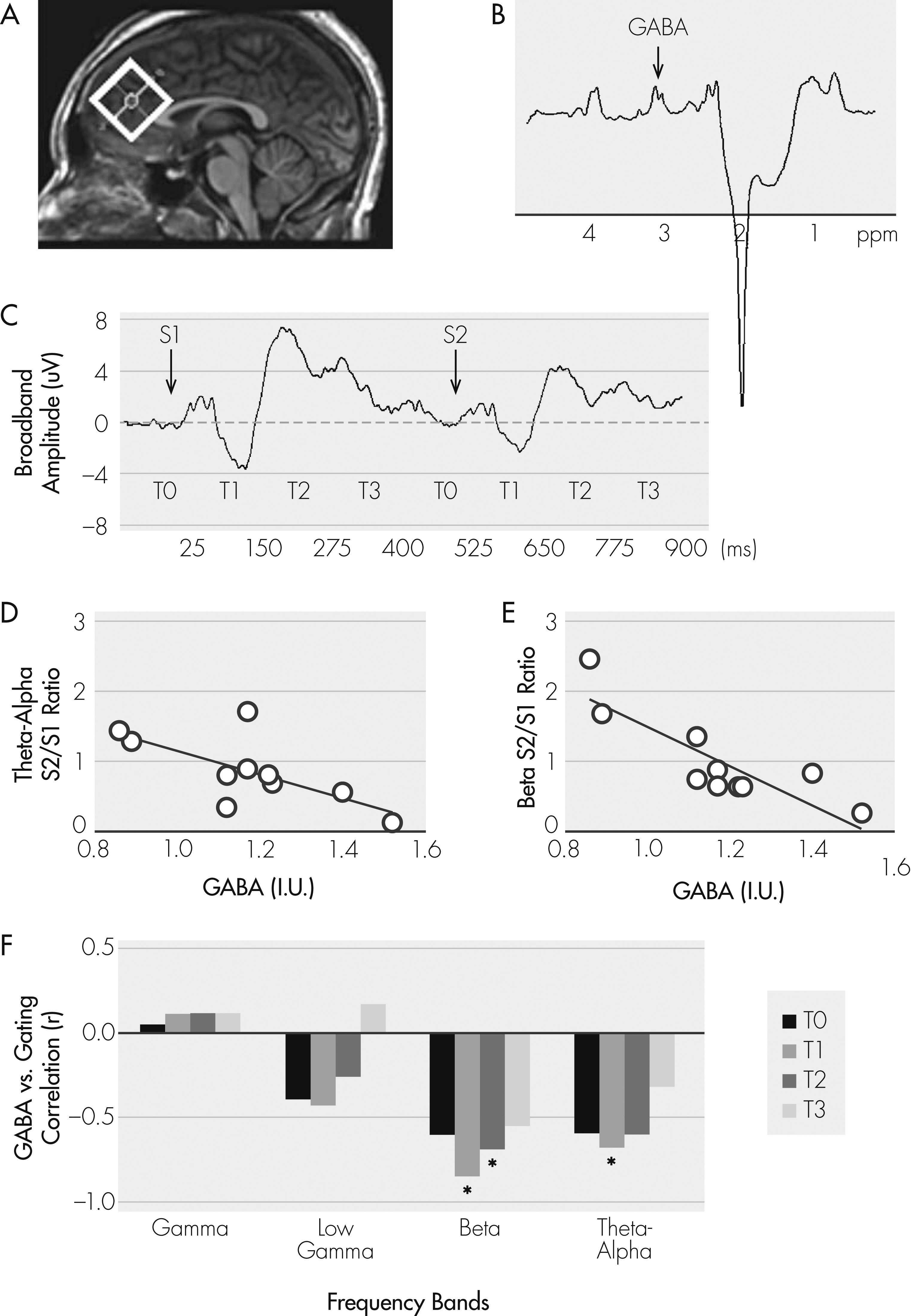
Ten subjects with a DSM-IV-TR diagnosis of schizophrenia (8 men, mean age 42.6 [SD: 11.8]) participated in MRS and electrophysiology sessions, recorded separately. MRS was conducted on a 3T Achieva scanner (Philips Healthcare; Best, The Netherlands) equipped with an 8-channel SENSE head coil. Anatomical T1-weighted images were acquired for spectroscopic voxel placement with a MP-RAGE sequence (SENSE factor 2, 1-mm isotropic voxels, 256 × 256 mm FOV, TR/TE/TI=8/3.8/842.5 msec, flip angle: 8°). Subjects were clinically stable and treated with antipsychotics (9 atypicals), but were not treated with anticonvulsants or benzodiazepines. GABA spectra were acquired from a 3.5 × 3.5 × 3.5 cc voxel in the medial prefrontal region with a MEGA-PRESS11,12 sequence optimized for detection of GABA (TR: 2.0 sec; TE: 68 msec; 14 msec editing pulse applied at 1.9 ppm and 7.5 ppm; 256 averages; Figure 1A, B). The rationale for the frontal region for GABA is that generators of the P50 auditory sensory response have been localized at the auditory cortical areas, but the gating mechanism for P50 has been localized primarily to the frontal lobe.13 The integral of the GABA peak was referenced to the integral of the unsuppressed water peak recorded from the same region,12 and GABA levels were corrected for the proportion of cerebrospinal fluid in the spectroscopic voxel. Electroencephalograms (EEG) were collected during a paired-click paradigm, whereby subjects listened to 150 paired-click stimuli (1-msec duration; 72 dB; 500-msec interclick interval; 10-sec intertrial interval). EEGs were recorded with a 32-channel electrode cap at 1 kHz, using a 0.1–200 Hz filter, mastoid reference, and skin impedance <5 Kohms. Data epochs were thresholded at ±75 μV, followed by visual inspection to remove artifacts, filtered at 10–100 Hz, 24-dB slopes, and averaged to obtain P50. P50 gating was measured at electrode site CZ, calculated as the ratio of the AEP P50 peaks of the second click (S2)/first click (S1).6 Discrete wavelet transform was applied to extract single-trial responses at theta-alpha (5–12 Hz), beta (12–20 Hz), low gamma (20–40 Hz), gamma (40–85 Hz), and very high gamma (>85 Hz) bands in response to S1 and S2. The temporal course of the response was broken into 125-msec windows in relationship to S1 and S2 (Figure 1C). The S2/S1 ratio of each frequency band/temporal window was computed as a measure of frequency/time-specific gating.6 Single-trial signal gating occurs primarily at the 25–150 msec (T1) and 150–275 msec (T2) post-stimulus windows at theta-alpha and beta bands.6 Therefore, we expected GABA levels to predict gating of the theta-alpha and beta oscillations during T1 and T2.
FIGURE 1. Relationship Between Gating of Frequency Oscillations and the Paired-Click Paradigm and GABA Levels
[A]: Location of medial frontal spectroscopic voxel; [B]: GABA spectrum; [C]: Grand average of broadband (1–100 Hz) averaged evoked potentials from the 10 subjects, showing temporal windows in relationship to first stimulus (S1) and second stimulus (S2) that were used in single-trial analyses. T0: −100 to 25msec from the stimulus, T1: 26–150 msec, T2: 151–275 msec, and T3: 276–400 msec; [D]: Correlation of GABA level and gating of theta-alpha band at T1 (r = −0.68, p=0.03); E: Correlation of GABA level and gating of beta band at T1 (r = −0.85; p=0.002); [F]: Correlation coefficients (Y axis) of GABA level and gating across frequency bands (gamma, low gamma, beta, theta-alpha are shown here) and temporal windows (T0–T3). For example, gating of beta at T1 is the ratio of beta at T1 in response to S1 divided by beta at T1 in response to S2.
*Statistically significant.
The relationship between gating measures and GABA levels were analyzed with Pearson’s correlation and linear regression. Analyses were conducted with the Statistical Package for Social Sciences (SPSS), Version 12.0 software package. Alpha was set to 0.05 for a priori tests, and Bonferroni corrected for post hoc multiple comparisons.
Results

Results showed that P50 gating was not significantly associated with GABA level (r=0.28; NS). However, a strong association was found between gating of theta-alpha oscillation and GABA levels for the T1 (r = −0.68; p=0.03; Figure 1D), but not T2: higher GABA was associated with stronger inhibition (lower ratio) at 25–150 msec post-stimulus. GABA levels accounted for about 47% of the variation in gating of the theta-alpha activities. Correlation with the beta band gating was even more robust: higher GABA was associated with stronger gating at T1 (r = −0.85; p=0.002; 72% of variance explained; Figure 1E) and T2 (r = −0.69; p=0.03; 48% of variance explained). There were no significant correlations between GABA level and S2/S1 ratio in other bands or windows (Figure 1F).
Exploration of responses to S1 alone yielded no significant correlations with GABA except for post-S1 beta oscillation at T3 (p=0.048), although this was not significant after Bonferroni correction for multiple comparisons, and indicates that GABA levels are associated more with inhibitory than sensory electrophysiological responses.

Discussion

This study is the first to show a relationship between in-vivo brain GABA levels and auditory inhibitory electrophysiological measures in schizophrenia. The paired-click gating paradigm has been theoretically linked to GABAergic function.14 Auditory gating is a measure of inhibitory processing, whereby the first auditory stimulus evokes a large electrical response, while inhibited electrical activity is seen in response to the second stimulus. Our finding that higher GABA level is associated with greater suppression of theta appears consistent with the observation that theta oscillations are reduced with greater GABA input and increased with GABA-receptor blockade in animals.14,15 We also showed that the association with GABA levels and gating of the electrical response extends to the beta frequency range.
A clear link of auditory gating function to GABA levels has not been previously established in humans. Our findings indicate an association between GABA levels and gating of the theta-alpha and beta oscillation activities, but not the classical P50 measures, in schizophrenia. Auditory gating with the paired-click paradigm occurs mostly in the theta-to-beta frequency bands, and gating of these lower-frequency activities shows a stronger genetic contribution in schizophrenia patients’ families than the P50 measures.6 The convergence and divergence of the underlying neurochemistry controlling P50 gating versus gating of oscillatory activities requires additional study; the current data suggest that GABA level could be more relevant to the suppression of beta-to-theta range activities. The results also indicate that GABA levels are more specifically associated with inhibitory (ratio of S2/S1) than sensory (S1) electrophysiological responses.
These findings also appear consistent with human MRS research that demonstrates the relationship between GABA levels and behavioral and psychophysiological inhibitory processing. One study revealed a relationship between occipital region GABA/Cr and visual suppression function, a test of visual inhibitory processing, in a combined schizophrenia and healthy sample.15 Two magnetoencephalography (MEG) studies in healthy subjects demonstrated associations between gamma frequency level during a visual stimulation task and GABA levels,9 and beta power during a finger motor task and GABA to N-acetyl-aspartate (NAA) ratio.10 Our finding of an association between GABA levels and auditory sensory gating measures in schizophrenia converges with these observations and together demonstrates that MRS measurements of GABA may provide an important marker of the human brain’s capacity for a wide range of inhibitory processing in both healthy and disease states. Lower GABA levels could reflect lower GABA synthesis, reduced GABAergic interneuron density, and diminished dendritic inhibitory neuron arborization, resulting in poor inhibitory processing.
Without the inclusion of a control group, it cannot be determined whether impaired inhibition of evoked oscillations is due to a GABA deficit in schizophrenia. Other study limitations include small sample size and the possibility that antipsychotics may affect the correlations. However, even if the GABA levels and/or oscillatory measures were influenced by medications, it does not reduce the significance of a strong correlation between them, which supports the role of GABA in the inhibition of frequency oscillations at 5–20 Hz.

Acknowledgments

We thank Terri Brawner, Kathleen Kahl, and Ivana Kusevic for conducting the MR scans, Jef West for subject recruitment and data collection, and Ann Summerfelt for EEG experiments.

References

1.
Siegel C, Waldo M, Mizner G, et al.: Deficits in sensory gating in schizophrenic patients and their relatives: evidence obtained with auditory evoked responses. Arch Gen Psychiatry 1984; 41:607–612
2.
Lewis DA, Hashimoto T: Deciphering the disease process of schizophrenia: the contribution of cortical GABA neurons. Int Rev Neurobiol 2007; 78:109–131
3.
Benes FM, Berretta S: GABAergic interneurons: implications for understanding schizophrenia and bipolar disorder. Neuropsychopharmacology 2001; 25:1–27
4.
de Wilde OM, Bour LJ, Dingemans PM, et al.: A meta-analysis of P50 studies in patients with schizophrenia and relatives: differences in methodology between research groups. Schizophr Res 2007; 97:137–151
5.
Greenwood TA, Braff DL, Light GA, et al.: Initial heritability analyses of endophenotypic measures for schizophrenia: the consortium on the genetics of schizophrenia. Arch Gen Psychiatry 2007; 64:1242–1250
6.
Hong LE, Summerfelt A, Mitchell BD, et al.: Sensory gating endophenotype based on its neural oscillatory pattern and heritability estimate. Arch Gen Psychiatry 2008; 65:1008–1016
7.
Smythe JW, Colom LV, Bland BH: The extrinsic modulation of hippocampal theta depends on the coactivation of cholinergic and GABA-ergic medial septal inputs. Neurosci Biobehav Rev 1992; 16:289–308
8.
Bocian R, Konopacki J: Posterior hypothalamic GABAergic mediation of hippocampal theta in the cat. Brain Res Bull 2007; 73:289–300
9.
Muthukumaraswamy SD, Edden RA, Jones DK, et al.: Resting GABA concentration predicts peak gamma frequency and fMRI amplitude in response to visual stimulation in humans. Proc Natl Acad Sci U S A 2009; 106:8356–8361
10.
Gaetz W, Edgar JC, Wang DJ, et al.: Relating MEG measured motor cortical oscillations to resting γ-aminobutyric acid (GABA) concentration. Neuroimage 2011; 55:616–621
11.
Mescher M, Merkle H, Kirsch J, et al.: Simultaneous in-vivo spectral editing and water suppression. NMR Biomed 1998; 11:266–272
12.
Edden RA, Barker PB: Spatial effects in the detection of gamma-aminobutyric acid: improved sensitivity at high fields using inner volume saturation. Magn Reson Med 2007; 58:1276–1282
13.
Korzyukov O, Pflieger ME, Wagner M, et al.: Generators of the intracranial P50 response in auditory sensory gating. Neuroimage 2007; 35:814–826
14.
Eccles JC: The Inhibitory Pathways of the Central Nervous System (Sherrington Lecture). Liverpool, UK, University Press, 1969
15.
Yoon JH, Maddock RJ, Rokem A, et al.: GABA concentration is reduced in visual cortex in schizophrenia and correlates with orientation-specific surround suppression. J Neurosci 2010; 30:3777–3781

Information & Authors

Information

Published In

Go to The Journal of Neuropsychiatry and Clinical Neurosciences
The Journal of Neuropsychiatry and Clinical Neurosciences
Pages: 83 - 87
PubMed: 23487198

History

Received: 21 December 2011
Revision received: 3 August 2012
Accepted: 4 August 2012
Published online: 1 January 2013
Published in print: Winter 2013

Authors

Details

Laura M. Rowland, Ph.D.
Maryland Psychiatric Research Center, Department of Psychiatry, University of Maryland School of Medicine, Baltimore, MD (LMR), Russell H. Morgan Department of Radiology and Radiological Science, Johns Hopkins University School of Medicine, Baltimore, MD (HZ, PBB), F.M. Kirby Research Center for Functional Brain Imaging, Kennedy Krieger Institute, Baltimore, MD (HZ, PBB).
Richard A.E. Edden, Ph.D.
Maryland Psychiatric Research Center, Department of Psychiatry, University of Maryland School of Medicine, Baltimore, MD (LMR), Russell H. Morgan Department of Radiology and Radiological Science, Johns Hopkins University School of Medicine, Baltimore, MD (HZ, PBB), F.M. Kirby Research Center for Functional Brain Imaging, Kennedy Krieger Institute, Baltimore, MD (HZ, PBB).
Kimberly Kontson, B.S.
Maryland Psychiatric Research Center, Department of Psychiatry, University of Maryland School of Medicine, Baltimore, MD (LMR), Russell H. Morgan Department of Radiology and Radiological Science, Johns Hopkins University School of Medicine, Baltimore, MD (HZ, PBB), F.M. Kirby Research Center for Functional Brain Imaging, Kennedy Krieger Institute, Baltimore, MD (HZ, PBB).
He Zhu, Ph.D.
Maryland Psychiatric Research Center, Department of Psychiatry, University of Maryland School of Medicine, Baltimore, MD (LMR), Russell H. Morgan Department of Radiology and Radiological Science, Johns Hopkins University School of Medicine, Baltimore, MD (HZ, PBB), F.M. Kirby Research Center for Functional Brain Imaging, Kennedy Krieger Institute, Baltimore, MD (HZ, PBB).
Peter B. Barker, D.Phil.
Maryland Psychiatric Research Center, Department of Psychiatry, University of Maryland School of Medicine, Baltimore, MD (LMR), Russell H. Morgan Department of Radiology and Radiological Science, Johns Hopkins University School of Medicine, Baltimore, MD (HZ, PBB), F.M. Kirby Research Center for Functional Brain Imaging, Kennedy Krieger Institute, Baltimore, MD (HZ, PBB).
L. Elliot Hong, M.D.
Maryland Psychiatric Research Center, Department of Psychiatry, University of Maryland School of Medicine, Baltimore, MD (LMR), Russell H. Morgan Department of Radiology and Radiological Science, Johns Hopkins University School of Medicine, Baltimore, MD (HZ, PBB), F.M. Kirby Research Center for Functional Brain Imaging, Kennedy Krieger Institute, Baltimore, MD (HZ, PBB).

Notes

Send correspondence to Laura M. Rowland, Ph.D.; e-mail: [email protected]

Funding Information

This work is supported by National Institutes of Health (R21MH082322 to PBB, K01MH077230 to LMR, R01MH085646, R01DA027680, and R21MH079172 to LEH).

Metrics & Citations

Metrics

Citations

Export Citations

If you have the appropriate software installed, you can download article citation data to the citation manager of your choice. Simply select your manager software from the list below and click Download.

For more information or tips please see 'Downloading to a citation manager' in the Help menu.

Format
Citation style
Style
Copy to clipboard

View Options

View options

PDF/EPUB

View PDF/EPUB

Get Access

Login options

Already a subscriber? Access your subscription through your login credentials or your institution for full access to this article.

Personal login Institutional Login Open Athens login
Purchase Options

Purchase this article to access the full text.

PPV Articles - Journal of Neuropsychiatry and Clinical Neurosciences

PPV Articles - Journal of Neuropsychiatry and Clinical Neurosciences

Not a subscriber?

Subscribe Now / Learn More

PsychiatryOnline subscription options offer access to the DSM-5-TR® library, books, journals, CME, and patient resources. This all-in-one virtual library provides psychiatrists and mental health professionals with key resources for diagnosis, treatment, research, and professional development.

Need more help? PsychiatryOnline Customer Service may be reached by emailing [email protected] or by calling 800-368-5777 (in the U.S.) or 703-907-7322 (outside the U.S.).

Media

Figures

Other

Tables

Share

Share

Share article link

Share