To the Editor: Virchow-Robin spaces (VRS) refers to vascular surrounding areas that can be seen along the trajectory of cerebral vessels inside brain parenchyma and subarachnoid space.
1,2 VRS are very commonly found on brain MRI, particularly in elderly persons, as small foci, occasionally confluent, CSF-isointense on all sequences, without gadolinium enhancement.
1–3 They are usually benign and typically asymptomatic.
1,3 However, when located in midbrain or thalamic regions, and expanded more than usual, they can cause hydrocephalus and brainstem syndromes.
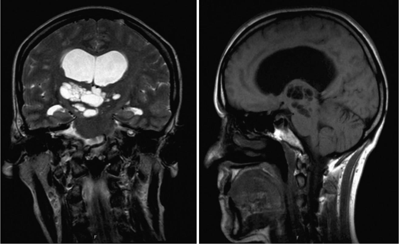
4 A 52-year-old healthy woman was seen for a 1-month history of forgetfulness and difficulties in organizing her work, without other cognitive complaints or functional impairment. Six months earlier, she had a transient episode highly suggestive of transient global amnesia (TGA): at a religious ceremony, without physical exertion, she became suddenly incapable of retaining new information, repeating the same questions, while adequately able to perform complex functions, for a period of 10 hours, with no persistent memory deficit over the following 4–5 months. Brain MRI showed enlarged meso-diencephalic VRS, with secondary hydrocephalus (
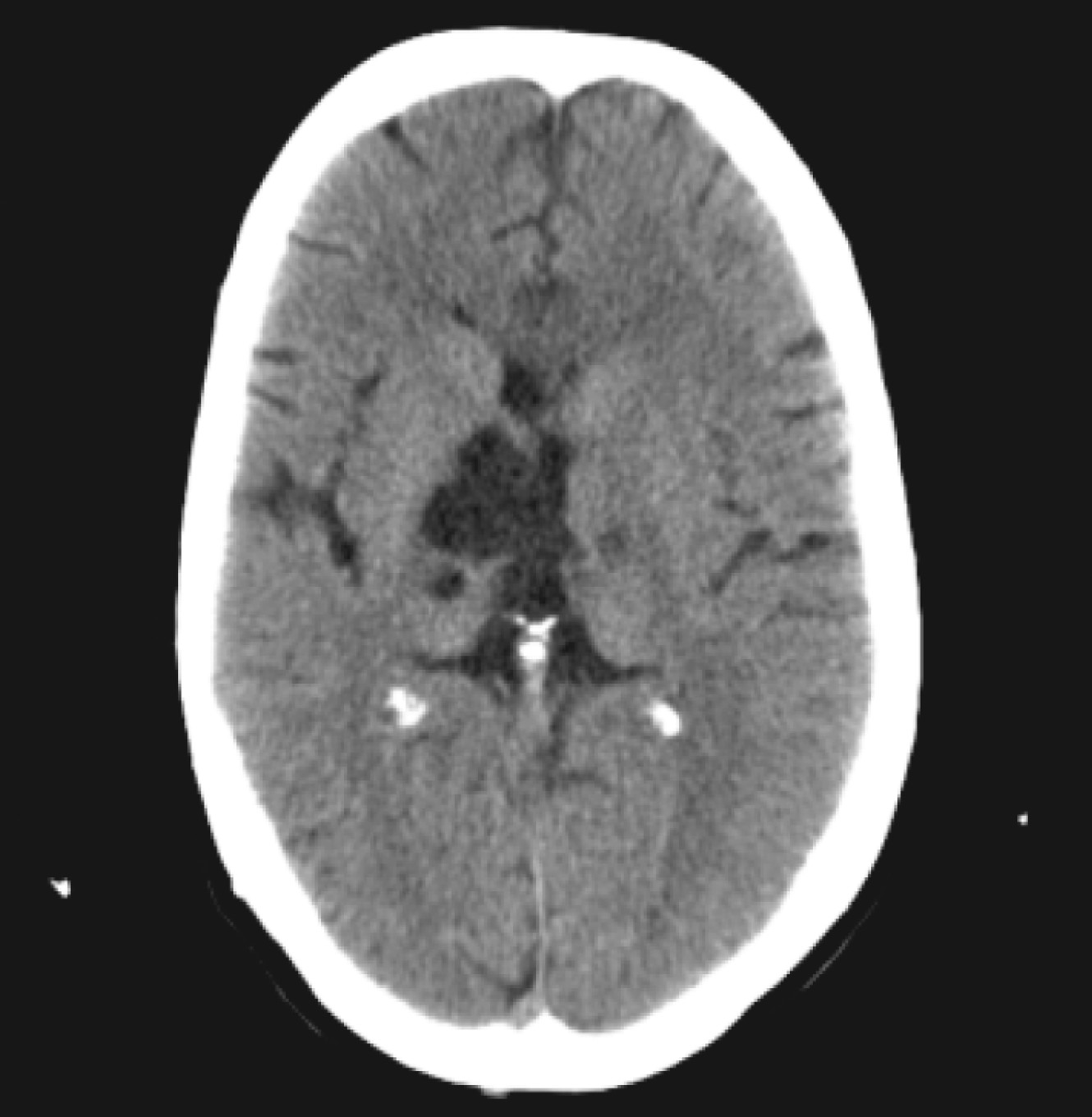
Figure 1). Neuropsychological study (NPS) revealed amnesic mild cognitive impairment. After neurosurgical evaluation, clinical and imaging surveillance was recommended. Over the next year, noticeable clinical deterioration was documented, with worsening cognitive difficulties, gait imbalance with occasional falls, and urinary urgency. Control MRI and NPS were similar to the previous ones. It was then decided to place a ventricle-peritoneal shunt. By the second day after surgery, significant clinical improvement was seen. At 12-month follow-up, she is completely symptom-free, with normal NPS. Cerebral CT still shows enlarged VRS, but without hydrocephalus (
Figure 2).
In rare cases, as seen in our patient, enlarged VRS can behave like a real space-occupying mass, leading to serious complications, such as hydrocephalus.
1 In these circumstances, differential diagnosis includes cystic gliomas, parasitic infections, and ependymal, neuroepithelial, and arachnoid cysts.
4 VRS MRI typical appearance can, in most cases, prevent invasive procedures such as biopsy.
2 When incidentally found, clinical and imaging surveillance is recommended.
1 At any point over the follow-up, the development of symptomatic hydrocephalus requires surgical resolution.
2 Ventriculo-peritoneal shunt, ventriculo-cisternostomy, and cysto-cisternostomy are then generally effective, although frequently having no effect on lesion size.
4 For some practitioners, direct endoscopic fenestration, further allowing histopathological diagnosis, represents the first treatment choice.
4Transient global amnesia (TGA) is defined as a transient episode of acute anterograde, and variable retrograde, memory loss, without changes in behavior or psychomotor skills, including ability to perform complex functions.
5 The TGA etiology is not well known, but the disorder has been regarded as benign, usually leaving no residual deficit, although slight changes in memory fixation have been reported to occur. We could find few descriptions of TGA with diagnosed hydrocephalus.
5 It is thought that the increased volume of the third ventricle may either disturb neurotransmission in the limbic system or cause blood flow decrease in central arteries.
5Rarely, enlarged VRS may produce obstructive hydroceph-alus. TGA in our patient can be cautiously seen as the very first manifestation of her VRS-induced hydrocephalus.


