Toxoplasmosis gondii (T. gondii), an intracellular parasite discovered in 1908, is present in approximately one-third of the earth’s population.
9,10 Its presence in humans was first recognized in 1923.
11 Once infected, humans remain seropositive for life. Studies focused on likely transmission vectors and likely subpopulations more at-risk have confirmed previously-known exposure risks of feline feces, eating raw or undercooked meats, and drinking unpasteurized goat’s milk.
12 Other exposure risks include eating raw oysters, clams, or mussels, and unwashed raw fruits and vegetables.
13–17 There are well-documented effects of acute infection in immunocompromised patients (i.e., encephalitis and large intracranial cysts) and in infants with congenital ocular disease and/or mental retardation secondary to in-utero exposure. Latent chronic infection in immunocompetent (healthy) adult persons was thought to be benign. However, recent evidence has challenged this assumption. Within healthy immunocompetent populations, there have been increasing numbers of reported associations between
T. gondii seropositivity and suicide, schizophrenia, psychiatric hospitalizations, depression, personality changes, and even, in very preliminary investigations, Alzheimer’s disease (AD).
In brief,
T. gondii has three infectious stages: sporozoites (in oocysts), bradyzoites (in tissue cysts), and tachyzoites.
9,10,14,17 The transmission life-cycle for humans usually begins with ingestion of the oocysts (found in soil, water, or feline feces) or tissue cysts (from undercooked meat). In either case, cysts open, and tachyzoites are quickly formed. These cross the gastrointestinal barrier, rapidly divide, and disseminate widely (can cross both placental and blood–brain barriers). In immunocompetent individuals, tachyzoites transform into the slow-growing bradyzoite stage, forming microscopic intracellular cysts in brain and muscle.
11,14,15 Key for the parasite to continue in this latent, asymptomatic stage is to achieve a balance between coexistence in its host cells without destruction of the host’s tissues. The immune cascade is necessary for this coexistence and includes release of many immune components, including macrophages, interleukins, cytokines, and other factors.
11,15,18The feline (including the domestic cat) is the definite host for this parasite to undergo gametogenesis and sexual reproduction.
9,10,15,19 Interestingly,
T. gondii can use many mammalian species (e.g., livestock, rodents, humans, sea otters) as intermediate hosts in which it undergoes asexual reproduction.
20 Long-standing evidence suggests a remarkable ability to manipulate a current host’s behavior to advance survivability and promote its propagation. This theory is termed “behavioral manipulation.”
4,21–24 There are some highly specific behavioral changes consistently found over decades of studies in various laboratory and naturalistic experimental conditions in
T. gondii-infected rodents. These include increased approach/decreased avoidance to feline urinary odors; decreased defensive behaviors, including spending more time in vulnerable open areas and decreased fear of novel stimuli; increased psychomotor activity; decreased climbing and rearing; and increased rate of entrapment.
4,24–27 As noted by many authors, these are all behaviors that could lead to increased probability of predation. Studies indicate that the behaviors are not due to generalized illness/sickness. Many areas of behavior, learning, and social interactions are not affected, indicating the very focused nature of the behavioral changes. A multifaceted study that included magnetic resonance imaging (MRI), neuropathologic, immunologic, neurologic, behavioral, and appearance measures of mice infected at the equivalent of early or mid-adult years and then evaluated 1 year later (equivalent to middle-to-early elderly human years), reported significant differences in grooming and body position, increased urination/defecation while being held, decreased exploratory movements, and impairments of sensorimotor function.
28 The seropositive group had many changes that echo human neurodegenerative disease patterns, including significantly enlarged ventricles on MRI (particularly near the aqueduct of Sylvius), contrast uptake asymmetries, increased expression of genes that can modify immune responses, and microscopic presence of inflammatory cells (particularly perivascular, hippocampal, and leptomeningeal, and in proximity to the aqueduct of Sylvius).
In view of the rodent evidence, scientists are beginning to examine whether the latent stage of
T. gondii infection in healthy persons is as asymptomatic as previously thought. Although there has been no suggestion that the parasite by itself is the direct and single cause of neuropsychiatric illness in immunocompetent individuals, there are several significant correlations that are noteworthy. A series of 16 studies over 15 years elucidated
T. gondii-related personality and psychomotor differences in healthy Czech citizens and military.
29,30 In total, over 3,700 subjects had antibody testing and completed personality inventory examinations. Nine of eleven studies using the Cattell’s 16-Personality Factor self-report questionnaire found significant and consistent results for both genders. Seropositive men overall had lower regard for rules and higher vigilance (suspicious, jealous, rigid/inflexible) than seronegative men. In contrast, seropositive women had greater regard for rules and higher warmth than seronegative women. Both seropositive genders were more anxious than matched healthy-comparison subjects. Three of five studies using the Cloninger Temperament and Character Inventory found both seropositive women and men to have lower novelty-seeking behavior scores than healthy-comparison subjects. Behavioral observations and interviews were completed to ascertain whether the gender differences found in self-report measures were replicated by objective measures. Seropositive men scored significantly lower than seronegative men on Self-Control, Clothes Tidiness, and Relationships. The differences were less impressive for the seropositive women, with only trends toward higher scores on Self-Control and Clothes Tidiness as compared with seronegative women. The authors view the study results as objective confirmation that
T. gondii presence can change a human host’s behaviors. In a later study, they presented evidence linking the observed differences to gender-specific coping styles (individualistic versus prosocial) under stress.
31 This group also found slightly slower reaction times and decreased concentration in
T. gondii-positive subjects and a higher relative risk for an at-fault traffic accident.
29,30 In a later prospective study (3,890 male military conscripts), they confirmed a higher risk for traffic accidents in the seropositive group and found that risk was mitigated by RhD positivity.
32 Such higher risk for traffic accidents has also been reported in Turkish populations.
33,34Higher rates of
T. gondii seropositivity have been reported for several psychiatric conditions in multiple parts of the world. A series of studies comparing the prevalence rates of the parasite with the national suicide rates in 20 European countries found that suicide rates were higher in countries with greater
T. gondii prevalence, with no effect of wealth/gross domestic product.
17,35,36 Both presence of the Finno-Ugrian gene (previously associated with suicide) and
T. gondii predicted higher suicide rates in men and women. When the data for women were stratified by age, they found strong statistical significance for higher suicide rates in
T. gondii-seropositive postmenopausal women. A study from Turkey of emergency room patients with suicide attempts found that 41% of the patient group was seropositive, as compared with 28% of the controls.
37 In the United States, significantly higher
T. gondii antibody titers were found in mood-disorder patients with previous suicide attempts than in mood-disorder patients without previous suicide attempts or in controls.
38 The same group reported that seropositivity was associated with suicide attempts in patients with schizophrenia younger than 38 years of age, but not in those older than 38.
39 Several groups have reported elevated rates of seropositivity in inpatient psychiatric hospitals, as compared with community controls. One group examined 15 surveys across different areas of China, comparing rates of seropositivity in psychotic inpatients versus healthy individuals from the same provinces; 14 of the 15 reports indicated significantly higher rates of
T. gondii antibodies in the psychotic inpatients.
40,41 A study of German psychiatric inpatients reported a significant association between comorbid personality disorder and antibody titer.
42 A study from Mexico found 18.2% of 137 inpatients to be seropositive, as compared with 8.9% of controls.
13 The rate for patients with schizophrenia was 26.3%.
There have been a multitude of studies over decades looking for underlying causes of chronic schizophrenia, including many theories of infection;
T. gondii has been considered since 1953.
16 A very recent metaanalysis reviewing 38 published studies of
T. gondii antibody seropositivity in patients previously diagnosed with schizophrenia compared with healthy controls (total: 6,058 patients; 8,715 controls) found that the patients were more likely to be seropositive (odds ratio: 2.71), confirming their previous study.
43,44 As compared with other risk factors for development of the illness (including having first-degree relatives with the condition),
T. gondii antibody positivity was considered an intermediate risk factor. To begin to ascertain whether having schizophrenia predisposes patients to intake of
T. gondii oocytes (i.e., poor hygiene habits) or
T. gondii is present before schizophrenia diagnosis, one group examined banked blood specimens from the U.S. military to determine rates of previous infection in new-onset schizophrenia within the military.
45 Although it was a small study (180 patients, 8% positive; 532 controls, 7% positive), there was a significant association between antibody level and risk of schizophrenia (hazard ratio: 1.24), indicating that
T. gondii could be considered one factor contributing to the illness.
A related line of study suggests the possible antiparasitic effects of select antipsychotic medications. The in-vitro antiprotozoan effects of phenothiazine-related compounds have been documented since the late 1800s.
46 More recently, several studies have used human fibroblast cells infected with
T. gondii tachyzoites to assess in-vitro anti-parasitic activity of psychiatric medications. One study reported modest anti-parasitic activity for sodium valproate; the other reported more robust activity for haloperidol and valproic acid.
46,47 The second study also reported modest results for seven other antipsychotic compounds. A third study of five antipsychotics produced mixed results.
48 Fluphenazine and thioridazine had a robust effect; trifluoperazine a modest effect; and haloperidol and clozapine, no effect. In-vivo studies have also produced mixed results. One study reported that haloperidol and valproic acid protected
T. gondii-infected rats from developing detrimental behavioral changes (equal to the effects of standard anti-parasitic agents), whereas another found no anti-parasitic advantages on either preventing acute infection or reducing cyst burden in chronic infection.
49,50A very new area of possible linkage is that of
T. gondii to Alzheimer’s disease (AD). In the inpatient study from Mexico (above), the authors mention that both patients in the study hospitalized for AD were seropositive for
T. gondii.
13 A study from Turkey compared rates of
T. gondii IgG seropositivity in 34 patients with AD versus well-matched healthy individuals.
51 The rates were significantly different: 44.1% in the AD group and 24.3% in the healthy-comparison subjects. Efforts to examine this linkage have been undertaken in a rodent model, using mice bred to carry the rodent equivalent of AD, with and without
T. gondii infection.
52 Mice infected with
T. gondii had significantly lower rates of β-amyloid plaque formation in cortex and hippocampus, and maintained learning and memory skills that declined in the
T. gondii seronegative group. The authors felt that these results were due to the increased presence of some anti-inflammatory processes in the seropositive group. They reaffirmed the importance of inflammation in the cascade of events necessary for AD progression and echoed a previous theme in the literature of a host–parasite balance with
T. gondii. These early studies and observations may open new lines of study in neurodegenerative diseases.
The neuropathology of chronic latent
T gondii infection is quite different from acute cerebral toxoplasmosis, in which necrosis and inflammation are widespread; infecting organisms may be present outside of host cells; and multiple types of brain cells may harbor parasites.
53–56 Most studies are in animal models, but a case report of incidental findings at autopsy of an immunocompetent patient indicated a similar pattern in humans.
56 An early study of chronic latent infection in mice (examined at 3, 6, and 12 months post-infection) reported that infected brains appeared normal on gross inspection.
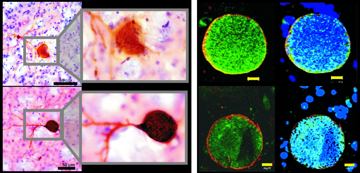
57 Parasite-containing cysts were found primarily in gray matter (90%). Ultrastructural examination of 50 cysts established that all were contained within intact host cells, most of which were positively identified as neurons by presence of synapses. Cysts were present in all parts (dendrite, axon, soma) of neurons, some of which were severely distended by the presence of a large cyst (
Figure 1). A study of latent congenital infection in mice also reported that all cysts were contained within intact host cells.
58 In that study, all host cells were identified as neurons on the basis of immunohistochemical staining of neurofilament protein. Later studies have consistently reported that cysts were found only in neurons in animals with chronic infection, although astrocytic processes could be in close association.
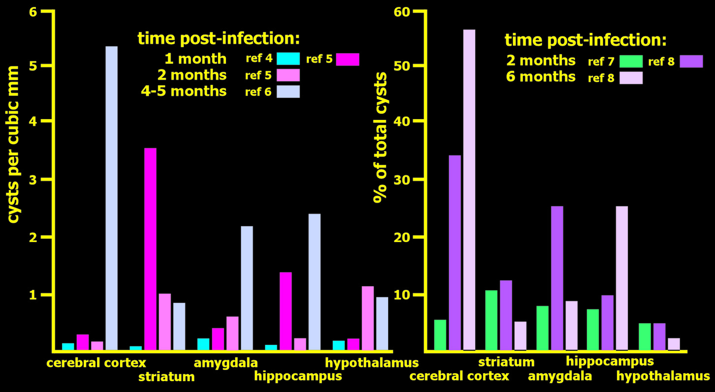
2,5,28 Studies that assessed cyst burden at two times after infection (1 month versus 2 months; 2 months versus 6 months) reported an overall decrease (approximately half in both studies) at the later time-point, indicating that some clearance of cysts can occur (
Figure 2).
2,5 Although both studies noted areas in which the burden of cysts did not decline, the areas were not the same. Several studies have quantified the relative burden of cysts across brain areas at various times post-infection (
Figure 2). At 1–2 months post-infection, there is a general pattern of a higher burden of cysts in subcortical areas than in cerebral cortex or cerebellum.
2,4,5,7 However, there were considerable differences across studies in the relative ranking of the most commonly involved structures (e.g., amygdala, hippocampus, hypothalamus, striatum, thalamus). At 4–6 months post-infection, the general pattern was a higher burden of cysts in areas of cerebral cortex than in subcortical areas.
5,6 The significance of these findings is difficult to assess, as studies varied considerably in many aspects of methodology, and most also reported considerable individual differences. As noted in one study, the finding of major differences across individuals does not support the presence of preferential targeting of specific areas (e.g., amygdala), as has been suggested as the basis for the induced behavioral changes.
6The neurobiological origins of the behavioral changes in rodents induced by chronic latent infection with
T gondii are presently not known, but several intriguing possibilities have been identified. The possible role of dopamine has been actively considered since a study found an increased level of dopamine, but not serotonin or norepinephrine, in brains of mice with chronic latent infection at 5 weeks post-infection.
59 In contrast, dopamine level was not found to be affected in a study of congenitally-infected mice.
60 Recent studies indicate that
T gondii cysts may be able to directly influence synthesis of dopamine, as this parasite has two genes encoding tyrosine hydroxylases (the rate-limiting step in dopamine synthesis).
61 Although they differed in other regions, both genes were very similar to the catalytic domain of mammalian tyrosine and phenylalanine hydroxylases. Tests demonstrated that expressed proteins were able to catabolize both tyrosine and phenylalanine, with a two-to-threefold preference for tyrosine. In cell cultures, expression of one of these genes was present at similar levels during all stages of infection, whereas the other increased greatly during the cyst-forming stage.
61 In a follow-up study, immunofluorescence assay of brain sections from mice at 6–8 weeks post-infection showed intense staining within cysts for antibodies to both dopamine and
T gondii tyrosine hydroxylase (
Figure 1).
3 Dopamine release was much higher (350%) in cultures of dopaminergic neurons that were infected, and the level correlated with parasite burden.
3 These results raise the possibility that dopamine signaling may be altered in the vicinity of
T gondii cysts, with both synaptic and volume transmission potentially affected.
62 Direct impairment of the functioning of neurons containing cysts is another potential mechanism of action recently identified in a study in mice that compared behavioral, neuropathological, and functional measures at 30 days and 60 days post-infection.
2 Although the burden of cysts was lower at the later time-point, changes in behavior were present at 60 days but not at 30 days. Also, at 60 days, more neurons had reduced or absent uptake of thallium (which acts as a potassium analog), indicating impaired functioning. As noted by the authors, these results indicate that duration of chronic infection may be an important factor and functional silencing of neurons may contribute to behavioral changes. There is also evidence for alterations in both focal brain volume and evoked activity in limbic areas in male rats infected with this parasite.
63 Of potentially high relevance to the known changes in behavior associated with chronic infection, brain activation evoked by exposure to cat urine (assessed by c-Fos) was found not only in the expected areas important for defensive behaviors, but also in nearby areas associated with reproductive behaviors.
Unlike chronic latent infection, imaging in acute toxoplasma encephalitis has been well documented over the years. Gadolinium (Gad)-enhanced T
1 MRI is the standard imaging procedure to identify the “target sign” of a ring-enhancing lesion containing a small area of hyperintense signal in acute toxoplasma encephalitis in immunocompromised patients.
64,65 Gad contrast highlights breaks in the blood–brain barrier, with hyperintensities in abnormal areas. It may not be positive in “preclinical” or early-stage infections.
65,66 Ultra-small superparamagnetic particles of iron oxide (USPIO), a new contrast agent for MRI, has been used recently to study neuroinflammatory diseases. Circulating monocytes in the bloodstream internalize these particles, which travel within the inflammatory cells into areas of phagocytosis. A study of 30 mice acutely infected with
T. gondii (by intracerebral injection of tachyzoites) were imaged with Gad and/or USPIO at various time-periods in the acute (1 week) infectious process, followed by microscopic examination.
66 The USPIO imaging (post-contrast T
2 and T
2* weighted images, decreased signal) identified several
T. Gondii lesions not visualized on either unenhanced T
2 and T
2*, or Gad-enhanced T
1-weighted MRI. However, the Gad imaging identified breaks in the blood–brain barrier more effectively than did the USPIO. The authors suggest that USPIO might be an additional imaging contrast to use in combination with Gad for earlier detection of subclinical neuroinflammatory processes. Perhaps, in the future, USPIO could be used to identify latent toxoplasma, as well.
Voxel-based morphometry (VBM), a method of measuring volumes of brain areas, was used to compare patients with schizophrenia (12
T. gondii seropositive; 32 seronegative) to a control group of matched (age, sociodemographic) individuals (13
T. gondii seropositive; 43 seronegative).
8 As previously seen in multiple studies, the patients with schizophrenia had reduced overall gray-matter volume in cortical areas, hippocampus, and caudate. The novel aspect of this study is that they went on to examine the influence of
T. gondii status. Patients with schizophrenia who were seropositive had significantly reduced gray-matter volume in left cerebellum and bilaterally in the caudate, thalamus, occipital cortex, and medial cingulate cortex, as compared with seronegative patients. When each patient group was compared with their respective controls, gray-matter reductions were much greater in the seropositive group (
Figure 3). No differences related to
T. gondii status were found in the control group. There were no differences in white-matter volume among any of the comparison groups. The authors proposed that
T. gondii triggered release of immune factors leading to downstream changes in glutamate NMDA, and/or that effects on dopamine (described above) were possible contributing factors to the imaging differences.
Conclusion
Toxoplasmosis gondii has been recognized as a human parasite for almost a century, yet much is still unknown about host reactions to its chronic presence. Recent evidence from both human and animal studies has led to new theories regarding how this parasite may change or manipulate a host’s behavior to gain advantage. Particularly fascinating are the possible links to neuropsychiatric disorders and/or personality changes that might occur or be exacerbated in individuals with chronic latent infection. These require more study to understand the possible interplay of this parasite with genetic predisposition and other factors resulting in neuropsychiatric illness.


