Full access
Letters
Published Online: 1 October 2013

Severe Transient Suicidality Due to Hemispheric Hyperperfusion After Successful Acute Stroke Thrombolysis

Publication: The Journal of Neuropsychiatry and Clinical Neurosciences
To the Editor: Intravenously administered tissue plasminogen activator (IV-tPA) remains the only approved drug therapy for achieving arterial recanalization in acute ischemic stroke. Timely administration of IV-tPA often results in rapid recanalization and rapid clinical recovery. However, rapid recanalization of an occluded intracranial artery rarely can lead to cerebral hyperperfusion syndrome, with variable presentations. We present a case that showed an excellent early recovery after IV-tPA for severe acute ischemic stroke. However, the patient developed severe suicidal ideation on Day 3. We present clinical and various imaging studies to elucidate the underlying pathophysiological mechanisms.
A 73-year-old man presented with sudden left-sided weakness for 140 minutes. Clinical evaluation revealed blood pressure 157/75mmHg and severe left hemiplegia (Power Medical Research Council Grade 0/5). The National Institutes of Health Stroke Scale (NIHSS) score was 19 points.
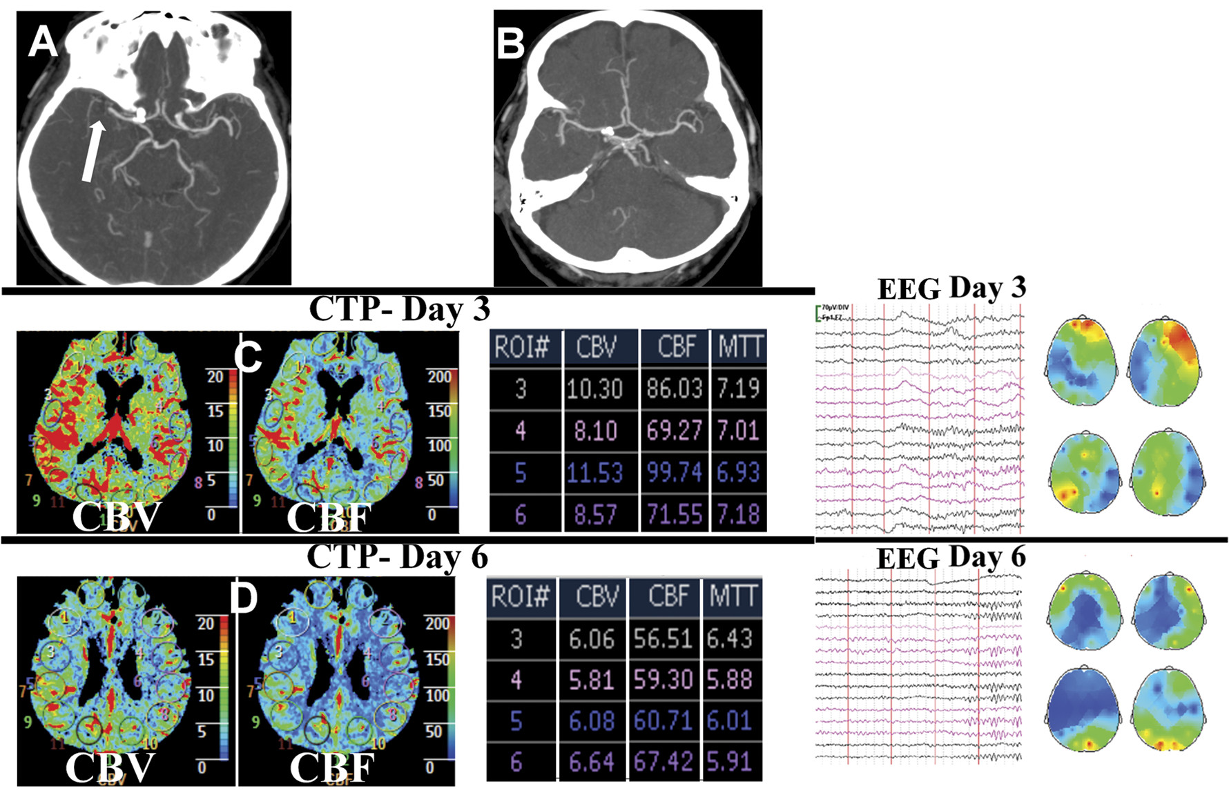
No abnormalities were noted on the emergently performed brain computerized tomography (CT). However, CT angiography (CTA) revealed right middle cerebral artery (MCA) occlusion (Figure 1 [A]). Intravenous thrombolysis and continuous transcranial Doppler (TCD) monitoring, commenced at 180 minutes from symptom-onset, resulted in complete recanalization of right MCA at 14 minutes after treatment onset. This was accompanied by rapid and persistent clinical recovery (NIHSS score 1 point at 1 hour). Neurological examination, brain CT, and CTA (Figure 1 [B]) performed on Day 2 were unremarkable.
FIGURE 1. Brain CT of Patient With Suicidal Ideation After Successful Acute Stroke Thrombolysis
Pre-thrombolysis brain CT angiography shows occluded right middle cerebral artery [A] that recanalized completely on Day 2 [B]. Significantly elevated cerebral blood flow (CBF) and volume (CBV) are evident in right frontal region (region 3 and 5; reference 4 and 6 on left hemisphere) on CT perfusion (CTP) performed on Day 3. Day-6 CTP shows normalization of various cerebral perfusion parameters. Corresponding raw electroencephalography (EEG) data and spectral brain mapping on Days 3 and 6 are shown. EEG performed on Day 3 shows considerable right fronto-temporal slowing, seen as red/yellow areas on spectral brain mapping. EEG and spectral maps show normalization of cerebral activity on Day 6.
Despite brilliant clinical recovery, our patient displayed severe suicidal ideation on Day 3. He had never suffered from depression or any psychiatric illness in past. Brain magnetic resonance imaging showed an acute lacunar infarction in the putamen. Repeat TCD showed considerably elevated flow velocities in right MCA (3 times more than the left). Further evaluation of cerebral hemodynamics by CT-perfusion (CTP) revealed relative right MCA territory hyperperfusion (Figure 1). Suspecting that regional hyperperfusion was responsible for his suicidal ideation, we refrained from initiating any medications. Head-up position, fluid restriction, and aggressive blood pressure control (to 120/80 mmHg) resulted in rapid improvement in psychiatric features during the next 2 days. CTP performed on Day 6 revealed normalization of cerebral perfusion parameters (Figure 1). Since his hospital discharge, on Day 7, he has remained well during the past 1 year.
“Reperfusion injury” after reversible ischemia may result in myocardial stunning in some cases.1 Similar “ischemic stunning” of the brain has been proposed in acute ischemic stroke when clinical recovery does not accompany successful systemic thrombolysis.2 Acute suicidal ideation, despite brilliant early recovery in our patient was unexpected. We hypothesized that the regional hyperperfusion led to the reduced neuronal activity (neurovascular uncoupling), manifesting as severe psychiatric disturbance. Our hypothesis was supported by the significant right fronto-temporal slowing on electroencephalography (EEG), performed on Day 3 (Figure 1). Rapid clinico-psychiatric recovery accompanied normalization of cerebral perfusion (Day 6 CTP in Figure 1) and neuronal activity (Day 6 EEG in Figure 1).
Footnote

Contributorship statement: Conception and acquisition of data: Leonard Yeo, Rahul Rathkrishnan, Roger Ho, Vijay K Sharma; interpretation of data and drafting the article: Leonard Yeo, Rahul Rathkrishnan; critical revision: Roger Ho, Vijay K Sharma; final approval of the version: Vijay K. Sharma, Rahul Rathkrishnan, Roger Ho, Leonard Yeo.

References

1.
Bolli R, Marbán E: Molecular and cellular mechanisms of myocardial stunning. Physiol Rev 1999; 79:609–634
2.
Alexandrov AV, Hall CE, Labiche LA, et al.: Ischemic stunning of the brain: early recanalization without immediate clinical improvement in acute ischemic stroke. Stroke 2004; 35:449–452

Information & Authors

Information

Published In

Go to The Journal of Neuropsychiatry and Clinical Neurosciences
The Journal of Neuropsychiatry and Clinical Neurosciences
Pages: E33 - E34
PubMed: 24247882

History

Published online: 1 October 2013
Published in print: Fall 2013

Authors

Affiliations

Vijay K. Sharma, M.D., RVT
Leonard L.L. Yeo, M.R.C.P.
Roger C. Ho, M.R.C.Psych.
Rahul Rathakrishnan, M.R.C.P.
Division of Neurology, National University Hospital, Singapore
Yong Loo Lin School of Medicine, National University of Singapore, Singapore
Division of Psychological Medicine, National University Hospital, Singapore

Notes

Correspondence: Dr. Vijay K Sharma; e-mail: [email protected]

Metrics & Citations

Metrics

Citations

Export Citations

If you have the appropriate software installed, you can download article citation data to the citation manager of your choice. Simply select your manager software from the list below and click Download.

For more information or tips please see 'Downloading to a citation manager' in the Help menu.

Format
Citation style
Style
Copy to clipboard

There are no citations for this item

View Options

View options

PDF/ePub

View PDF/ePub

Get Access

Login options

Already a subscriber? Access your subscription through your login credentials or your institution for full access to this article.

Personal login Institutional Login Open Athens login
Purchase Options

Purchase this article to access the full text.

PPV Articles - Journal of Neuropsychiatry and Clinical Neurosciences

PPV Articles - Journal of Neuropsychiatry and Clinical Neurosciences

Not a subscriber?

Subscribe Now / Learn More

PsychiatryOnline subscription options offer access to the DSM-5-TR® library, books, journals, CME, and patient resources. This all-in-one virtual library provides psychiatrists and mental health professionals with key resources for diagnosis, treatment, research, and professional development.

Need more help? PsychiatryOnline Customer Service may be reached by emailing [email protected] or by calling 800-368-5777 (in the U.S.) or 703-907-7322 (outside the U.S.).

Media

Figures

Other

Tables

Share

Share

Share article link

Share