Full access
Letters
Published Online: 1 January 2014

Cerebral Cryptococcoma in an HIV-Negative Patient: Experience Learned From a Case

Publication: The Journal of Neuropsychiatry and Clinical Neurosciences
To the Editor: Cryptococcosis is the most common mycotic infection of the CNS and primarily manifests as meningitis. Cryptococcal infection of the nervous system was first described early in the beginning of the last century. Thus far, few cryptococcal infection cases have been reported to be associated with HIV-positive patients. Here, the authors report a rare case of a 75-year-old male patient with no previous history of any systemic diseases such as diabetes, hypertension, or another immunocompromised condition, but with a cryptococcoma in his brain.
Cyptococcosis, the most common mycotic infection of the CNS, primarily manifests as meningitis. This ubiquitous organism typically enters the human body via the respiratory tract and then spreads hematogenously from the lung to the CNS.1 According to the analysis of the French Cryptococcosis study group, of the 1057 cryptococcosis cases, 86% involved patients with AIDS. Malignancies (32%), organ transplantation (19%), and corticosteroid therapy (33%) were the main predisposing conditions in 163 patients without AIDS.2
Intracranial cryptococcosis primarily manifests as meningitis, headache, fever, and nuchal stiffness. In some cases, mass lesions can also develop.3 Here the authors report a case of cerebral cryptococcoma found in a 75-year-old male patient who had no diabetes, hypertension, or AIDS, nor received immunosuppressive medications, and he did not present any manifestations of meningioencephalitis.

Case Report

A 75-year-old man was admitted to our hospital because he had right side weakness. The symptom had been progressive for several days prior to his admission. The patient had no severe headache or fever. He exercised regularly and did not drink, smoke, or take drugs. Physical examination showed weakness in his right arm, as well as in his right leg to lesser degree, and mildly diminished touch sense in the right limbs compared with the left ones. Physical examination showed no choking, hemianopia, ataxia, or dysarthria.
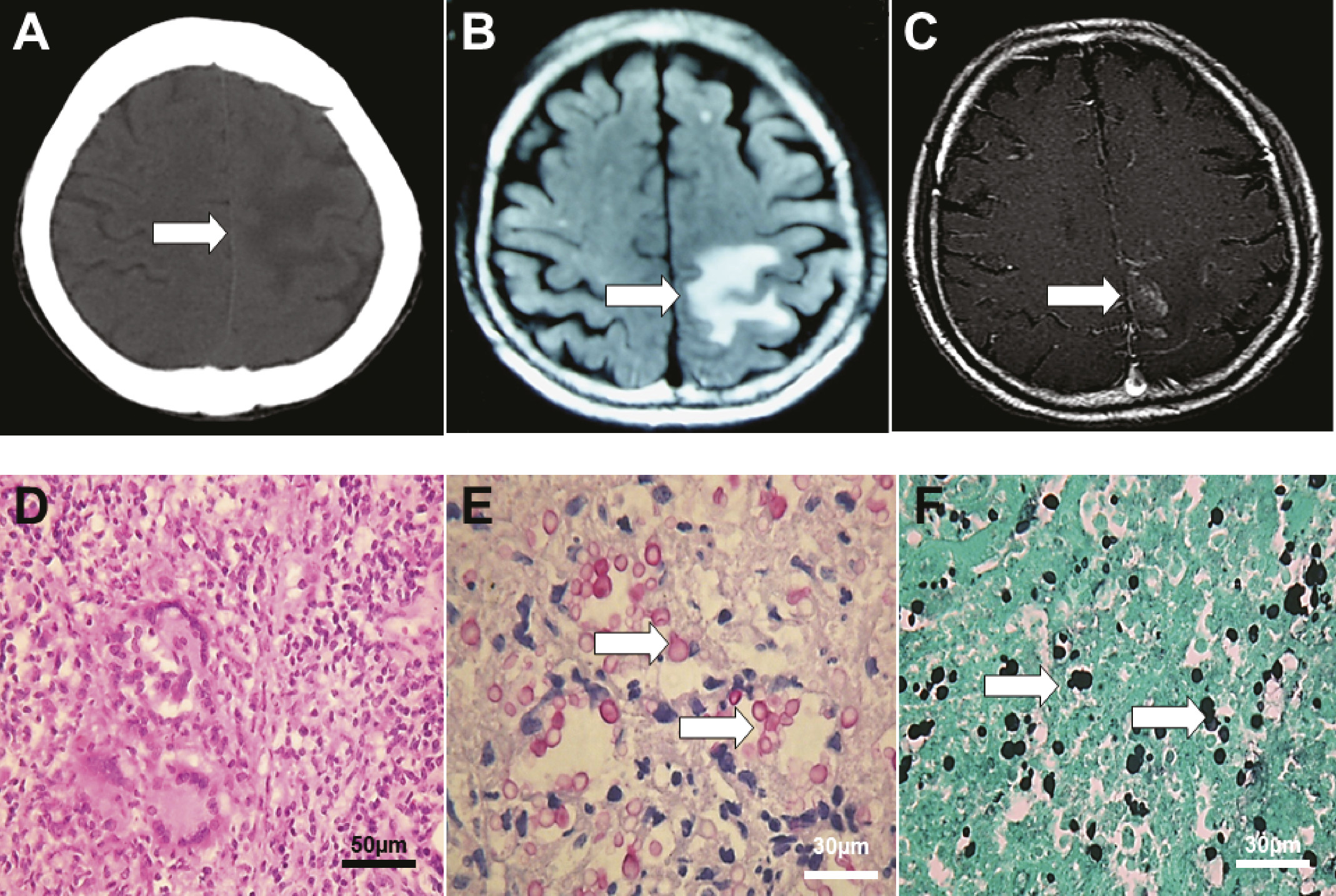
Laboratory tests showed that the patient’s hematocrit was 0.37, and the white cell count was 6600/mL, with 68% neutrophils, 2% eosinophils, and 24% lymphocytes. Repeated tests for HIV were negative. CT scans (Figure 1A) and MRI scan showed a single mass with focal white matter edema located at the left parietal lobe adjacent to the falx cerebri (Figure 1, B and C). A brain tumor was suspected, and a neurosurgeon was then consulted for excision at the sixth day after admission. Histopathology results showed acute and chronic inflammation with necrosis and granulomatous inflammation (Figure 1D); Periodic acid-Schiff stain and Grocott's methenamine silver stain were positive for Cryptococcus elements (Figure 1, E and F). Intravenous fluconazole therapy was started after surgery. The patient died from systemic sepsis at the 17th day after surgery.
FIGURE 1. Imaging Scan and Histological Results
[A]: Noncontrast brain CT scan showed abnormal white matter edema over left hemisphere (arrow). [B]: Corresponding area revealed diminished cortical gyrus in subsequent MRI scans as T2-weighted image. [C]: Postgadolinium T1-weighted image indicated a central necrotic mass adjacent to the falx cerebri. [D]: Acute inflammation with granuloma formation (hematoxylin and eosin stain; scale bar, 50 μm). [E]: Microabscesses containing many cryptococci (Periodic acid-Schiff stain). Cryptococci are ovoid to single spherical budding, thick-wall yeasts (arrows). [F]: Cryptococci in inflammatory debris (arrows, Grocott's methenamine silver stain). Scale bar for E and F, 30 μm.

Discussion

Cryptococci prey on individuals with abnormal cell-mediated immunity caused by a variety of debilitating conditions. The major cause of immunocompromise is AIDS in both western3 and eastern countries.4 Majors symptoms are headache (83%), vomiting (65%), proptosis (48%), and visual disturbance (48%).5 Yu et al.4 reported that two-thirds of patients presented with the triad of fever, headache, and neck rigidity.
This case is very unique because the patient never complained about headache, fever, or visual problems except for weakness in his right limbs and his brain scan showed a locus resembling a malignant or metastatic tumor that mimics that of a central necrotic brain tumor rather than a pygenic brain abscess. The patient’s living space was free from pigeons and domestic fowl. The laboratory tests showed no HIV antibody in his serum. He had no diabetes, malignancies, or hematological disorders.
In conclusion, cryptococcosis is easily misdiagnosed in HIV-negative patients because cerebral cryptococcoma is rare in HIV-negative patients, and there is a misconception that the disease affects mainly immunocompromised individuals.
References

1.
Mathews VP, Alo PL, Glass JD, et al: AIDS-related CNS cryptococcosis: radiologic-pathologic correlation. AJNR Am J Neuroradiol 1992; 13:1477–1486
2.
Dromer F, Mathoulin S, Dupont B, et al: French Cryptococcosis Study Group: Epidemiology of cryptococcosis in France: a 9-year survey (1985-1993). Clin Infect Dis 1996; 23:82–90
3.
Garcia CA, Weisberg LA, Lacorte WS: Cryptococcal intracerebral mass lesions: CT-pathologic considerations. Neurology 1985; 35:731–734
4.
Yu YL, Lau YN, Woo E, et al: Cryptococcal infection of the nervous system. Q J Med 1988; 66:87–96
5.
Dubey A, Patwardhan RV, Sampth S, et al: Intracranial fungal granuloma: analysis of 40 patients and review of the literature. Surg Neurol 2005; 63:254–260, discussion 260

Information & Authors

Information

Published In

Go to The Journal of Neuropsychiatry and Clinical Neurosciences
The Journal of Neuropsychiatry and Clinical Neurosciences
Pages: E34 - E35
PubMed: 26037882

History

Published online: 1 January 2014
Published in print: Fall 2014

Authors

Details

Chung-hsin Yeh, M.D., Ph.D.
Dept. of Neurology, Show-Chwan Memorial Hospital, Changhua, Taiwan (C-hY, S-fML, M-cC); Dept. of Nursing, College of Medicine and Nursing, HungKuang University, Taichung City, Taiwan (ROC) (C-hY); and Dept. of Pathology (C-lK), Dept. of Radiology (H-tH), and Dept. of Neurosurgery (H-mS), Show-Chwan Memorial Hospital, Changhua, Taiwan
Shung-fen Maggie Lin, M.S.
Dept. of Neurology, Show-Chwan Memorial Hospital, Changhua, Taiwan (C-hY, S-fML, M-cC); Dept. of Nursing, College of Medicine and Nursing, HungKuang University, Taichung City, Taiwan (ROC) (C-hY); and Dept. of Pathology (C-lK), Dept. of Radiology (H-tH), and Dept. of Neurosurgery (H-mS), Show-Chwan Memorial Hospital, Changhua, Taiwan
Mei-chun Chiu, M.S.
Dept. of Neurology, Show-Chwan Memorial Hospital, Changhua, Taiwan (C-hY, S-fML, M-cC); Dept. of Nursing, College of Medicine and Nursing, HungKuang University, Taichung City, Taiwan (ROC) (C-hY); and Dept. of Pathology (C-lK), Dept. of Radiology (H-tH), and Dept. of Neurosurgery (H-mS), Show-Chwan Memorial Hospital, Changhua, Taiwan
Chien-long Kuo, M.D.
Dept. of Neurology, Show-Chwan Memorial Hospital, Changhua, Taiwan (C-hY, S-fML, M-cC); Dept. of Nursing, College of Medicine and Nursing, HungKuang University, Taichung City, Taiwan (ROC) (C-hY); and Dept. of Pathology (C-lK), Dept. of Radiology (H-tH), and Dept. of Neurosurgery (H-mS), Show-Chwan Memorial Hospital, Changhua, Taiwan
Hua-tzu Huang, M.D.
Dept. of Neurology, Show-Chwan Memorial Hospital, Changhua, Taiwan (C-hY, S-fML, M-cC); Dept. of Nursing, College of Medicine and Nursing, HungKuang University, Taichung City, Taiwan (ROC) (C-hY); and Dept. of Pathology (C-lK), Dept. of Radiology (H-tH), and Dept. of Neurosurgery (H-mS), Show-Chwan Memorial Hospital, Changhua, Taiwan
Hai-ming Shoung, M.D.
Dept. of Neurology, Show-Chwan Memorial Hospital, Changhua, Taiwan (C-hY, S-fML, M-cC); Dept. of Nursing, College of Medicine and Nursing, HungKuang University, Taichung City, Taiwan (ROC) (C-hY); and Dept. of Pathology (C-lK), Dept. of Radiology (H-tH), and Dept. of Neurosurgery (H-mS), Show-Chwan Memorial Hospital, Changhua, Taiwan

Notes

Correspondence: Hai-ming Shoung, M.D.; e-mail: [email protected]

Metrics & Citations

Metrics

Citations

Export Citations

If you have the appropriate software installed, you can download article citation data to the citation manager of your choice. Simply select your manager software from the list below and click Download.

For more information or tips please see 'Downloading to a citation manager' in the Help menu.

Format
Citation style
Style
Copy to clipboard

View Options

View options

PDF/EPUB

View PDF/EPUB

Login options

Already a subscriber? Access your subscription through your login credentials or your institution for full access to this article.

Personal login Institutional Login Open Athens login
Purchase Options

Purchase this article to access the full text.

PPV Articles - Journal of Neuropsychiatry and Clinical Neurosciences

PPV Articles - Journal of Neuropsychiatry and Clinical Neurosciences

Not a subscriber?

Subscribe Now / Learn More

PsychiatryOnline subscription options offer access to the DSM-5-TR® library, books, journals, CME, and patient resources. This all-in-one virtual library provides psychiatrists and mental health professionals with key resources for diagnosis, treatment, research, and professional development.

Need more help? PsychiatryOnline Customer Service may be reached by emailing [email protected] or by calling 800-368-5777 (in the U.S.) or 703-907-7322 (outside the U.S.).

Media

Figures

Other

Tables

Share

Share

Share article link

Share