Full access
Clinical and Research Reports
Published Online: 28 March 2016

The Effect of Intravenous Citalopram on the Neural Substrates of Obsessive-Compulsive Disorder

Publication: The Journal of Neuropsychiatry and Clinical Neurosciences

Abstract

This study investigated the effect of an intravenous serotonin reuptake inhibitor on the neural substrates of obsessive-compulsive disorder (OCD), as intravenous agents may be more effective in treating OCD than conventional oral pharmacotherapy. Eight OCD subjects and eight control subjects received alternate infusions of citalopram and placebo during functional magnetic resonance imaging, in a randomized, symptom-provocation, crossover design. Compared with baseline, OCD subjects displayed significant changes in prefrontal neural activity after the citalopram infusion relative to placebo, and these changes correlated with reductions in subjective anxiety.
The cortico-striatal-thalamo-cortical circuits arising from the orbitofrontal cortex (OFC) and anterior cingulate cortex, among others, have commonly been implicated in the pathophysiology of obsessive-compulsive disorder (OCD).1,2 These regions, which are often reported as being hyperactive in OCD, have been shown to normalize after prolonged treatment with oral selective serotonin reuptake inhibitors (SSRIs).35 However, the effects of intravenous (IV) antiobsessive agents on the neural substrates of OCD have not previously been investigated, even though they are effective at treating refractory patients, are safe and well-tolerated, produce greater symptom reductions, and have faster onsets of action than their oral counterparts.6 To better understand the mechanism of IV antiobsessive agents, the present study evaluated the effect of an intravenously administered SSRI, compared with placebo, on the neural activity of OCD patients and matched controls.

Methods

Participants

Eight right-handed patients between the ages of 18 and 50, who met DSM-IV-TR7 criteria for OCD, participated in the study after being recruited from the Centre for Addiction and Mental Health in Toronto, Canada. OCD patients with lifetime comorbid diagnoses, as determined by the Structured Clinical Interview for DSM-IV Disorders,8 were excluded. However, patients with comorbid major depressive disorder and other anxiety disorders were allowed to participate, providing OCD was the primary diagnosis. All OCD subjects must have had contamination or safety-doubting as their primary obsessions, and washing-cleaning or checking behaviors as their primary compulsions. The minimum Yale-Brown Obsessive-Compulsive Scale9 score for enrollment was 17. Subjects taking medication were weaned off so that they were medication free for 2 weeks prior to each scan. However, patients who were being treated with fluoxetine or behavior therapy at the time of the study were excluded. Eight age-, gender-, and education-matched healthy controls with no personal or family history of psychiatric illness were recruited from the community. For the variables of age and education, patient and control subjects were matched one-to-one within 3 years (see Table 1).10,11
TABLE 1. Demographic and Clinical Characteristics of the Samplesa
VariableOCDControls
Age (mean, SD)27.6 (2.3)25.5 (2.9)
Years of education (mean, SD)15.9 (2.4)17.3 (1.4)
Male/female4/44/4
Q-LES-Q (mean, SD)1044.5 (12.9)59.1 (5.2)
BDI-II (mean, SD)11,*9.8 (4.8)1.0 (.9)
Y-BOCS (mean, SD)21.8 (4.4)N/A
Illness duration, years (mean, SD)10.3 (7.5)N/A
Age of onset, years (mean, SD)16 (6.2)N/A
Lifetime comorbid diagnosesMDD: N=2N/A
 Panic disorder: N=1
MedicationsSertraline: N=1N/A
 Citalopram: N=1
a
BDI-II, Beck Depression Inventory II; Q-LES-Q, Quality of Life Enjoyment and Satisfaction Questionnaire; Y-BOCS, Yale-Brown Obsessive-Compulsive Scale
*
BDI-II scores were not significantly different between the two groups (U=27, p=.18).

Procedures

Informed consent was obtained from all participants after procedures were fully explained. All participants completed two functional magnetic resonance imaging (fMRI) scans, which were separated by a minimum 2-week period. Participants were randomized to receive either IV citalopram or placebo during their first fMRI scan and the alternate infusion during their second scan. They were blind to the infusion order. At each scan, after two 6-minute symptom-provocation baseline (preinfusion) runs were completed, a 30-minute infusion of either placebo (250 ml of 0.9% sodium chloride) or citalopram (20 mg in 250 ml 0.9% saline) was initiated. Four additional runs were conducted, two during infusion administration (midinfusion) and two immediately after the infusion ended (postinfusion), for a total of six functional runs per scan.
Each symptom-provocation run was composed of seven picture blocks presenting either aversive scenes, neutral scenes, or scenes corresponding to common OCD symptoms (i.e., checking, washing, or hoarding). Picture blocks were grouped by category and consisted of five scenes, each presented for 3.2 seconds on a black background. Immediately following the presentation of each block, participants rated how anxious the pictures made them feel using a 7-point Likert scale and a response button box. In total, 210 color pictures were used for the provocation task (45 scenes for each of the washing, checking, neutral, and aversive picture categories, and 30 scenes for the hoarding category). Pictures were chosen from the International Affective Picture System12 to represent the neutral and aversive blocks. Scenes depicting the OCD symptoms of washing, checking, and hoarding were obtained with a standard digital camera and from an Internet search. A psychiatrist confirmed the anxiety-arousing nature of the symptom-related pictures.
Arrow blocks separated each picture block and served as a buffer to minimize potential carry-over effects. Arrows pointing either left or right were randomly displayed on the screen, and participants were asked to indicate which way they were pointing as quickly and accurately as possible by pressing the appropriate buttons. Each arrow block lasted 20 seconds and consisted of ten white arrows on a black background.

Image Acquisition and Data Analysis

Neuroimaging (fMRI) scans were conducted at Baycrest Hospital on a 3T scanner (Siemens Medical Solutions, Erlangen, Germany). Stimulus presentation was performed using Visual Basic (Visual Studio 2005; Microsoft, Redmond, Washington) on a standard computer, which also collected subjects’ anxiety ratings. The stimuli were projected on a screen placed in the bore of the magnet and seen by the participant by means of a mirror attached to the head coil. Functional images were acquired using T2* weighted gradient echo planar imaging sequences covering 30 slices (5.00 mm thick, zero gap), aligned oblique axially, and encompassing the entire cerebral cortex (repetition time=2,000 ms, echo time=30 ms, field of view=200 mm, flip angle=70°, matrix=64×64, interleaved acquisition). A high T1 weighted anatomical scan (repetition time=2,000 ms, echo time=2.63 ms, field of view=256, slice thickness=1 mm, 160 slices) was acquired after the infusions started.
Spatial preprocessing and whole-brain image analysis of MRI images were performed using Statistical Parametric Mapping, version 8 (University College London, London, United Kingdom; http://www.fil.ion.ucl.ac.uk/spm/software/spm8). The first three images from each run were excluded from the analyses to eliminate any T2*-equilibrium effects. To correct for subject motion, images were realigned to the mean image obtained for each participant and co-registered with their T1-weighted structural image. The T1 image was segmented using template (International Consortium for Brain Mapping) tissue probability maps for gray and white matter and cerebrospinal fluid. Parameters obtained from this step were subsequently applied to the functional and structural data during normalization into a standard stereotactic space (Montreal Neurological Institute template) using a 12-parameter affine model. Images were then smoothed with a Gaussian filter, set at 6 mm full-width-half-maximum.
Single subject data were analyzed at the first level with general linear statistical models, and the time series of the images were convolved with a canonical hemodynamic response function. Each model included high-pass filtering to remove low-frequency signal drift (period=128 seconds).
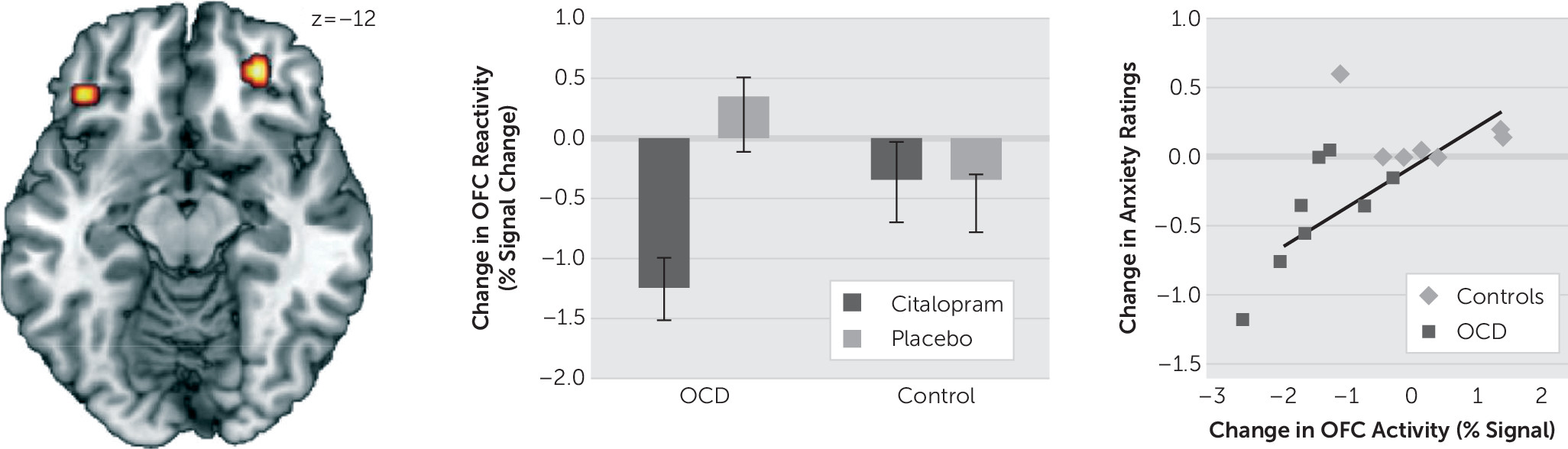
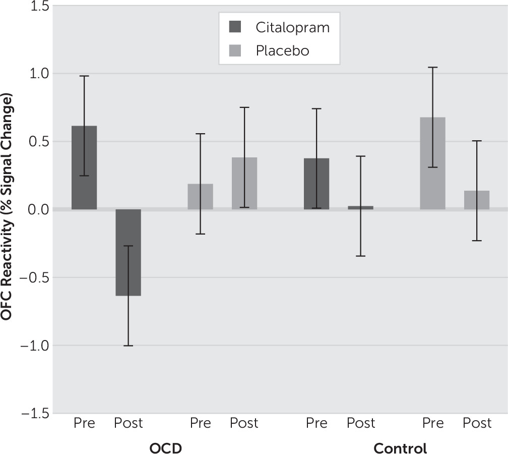
Our analysis was simplified to reflect the subtypes of the clinical sample, which featured washing and checking subtypes of OCD. Signal estimates of the washing and checking blocks were averaged together (termed “symptom” blocks) and contrasted against the neutral blocks as an index of neural reactivity. Neural reactivity was calculated for each subject at each of the pre-, mid-, and postinfusion time points. Independent samples t tests were then used to characterize reactivity differences between OCD subjects and controls at baseline. To examine the distinct effects of citalopram, mixed-model analyses of variance (ANOVAs) were performed. First, to examine initial infusion effects, a Time (Preinfusion versus Midinfusion) × Group (OCD versus Control) × Drug (Citalopram versus Placebo) ANOVA was performed. To examine full-infusion effects, a Time (Preinfusion versus Postinfusion) × Group × Drug ANOVA was performed. While the main analyses involved separately comparing the midinfusion and postinfusion scans to baseline (i.e., preinfusion scans), an additional ANOVA was conducted comparing changes in infusion-related activity across all three time points [Time (Preinfusion versus Midinfusion versus Postinfusion) × Group × Drug]. All statistical maps were set at a threshold of p<.001, uncorrected for cluster sizes greater than ten contiguous voxels, which is equivalent to a familywise error rate of PFWE<0.05 in a Monte Carlo simulation (AlphaSim; http://afni.nih.gov/afni/docpdf/AlphaSim.pdf).
To investigate the behavioral relevance of observed changes in neural reactivity, anxiety ratings from the checking and washing blocks were averaged to create composite symptom anxiety ratings and combined in a Group × Drug × Time ANOVA. Changes in anxiety ratings were regressed on observed changes in brain activity; correlations surviving an α<.05 Bonferroni correction were considered significant.

Results

Anxiety Ratings

For the symptom blocks, OCD subjects had significantly higher anxiety ratings than controls at all time points (F2, 28=11.08, p<.001). Significant interactions were found between Time × Group (F2, 28=10.40; p<.001) and for Group × Drug × Time (F2,28=3.64; p=.039). Post hoc tests revealed that post-citalopram-infusion ratings were uniquely lower than preinfusion (p<.001) and midinfusion (p=.003) ratings in the patient group (Figure 1).
FIGURE 1. Subjective Anxiety Ratings for the Symptom-Provoking Picture Blocks

Baseline

For the symptom contrast, OCD subjects exhibited greater activity in the left middle frontal gyrus and decreased activity in the right inferior temporal gyrus, compared with controls (Table 2).
TABLE 2. Main fMRI Findings for the Symptom Versus Neutral Contrasts
Time Points ContrastedBrain RegionsBrodmann AreaSideMNI coordinatesCluster sizeZ value
xyz
Baseline OnlyOCD>controls       
Middle frontal10L–30537153.83
Controls>OCD       
Inferior temporal37R57–58–2113.78
         
Pre versus PostDrug × group       
 Orbitofrontal11R2744–14194.15
 Orbitofrontal47L–3932–14143.86
         
Pre versus Mid versus PostDrug × group       
CerebellumL–9–61–32214.16
Calcarine sulcus18R24–971313.99
 Superior occipital18L–18–914383.77

Preinfusion Versus Midinfusion

There were no significant interactions between drug and group when comparing baseline to midinfusion scans.

Preinfusion Versus Postinfusion

Significant interactions between drug condition and diagnostic group were found bilaterally in the OFC; the OCD group exhibited reductions in OFC activity after the citalopram infusion compared with baseline (Figure 2, Figure 3). Pre-post changes in subjective anxiety ratings correlated with the size of the interaction effects in the OFC. Specifically, reductions in left OFC activity correlated with reductions in anxiety ratings [r(14)=.66, p=.006].
FIGURE 2. Preinfusion Versus Postinfusion Model: Interaction Between Group, Time and Drug Condition Bilaterally in the OFC; the Correlation Between Changes in Subjective Anxiety Ratings and Changes in Left OFC Activity
FIGURE 3. Pre- Versus Postinfusion Model: OFC Reactivity Values for Each of the Conditions in the Significant Group × Time × Infusion Interaction

Preinfusion Versus Midinfusion Versus Postinfusion

Significant interactions were found in the cerebellum, calcarine sulcus, and superior occipital gyrus (Table 2); however, the magnitude of these changes did not correlate with changes in subjective anxiety.

Discussion

To our knowledge, this is the first study to evaluate the effect of an IV antiobsessive agent on the neural substrates of an OCD sample. While replication studies are needed to confirm our findings, the present study provides evidence that IV citalopram has immediate effects on the neuronal substrates implicated in OCD, as well as on the anxiety associated with OCD-related stimuli. The reduced OFC activity observed in the OCD group after the citalopram infusion is similar to those previously reported with long-term oral SSRI treatment.35,13,14 This altered prefrontal activity supports the notion that compromised cortico-striatal-thalamo-cortical circuitry in OCD2 may normalize after successful treatment with SSRIs. Moreover, reductions in anxiety ratings correlated with changes in OFC activity, adding to the evidence of the role of this region in the pathophysiology of OCD and the potential mechanism of the anxiolytic effect of SSRIs. Of note, the results of the pre- versus mid- versus postinfusion model were different from the pre- versus postinfusion model, demonstrating the need to look at longer time courses to view citalopram-based down-regulation of OFC activity in OCD (whereas, at midinfusion, variability of treatment response between participants likely obfuscated such effects).
Intravenous serotonin reuptake inhibitors have been shown to be more effective and confer quicker benefits than oral medications when treating OCD symptoms (6–8 weeks for oral versus 4 days–3 weeks for IV treatment).6 This study represents the first, necessary step in studying the neurobiological mechanism for such benefits. Though the effects observed in the present study were most likely temporary, as participants only received a single dose of citalopram, it may be reasonable to suggest that these effects could persist with a prolonged trial of IV citalopram.15 Indeed, it would be interesting to see if longer courses of IV treatment produce lasting reductions in prefrontal neural activity, coupled with clinically meaningful reductions in OCD symptoms.
Although these findings may support the neurobiological model of OCD, the small sample sizes, the single-blind nature of the study, and the presence of comorbid conditions (although not primary) in the patient group limit the generalizability of our findings. Another possible confound is that citalopram may have direct effects on the cerebral vasculature, so that the effects observed in this study may not necessarily be due to alterations in neuronal activity; however, the vascular effects of citalopram-induced serotonin manipulations are not believed to be sufficient to alter blood oxygen level–dependent signals.16 Nevertheless, caution should be used when interpreting the impact of the results, as the acute effects of drugs, especially from a single dose, can be different from chronic treatment; and, even though the OCD subjects reported experiencing significantly more anxiety than controls did at baseline, they perceived the stimuli as only mildly arousing. It is recommended that future studies use more provocative stimuli, longer treatment trials, and standardized OCD rating scales to assess the clinical efficacy of IV treatments. Further systematic and larger investigations of IV antiobsessive agents are warranted.

References

1.
Rotge JY, Guehl D, Dilharreguy B, et al: Provocation of obsessive-compulsive symptoms: a quantitative voxel-based meta-analysis of functional neuroimaging studies. J Psychiatry Neurosci 2008; 33:405–412
2.
Saxena S, Rauch SL: Functional neuroimaging and the neuroanatomy of obsessive-compulsive disorder. Psychiatr Clin North Am 2000; 23:563–586
3.
Nakao T, Nakagawa A, Yoshiura T, et al: Brain activation of patients with obsessive-compulsive disorder during neuropsychological and symptom provocation tasks before and after symptom improvement: A functional magnetic resonance imaging study. Biol Psychiatry 2005; 57:901–910
4.
Kang DH, Kwon JS, Kim JJ, et al: Brain glucose metabolic changes associated with neuropsychological improvements after 4 months of treatment in patients with obsessive-compulsive disorder. Acta Psychiatr Scand 2003; 107:291–297
5.
Rauch SL, Shin LM, Dougherty DD, et al: Predictors of fluvoxamine response in contamination-related obsessive compulsive disorder: A PET symptom provocation study. Neuropsychopharmacology 2002; 27:782–791
6.
Ravindran L, Jung S, Ravindran A: Intravenous anti-obsessive agents: a review. J Psychopharmacol 2008, 24: 287–296.
7.
American Psychiatric Association: Diagnostic and Statistical Manual of Mental Disorders, 4th ed. Washington, DC, American Psychiatric Publishing, 2000
8.
First MB, Spitzer RL, Gibbon M, et al: . Structured Clinical Interview for DSM-IV-TR Axis I Disorders, Research Version, Patient Edition With Psychotic Screen. New York, Biometrics Research, New York State Psychiatric Institute, 2002
9.
Goodman WK, Price LH, Rasmussen SA, et al: The Yale-Brown Obsessive Compulsive Scale. I. Development, use, and reliability. Arch Gen Psychiatry 1989; 46:1006–1011
10.
Endicott J, Nee J, Harrison W, et al: Quality of Life Enjoyment and Satisfaction Questionnaire: A new measure. Psychopharmacol Bull 1993; 29:321–326
11.
Beck AT, Steer RA, Brown GK, et al: BDI-II, Beck Depression Inventory. San Antonio, TX, Psychological Corporation, 1996
12.
Lang P, Bradley M, Cuthbert B: International Affective Picture System (IAPS): Affective ratings of pictures and instruction manual. Gainesville, University of Florida, Center for Research in Psychophysiology, 1999
13.
Saxena S, Brody AL, Maidment KM, et al: Localized orbitofrontal and subcortical metabolic changes and predictors of response to paroxetine treatment in obsessive-compulsive disorder. Neuropsychopharmacology 1999; 21:683–693
14.
Hendler T, Goshen E, Tzila Zwas S, et al: Brain reactivity to specific symptom provocation indicates prospective therapeutic outcome in OCD. Psychiatry Res 2003; 124:87–103
15.
Pallanti S, Quercioli L, Koran LM: Citalopram intravenous infusion in resistant obsessive-compulsive disorder: an open trial. J Clin Psychiatry 2002; 63:796–801
16.
Anderson IM, McKie S, Elliott R, et al: Assessing human 5-HT function in vivo with pharmacoMRI. Neuropharmacology 2008; 55:1029–1037

Information & Authors

Information

Published In

Go to The Journal of Neuropsychiatry and Clinical Neurosciences
The Journal of Neuropsychiatry and Clinical Neurosciences
Pages: 243 - 247
PubMed: 27019066

History

Received: 1 September 2015
Revision received: 13 December 2015
Accepted: 28 January 2016
Published online: 28 March 2016
Published in print: Summer 2016

Authors

Affiliations

Tracy P. Bhikram, M.Sc.
From the University of Toronto, Toronto, Canada (TPB, NASF, LNR, DKC, BGP, AVR); the Centre for Addiction and Mental Health, Toronto (TPB, LNR, YGP); the Campbell Research Institute, Toronto (BGP, AVR); the Rotman Research Institute, Toronto (NASF); and the Baycrest Centre for Geriatric Care, Toronto (DKC).
Norman A.S. Farb, Ph.D.
From the University of Toronto, Toronto, Canada (TPB, NASF, LNR, DKC, BGP, AVR); the Centre for Addiction and Mental Health, Toronto (TPB, LNR, YGP); the Campbell Research Institute, Toronto (BGP, AVR); the Rotman Research Institute, Toronto (NASF); and the Baycrest Centre for Geriatric Care, Toronto (DKC).
Lakshmi N. Ravindran, M.D., F.R.C.P.C.
From the University of Toronto, Toronto, Canada (TPB, NASF, LNR, DKC, BGP, AVR); the Centre for Addiction and Mental Health, Toronto (TPB, LNR, YGP); the Campbell Research Institute, Toronto (BGP, AVR); the Rotman Research Institute, Toronto (NASF); and the Baycrest Centre for Geriatric Care, Toronto (DKC).
Yousef G. Papadopoulos, M.D., F.R.C.P.C.
From the University of Toronto, Toronto, Canada (TPB, NASF, LNR, DKC, BGP, AVR); the Centre for Addiction and Mental Health, Toronto (TPB, LNR, YGP); the Campbell Research Institute, Toronto (BGP, AVR); the Rotman Research Institute, Toronto (NASF); and the Baycrest Centre for Geriatric Care, Toronto (DKC).
David K. Conn, M.B., F.R.C.P.C.
From the University of Toronto, Toronto, Canada (TPB, NASF, LNR, DKC, BGP, AVR); the Centre for Addiction and Mental Health, Toronto (TPB, LNR, YGP); the Campbell Research Institute, Toronto (BGP, AVR); the Rotman Research Institute, Toronto (NASF); and the Baycrest Centre for Geriatric Care, Toronto (DKC).
Bruce G. Pollock, M.D., Ph.D., F.R.C.P.C.
From the University of Toronto, Toronto, Canada (TPB, NASF, LNR, DKC, BGP, AVR); the Centre for Addiction and Mental Health, Toronto (TPB, LNR, YGP); the Campbell Research Institute, Toronto (BGP, AVR); the Rotman Research Institute, Toronto (NASF); and the Baycrest Centre for Geriatric Care, Toronto (DKC).
Arun V. Ravindran, M.D., Ph.D., F.R.C.P.C.
From the University of Toronto, Toronto, Canada (TPB, NASF, LNR, DKC, BGP, AVR); the Centre for Addiction and Mental Health, Toronto (TPB, LNR, YGP); the Campbell Research Institute, Toronto (BGP, AVR); the Rotman Research Institute, Toronto (NASF); and the Baycrest Centre for Geriatric Care, Toronto (DKC).

Notes

Send correspondence to Dr. Ravindran; e-mail: [email protected]

Competing Interests

The authors report no financial relationships with commercial interests.

Metrics & Citations

Metrics

Citations

Export Citations

If you have the appropriate software installed, you can download article citation data to the citation manager of your choice. Simply select your manager software from the list below and click Download.

For more information or tips please see 'Downloading to a citation manager' in the Help menu.

Format
Citation style
Style
Copy to clipboard

There are no citations for this item

View Options

View options

PDF/ePub

View PDF/ePub

Get Access

Login options

Already a subscriber? Access your subscription through your login credentials or your institution for full access to this article.

Personal login Institutional Login Open Athens login
Purchase Options

Purchase this article to access the full text.

PPV Articles - Journal of Neuropsychiatry and Clinical Neurosciences

PPV Articles - Journal of Neuropsychiatry and Clinical Neurosciences

Not a subscriber?

Subscribe Now / Learn More

PsychiatryOnline subscription options offer access to the DSM-5-TR® library, books, journals, CME, and patient resources. This all-in-one virtual library provides psychiatrists and mental health professionals with key resources for diagnosis, treatment, research, and professional development.

Need more help? PsychiatryOnline Customer Service may be reached by emailing [email protected] or by calling 800-368-5777 (in the U.S.) or 703-907-7322 (outside the U.S.).

Media

Figures

Other

Tables

Share

Share

Share article link

Share