Full access
Letters
Published Online: 19 April 2016

Deep Dyslexia in Chinese Primary Progressive Aphasia of Semantic Variant

Publication: The Journal of Neuropsychiatry and Clinical Neurosciences
To the Editor: Deep dyslexia, characterized by semantic errors during reading, is perhaps the most extensively studied central dyslexia.1 The failure to produce a phonologically matched but semantically relevant response suggests impairment of processes mediating the access of stimuli to the visual word form system.2 The current literature mostly describes this phenomenon in alphabetic languages, with deep dyslexia mostly being observed in the left hemisphere, especially in relationship to large perisylvian lesions extending to the frontal lobe.1 Generally, deep dyslexia is rarely seen in patients with fluent aphasia. This case report highlights two important points. First, we describe acquired deep dyslexia in a Chinese-speaking neurodegenerative model. Second, deep dyslexia is observed in a fluent aphasia syndrome. Both conditions remain rare in the literature. The mechanisms underpinning this phenomenon and the brain-behavior correlation highlighted in this case are discussed.

Case Report

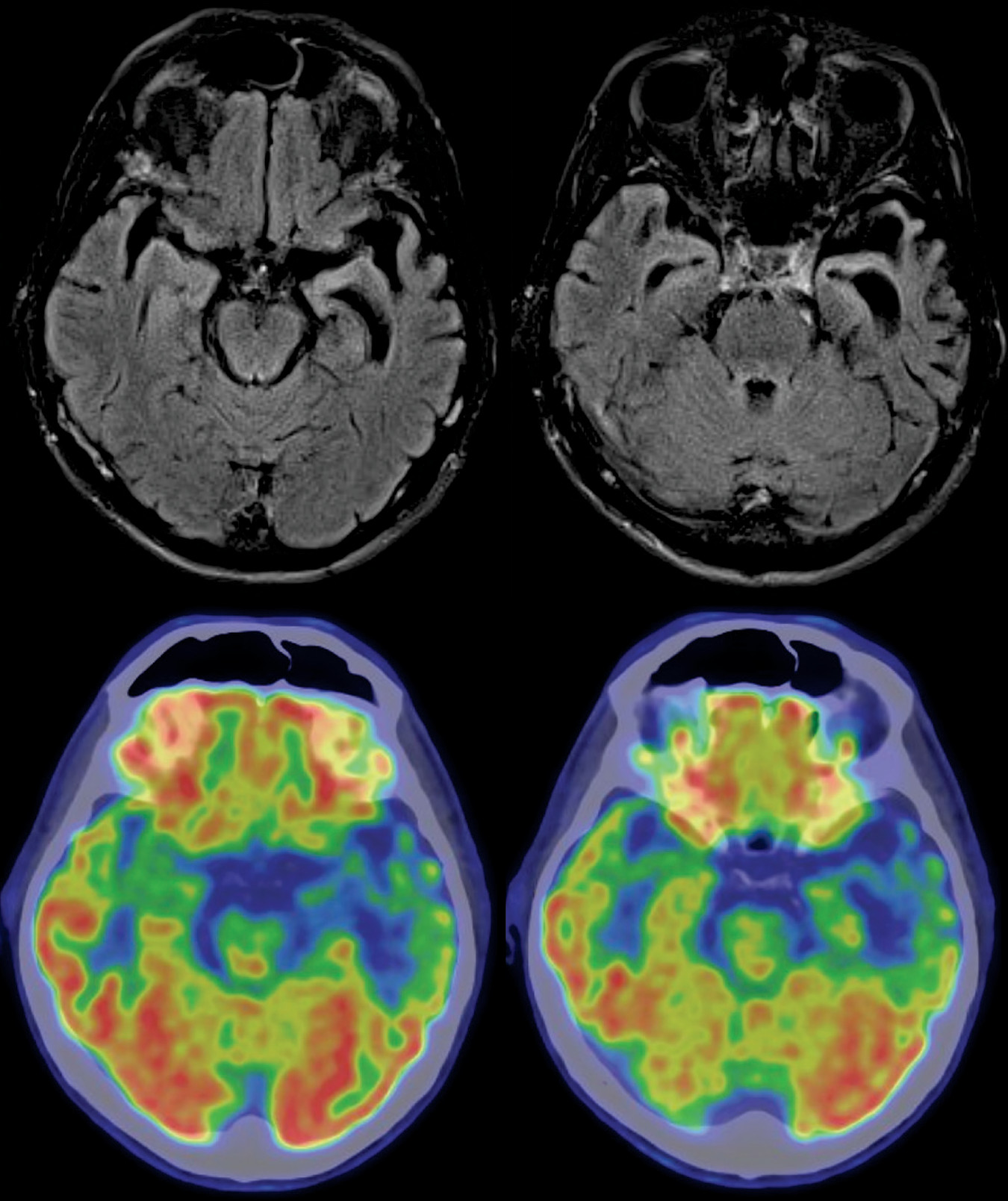
A right-handed 56-year-old Chinese Singaporean man had difficulty in comprehending sentences for 1 year. He received a Chinese-stream education up to secondary school. Neurobehavioral assessments showed fluent spontaneous speech and a Mini-Mental State Examination score of 19 of 30. The patient had a score of 3 of 15 on the modified version of the Boston Naming Test, naming a camel as “duck” and a bench as “table,” and he failed to comprehend 14 of 15 items after a 30-minute interval. Reading tasks of 10 words, 10 sentences, and a short passage from the standard modern Chinese version of the Bilingual Aphasia Battery were delivered. The patient committed a total of 21 reading errors, with 16 being deep dyslexic in nature. Examples of the responses are summarized in Table 1. An MRI scan of the brain showed left anterior temporal atrophy and a [18F]fluorodeoxyglucose–positron emission tomography scan showed predominant left anterior temporal hypometabolism (Figure 1).
TABLE 1. Reading Task Characters and Deep Dyslexic Semantic Errors in Our Patient
Print CharactersError Response
貓/mao1/cat鴨子/ya1zi3/duck
狼/lang2/wolf熊/xiong2/bear
敲/qiao1/knock刮/gua1/scratch
針/zhen1/needle夾子/jia2zi3/clip
咬/yao3/bite叫/jiao4/call
推/tui1/push拉/la1/pull
咬/yao3/bite親/qin1/kiss
鴿/ge1/pigeon雞/ji1/chicken
朋友/peng2you3/friend兄弟/xiong1di4/brother
FIGURE 1. MRI Brain Fluid-Attenuated Inversion Recovery Images (Top) and [18F]Fluorodeoxyglucose–Positron Emission Tomography Brain Images (Bottom) Showing Predominant Left Anterior Temporal Lobe Atrophy and Hypometabolism

Discussion

The literature describing acquired dyslexia in the Chinese language remains sparse to date. Although patients with English-speaking–acquired deep dyslexia are mostly described with larger left perisylvian lesions (e.g., by stroke),1 the rarity of literature describing this condition in Chinese-speaking individuals likely suggests a different mechanism underpinning such phenomenology. The Chinese language is ideographic and is generally believed to be lacking in grapheme-phoneme correspondence. Yin and Weekes3 proposed the triangle model of Chinese reading, where phonology output is directly imprinted into orthography in nonsemantic pathway reading. Other evidence agrees with the above notion that access to Chinese language phonology is distinct from alphabetic language.4 Shu et al.5 as well as Yin and Butterworth6 described cases of deep dyslexia that were either developmental, traumatic or vascular-related brain lesions, which were explained well by the triangle model. However, most patients suffered more diffuse and less well-localized lesions. Thus, more determining or precise brain-behavior correlation is challenging.
The current literature suggests the left temporal pole as the convergence region for semantic knowledge. In addition, a recent report by Busigny et al.7 suggested a case for lexico-facio-semantics disassociation, secondary to a dysfunction of the left anterior temporal lobe. Because Chinese is an ideographic language in which semantic data are tagged to each individual character, we speculate that this region also serves as a center of convergence of lexical semantics of the language. This most likely mediates the semantic relevance of orthographical representations, in this case, seen through an impaired nonsemantic reading pathway.
In conclusion, we propose the left anterior temporal region as a convergence point of Chinese lexical semantics; therefore, this region deserves further future studies.

References

1.
Coltheart M, Patterson K, Marshall JC (eds): Deep Dyslexia. London, Routledge and Kegan Paul, 1980
2.
Colangelo A, Buchanan L: Localizing damage in the functional architecture: the distinction between implicit and explicit processing in deep dyslexia. J Neurolinguist 2007; 20:111–144
3.
Yin WG, Weekes BS: Dyslexia in Chinese: clues from cognitive neuropsychology. Ann Dyslexia 2003; 53:255–279
4.
Tan LH, Laird AR, Li K, et al: Neuroanatomical correlates of phonological processing of Chinese characters and alphabetic words: a meta-analysis. Hum Brain Mapp 2005; 25:83–91
5.
Shu H, Meng X, Chen X, et al: The subtypes of developmental dyslexia in Chinese: evidence from three cases. Dyslexia 2005; 11:311–329
6.
Yin WG, Butterworth B: Deep and surface dyslexia in Chinese, in Language Processing in Chinese. Edited by Chen HC, Tzeng OJ. Amsterdam, Elsevier Science Publishers, 1992, pp 349–366
7.
Busigny T, de Boissezon X, Puel M, et al: Proper name anomia with preserved lexical and semantic knowledge after left anterior temporal lesion: a two-way convergence defect. Cortex 2015; 65:1–18

Information & Authors

Information

Published In

Go to The Journal of Neuropsychiatry and Clinical Neurosciences
The Journal of Neuropsychiatry and Clinical Neurosciences
Pages: e25 - e26
PubMed: 27093385

History

Published in print: Spring 2016
Published online: 19 April 2016

Authors

Details

Simon Kang Seng Ting, M.D.
Dept. of Neurology, National Neuroscience Institute, Singapore General Hospital Campus, Singapore.
Stephanie Pei Shi Chia, B.Sc. (Hons) (Psych)
Dept. of Neurology, National Neuroscience Institute, Singapore General Hospital Campus, Singapore.
Shahul Hameed, M.D.
Dept. of Neurology, National Neuroscience Institute, Singapore General Hospital Campus, Singapore.

Notes

Send correspondence to Simon Kang Seng Ting; e-mail: [email protected]

Funding Information

The authors report no financial relationships with commercial interests.

Metrics & Citations

Metrics

Citations

Export Citations

If you have the appropriate software installed, you can download article citation data to the citation manager of your choice. Simply select your manager software from the list below and click Download.

For more information or tips please see 'Downloading to a citation manager' in the Help menu.

Format
Citation style
Style
Copy to clipboard

View Options

View options

PDF/EPUB

View PDF/EPUB

Login options

Already a subscriber? Access your subscription through your login credentials or your institution for full access to this article.

Personal login Institutional Login Open Athens login
Purchase Options

Purchase this article to access the full text.

PPV Articles - Journal of Neuropsychiatry and Clinical Neurosciences

PPV Articles - Journal of Neuropsychiatry and Clinical Neurosciences

Not a subscriber?

Subscribe Now / Learn More

PsychiatryOnline subscription options offer access to the DSM-5-TR® library, books, journals, CME, and patient resources. This all-in-one virtual library provides psychiatrists and mental health professionals with key resources for diagnosis, treatment, research, and professional development.

Need more help? PsychiatryOnline Customer Service may be reached by emailing [email protected] or by calling 800-368-5777 (in the U.S.) or 703-907-7322 (outside the U.S.).
Media

Figures

Other

Tables

Share

Share

Share article link

Share