Full access
Case Reports
Published Online: 15 March 2017

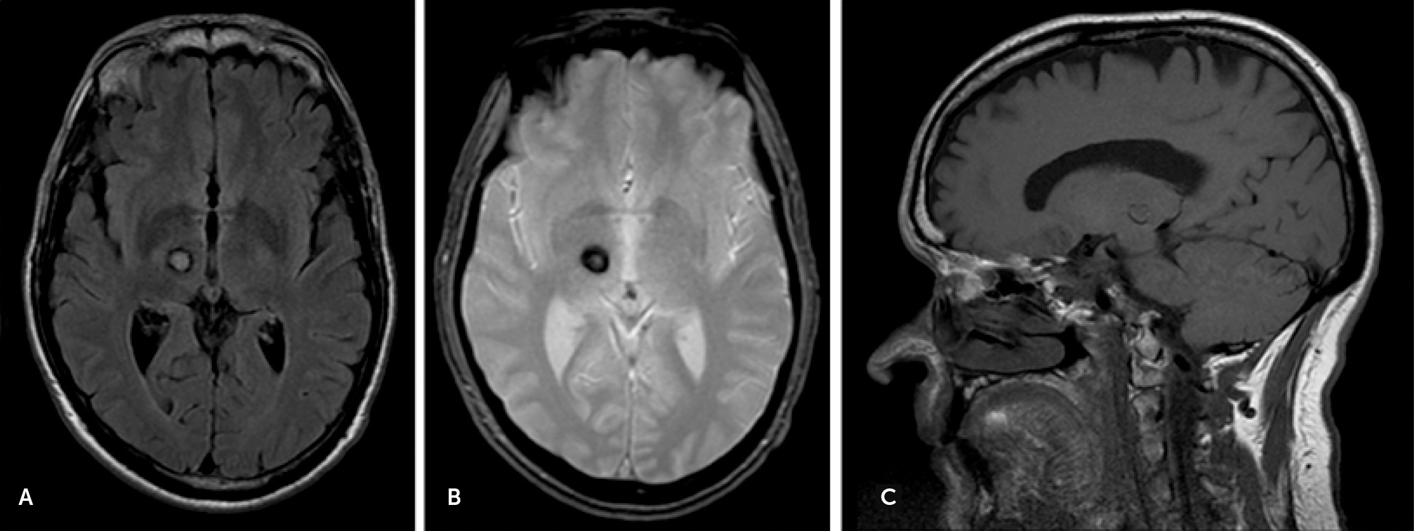
Behavioral Disconnection Syndrome Manifesting as Combined Mania and Visual-Auditory Hallucinations Secondary to Isolated Right Thalamic Hemorrhage

Publication: The Journal of Neuropsychiatry and Clinical Neurosciences
Damage to the thalamus can contribute to the phenomenon of diaschisis manifesting as various behavioral symptoms. We present the case of a 55-year-old man who presented with acute mania and late-onset combined visual and auditory hallucinations after right thalamic hemorrhage. This is the first report, to our knowledge, of a combination of mania and visual-auditory hallucination in a patient with isolated thalamic damage. Details of all previous case reports on isolated thalamic damage presenting with mania or hallucination are provided. We propose the networks of diencephalic damage causing mania and hallucinations, which represent the behavioral disconnection syndromes.
It is challenging to localize a single lesion when neuropsychiatric symptoms are the only manifestations. The thalamus is a complex structure connecting between the brainstem and various cortical areas. Damage to the thalamus can contribute to various behavioral conditions. We report the first case, to our knowledge, of a patient who suffered from right thalamic hemorrhage presenting with combined acute mania and visual-auditory hallucinations. The case broadens the clinical approach on the behavioral disturbance secondary to focal brain lesion on the thalamus. We also systematically review the previous reports on the cases of isolated thalamic damage with mania or hallucination. Finally, we provide the proposed networks of diencephalic damage causing mania and hallucinations.

Case Report

A 55-year-old man without a previous history of psychiatric conditions or substance abuse presented with a 1-week history of acute mania. He had irritability, grandiosity, combative speech, paranoid delusion, high energy levels, decreased need for sleep, pressurized speech, euphoria, and inappropriate cheerfulness. The patient had been a mild-mannered and composed person prior to this event. Neurological examination showed no focal neurologic deficits; urine drug screen was negative; MRI showed cavernous malformation with hemorrhage involving the dorsomedial and pulvinar nucleus of the right thalamus (Figure 1); and electroencephalography was normal. Risperidone was started. At 4 months follow-up, the manic symptoms resolved, and risperidone was discontinued.
FIGURE 1. MRI Head Showing the Cavernous Malformation Hemorrhage on the Right Thalamus Involving the Dorsomedial and Pulvinar Nucleia
a The image shows A) axial view on the fluid-attenuated inversion recovery sequence; B) axial view on the gradient-echo sequence; and C) sagittal view on the T1 sequence.
One year later, he presented with acute psychosis. He had vividly visual hallucination (transient texts on his phone, unknown people and colorful airplanes). He also reported the musical hallucination that he heard pleasant classical music in particular at night. He denied any commanding or voice hallucination. He had hypnagogic visual and musical hallucinations. He has insight to the hallucinations. MRI head demonstrated stable cavernous malformation (Figure 1). Risperidone was resumed and effectively improved hallucination within a week.

Discussion

Mania

Mania after thalamic lesions has been described in a small number of patients. Symptoms of mania secondary to focal brain lesions are clinically similar to the features of primary psychiatric mania. Some features may help localizing lesions to the thalamus or differentiating from primary mania, such as hemianesthesia, hemiparesis, hypersomnolence, abnormal movement, abnormal gaze, and amnesia.
From literature review, we found 18 cases of mania (including our case) caused by isolated thalamic lesions on various thalamic nuclei, including the dorsomedial, intralaminar, pulvinar, anterior, ventrolateral, and ventromedial nuclei (Table 1). It is interesting that, in all of these cases, the lesions involved the right thalamus, with the isolated right thalamus in 72% (N=13/18) of cases and both thalami in 28% (N=5/18) of cases. The majority of patients (80%) developed mania immediately or within a few days after the damage; however, late-onset mania may occur up to many months after the events. In cases with mild symptoms, patients usually spontaneously recover without medication. In more severe cases, mood stabilizers or atypical antipsychotics are effective treatment. Even though most of the reported patients did not have recurrent mania, a minority of the cases developed hypomania or cyclic depression-mania during the follow-up period.
TABLE 1. Cases With Isolated Thalamic Damage Causing Mania
StudySexAge (Years)LocationsHemisphereOnsetEtiologiesManic SymptomsAssociated SymptomsTreatment
Cummings et al.13Two casesNAUnspecifiedRightAcuteIschemic strokeCase 1: euphoria, talkativeness, hypersexuality, euphoric, irritable, and poor judgementHemisensory loss, amnesia, denial of illness, and depression (case 2)Lithium in one case with improvement; no medication in one case
Case 2: euphoric, decreased need for sleep, and preoccupied with religion
Gentilini et al.14Male66ParamedianRight and LeftAcuteIschemic StrokeHypersexuality, grandiosity, and cheerfulnessAmnesia, somnolence, and delusionNot reported
Female47ParamedianRight and LeftAcuteIschemic strokeHyperphagia, euphoria, and childish behaviorsHypersomnolence, abnormal eye movement, and depressionNot reported
Male35ParamedianRight and LeftAcuteIschemic strokeHyperphagia and irritabilityAmnesiaNot reported
Bogousslavsky et al.6Female72Paramedian, dorsomedial and ventral lateralRightAcuteIschemic strokeSmiling and inappropriate laughing and jokes, logorrhea, rapid switching of ideas, lack of spontaneity, and disinhibitionHypersomnolence, disorientation,Not reported
Starkstein et al.15Three casesNAUnspecifiedRightNAHemorrhage and ischemic strokeMania (unspecified symptoms in each case)NANA
Kulisevsky et al.16Female81Ventral lateral and ventromedialRightAcuteIschemic strokeEuphoria, talkativeness, grandiosity, flight of ideas, inappropriate jokes, and decreased need for sleepLeft hemichoreaHaloperidol with improvement after 4 weeks
McGilchrist et al.17Male43ParamedianRight and LeftAcuteIschemic strokeCyclic elation, hypersexuality, hyperphagia, flight of ideas, and decreased need for sleepHypersomnolence, cyclical apathy and depressionNot reported
Daum et al.18NA64AnteriorRightAcuteIschemic StrokeHypersexuality, talkativeness, irritability, mood fluctuation, and verbal aggressionAmnesiaNot reported
Vuilleumier et al.19Male63UnspecifiedRightAcuteIschemic strokeMania episode followed by hypomaniaProsopoaffective agnosiaNot reported
Leibson20Male53UnspecifiedRightAcuteHemorrhageTalkativeness, cheerfulness, hypersexuality, and increased energyAnosognosia, headache and left-sided numbness and weakness, gaze problem, and hemineglect,Resolved 10 weeks after the stroke
Inzelberg et al.21Male61PulvinarRightAcuteIschemic strokeEuphoria, talkativeness, disinhibition, flight of ideas, hypersexuality, decreased need for sleep, and increased goal-directed activityLeft hemichoreaHaloperidol with improvement after 8 weeks
Benke et al.7Male38Dorsomedial, intralaminar and anteriorRight and Left4 weeksIschemic strokeLogorrhea, restlessness, mood elevation, inflated self-esteem, reduced need for food and sleep, and hypersexualityAmnesia, vertical gaze paralysis, and dysarthriaLow-dose neuroleptic with improvement after 8 weeks
Lopez et al8Male63UnspecifiedRight5 monthsIschemic strokeCyclic maniaBipolar symptoms with cyclic depression, and melancholic stuporRisperidone, valproic acid, and quetiapine for mania
Routh and Hill22Male83PosteriorRight2 weeksHemorrhageAggressiveness, speech disinhibition, talkativeness, flight of ideas, and inflated self-esteemParanoid delusionsOlanzapine
Julayanont et al. (current case)Male55Dorsomedial and pulvinarRightAcuteHemorrhageIrritability, grandiosity, combative speech, high-energy levels, decreased need for sleep, pressurized speech, euphoria, and inappropriate cheerfulnessParanoid delusions and amnesiaRisperidone with improvement after 4 weeks

Peduncular Hallucinosis

Peduncular hallucinosis classically presents with vivid visual hallucination usually with preserved insight. Midbrain damage with or without thalamic involvement is the typical origin of hallucination. Isolated thalamic lesions were rarely reported to cause this condition (Table 2). From the literature review, we found 15 cases (including our case) of hallucinations secondary to isolated thalamic damage. Even though there is no specific thalamic nucleus consistently reported to cause peduncular hallucinosis, the dorsomedial (46%, N=6/13 cases), pulvinar (31%, N=4/13 cases), and anterior (15%, N=2/15 cases) nuclei are the common locations contributing to this condition (Table 2). Unlike mania, there is no relationship between the side of diencephalic lesions and hallucinations. From 15 case reports, the lesions were on the right side in 47% (N=7/15 cases), on the left side in 47% (N=7/15 cases), and on both sides in 6% (N=1/15 case). Similar to mania, the majority of peduncular hallucinosis (66%, N=10/15 cases) occurred within the first week after damages, but late-onset can present up to a year as reported in our case. Atypical antipsychotics are the drug of choice in severe cases.
TABLE 2. Cases With Isolated Thalamic Damage Causing Hallucinations
StudySexAge (Years)LocationsHemisphereEtiologiesOnsetHallucination FeaturesAssociated SymptomsTreatment
VisualAuditory
Feinberg and Rapcsak23Male83DorsomedialRightIschemic strokeAcuteAnimals in his house (flying birds and dogs), military marching, girls in an examination roomNoVertigo, ataxic gaitResolved in 1 week
Serra et al.24Male68PosteriorRightIschemic strokeAcuteAnimals and men’s headNoLeft hemiparesis, paraesthesiasNot reported
Inzelberg et al.25Female75UnspecifiedLeftIschemic stroke2–3 daysNoMusical hallucination of popular songs from her youthRight hemiparesis and dysphasiaImproved with no reported treatment
Noda et al.26Male72DorsomedialRightIschemic stroke3 daysVivid recollection of his old job, dish of sweet potatoes and cigarette between his fingersNoHypersomnolence and nocturnal insomniaResolved over 2 weeks with no reported treatment
Female46AnteriorLeftIschemic stroke4 daysParents who passed away 10 years ago, teapot and teacups on a tableCommanding voice: “Don’t eat”Memory impairment, right upper limb weaknessResolved over 2 weeks with no reported treatment
Manford and Anderson27Male58PulvinarRightIschemic stroke10 daysBall of light, a man in a suit, a black butterfly, dwarves, striped fish swimmingNoNoNot reported
Yoshida et al.28Female73DorsomedialLeftIschemic stroke2 monthsNoVoices telling her about misfortunes, voices of threats and commands,Anterograde and retrograde amnesiaRisperidone and olanzapine
Mollet et al.29Female61LateralRightIschemic strokeAcuteCollege-age boys in colorful Hawaiian shirts, men in black religious clothesConversation between college boysPain and numbness at the left hemibodyNot reported
Güzelcan et al.30Male46UnspecifiedrightTumorNot reportedParades of people, seeing spiders, mice runningNoNoRisperidone
Mittal and Khan31Female19VentroanteriorLeftIschemic strokeAcuteNoCommanding VoicesConfusion, flat affect, imaginary boyfriend, and paranoid delusionRisperidone
Lee et al.32Male20AnteriorleftIschemic strokeAcuteDangerous hands and snakesNoAmnesia, anomia, and depersonalizationNot reported
Fornazzari et al.12Male45Lateral and posteriorLeftHemorrhage9 monthsSound-tactile, sound-color, and grapheme-gustatory synesthesiasNoNot reported
Delgado and Bogousslavsky33Male61DorsomedialLeftIschemic strokeAcuteBody distortion above the waist, lights and mice running down the wallNoTransient perioral and left paresthesiasDisappeared without treatment
 Female48ParamedianRight and Left (right >left)Ischemic strokeAcuteNoVoice and noise distortion made by “dinosaurs”DisorientationDisappeared without treatment
Julayanont et al. (current case)Male55Dorsomedial and pulvinarRightHemorrhage12 monthsTransient texts on his phone, unknown people and colorful airplanesMusical hallucinationParanoid delusions and amnesiaRisperidone
Even though visual hallucination is the classic type of peduncular hallucinosis, auditory hallucinations also occur after diencephalic damage. To our knowledge, there have been only two case reports of combined visual and auditory hallucinations secondary to isolated thalamic lesions, which involved the anterior and lateral nuclei in each case. Our case is the first report of damage to the dorsomedial and pulvinar nuclei contributing to combined hallucinations.

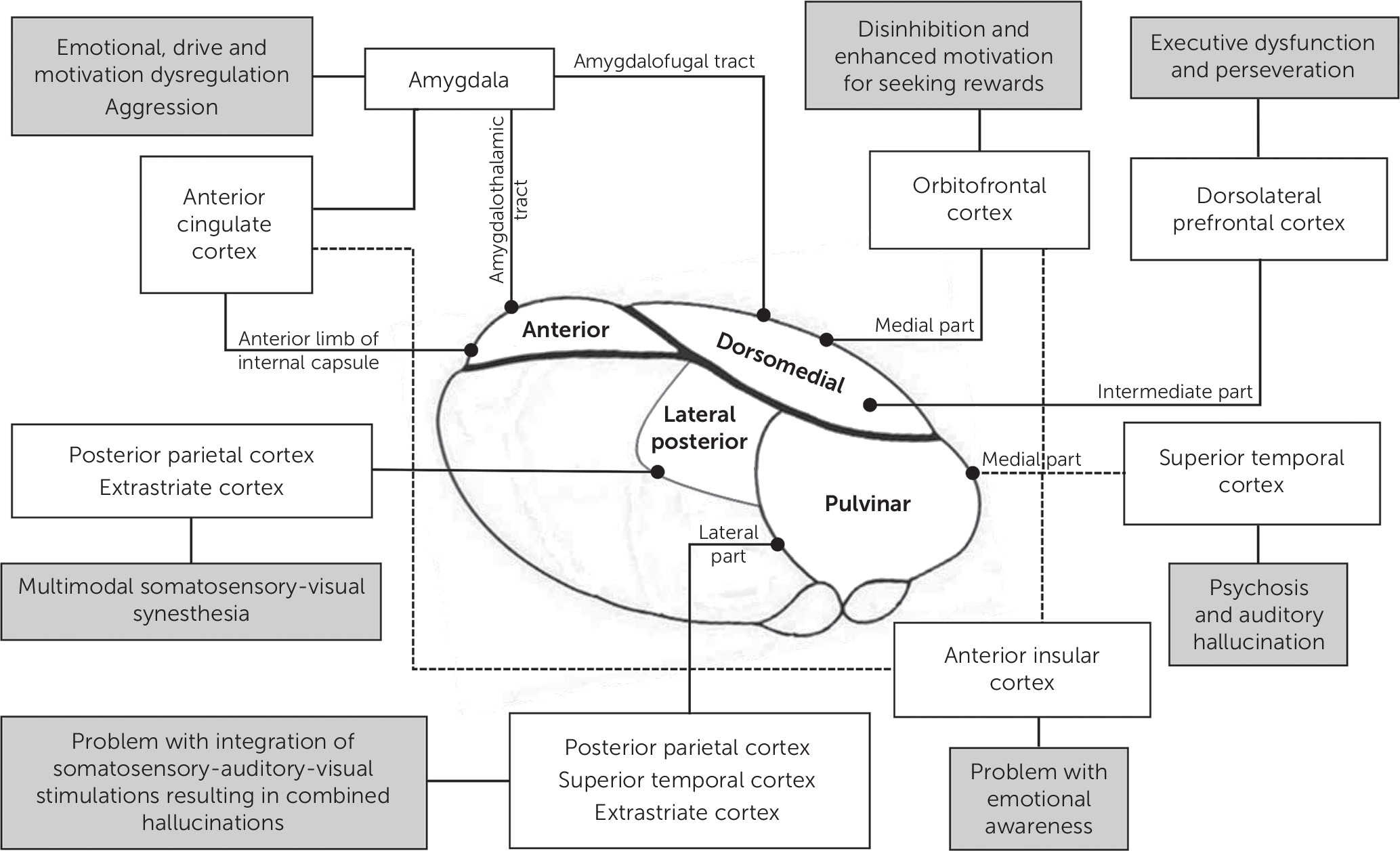
Network Model of Mania and Hallucinations Secondary to Thalamic Insults

Mania secondary to diencephalic lesions derives from the phenomenon of diaschisis that is caused by disconnection of the thalamus from the thalamo-prefrontal-limbic networks. The magnocellular (medial) part of the dorsomedial nucleus connects the thalamus with the limbic system via the orbitofrontal cortex.1 Damage to this network causes disinhibition of inappropriate behaviors and enhanced reward-seeking behaviors.2 The medial part of the dorsomedial nucleus also projects the neuronal network to the amygdala via the amygdalofugal tract, and thus a lesion on this nucleus can lead to emotional dysregulation, aggression, increased oral intake, and hypersexuality. In addition, damage to the intermediate part of dorsomedial nucleus, which links to the dorsolateral prefrontal cortex, can contribute to executive dysfunction.
Damage to the anterior nucleus, which is connected to the mammillary bodies and amygdala via the mamillothalamic and ventral amygdalothalamic tract, respectively, causes problem with emotional expression and dysregulation of motivation.1 The pulvinar nucleus is an associative nucleus connecting to multiple cortical areas.1 The medial part of the pulvinar nucleus sends some connections to the anterior insular cortex and functions in controlling emotional awareness that is required for emotional regulation.3 Some fibers from the intermediate part of the pulvinar nucleus also project to the dorsolateral prefrontal cortex and take part in executive performance.
The role of the right thalamus in the pathogenesis of mania is supported by previous studies demonstrating decreased right thalamic volume in patients with bipolar disorder compared with normal controls4,5 and reduced right thalamic and ipsilateral prefrontal/orbitofrontal perfusion in cases with diencephalic mania.68 It is hypothesized that emotional regulation is lateralized, with the right hemisphere controlling negative moods and the left hemisphere controlling positive emotions. The interhemispheric imbalance, with left-sided hyperfunction and right-sided hypofunction, on the prefrontal, orbitofrontal, and insular cortices, may contribute to the emotional releasing effect from the contralesional side and result in secondary mania.9,10
Similar to mania, the diaschisis of neural networks also explains the pathogenesis of peduncular hallucinosis. In the classic peduncular hallucinosis from mesencephalic lesions, the disruption along the pathway from the ascending reticular activating system in the brainstem to the thalamus may contribute to the hallucinations. While in the cases with thalamic lesions, dysregulation between the thalamus and the extrastriate cortex and superior temporal gyrus leads to visual and auditory hallucination, respectively. This hypothesis is supported by a study on the lesion-based resting-state networks, which showed the maximal network overlap for visual hallucinations on the extrastriate visual cortex and for auditory hallucinations on the superior temporal gyrus.11
The pulvinar nucleus is the multimodal integrative nucleus connecting to the posterior parietal, superior temporal, and extrastriate cortices. Damage to this structure may also cause problems with somatosensory-auditory-visual integration, resulting in multimodal hallucinations. The lateral posterior nucleus was reported to cause multimodal somatosensory-visual-auditory-gustatory synesthesia.12
We propose the network explaining the pathogenesis of mania and peduncular hallucinosis secondary to thalamic lesions (see Figure 2).
FIGURE 2. Proposed Network of Mania and Peduncular Hallucinosis Secondary to Thalamic Lesions

References

1.
Schmahmann JD: Vascular syndromes of the thalamus. Stroke 2003; 34:2264–2278
2.
Perry DC, Sturm VE, Seeley WW, et al: Anatomical correlates of reward-seeking behaviours in behavioural variant frontotemporal dementia. Brain 2014; 137:1621–1626
3.
Craig ADB: How do you feel--now? The anterior insula and human awareness. Nat Rev Neurosci 2009; 10:59–70
4.
Sani G, Chiapponi C, Piras F, et al: Gray and white matter trajectories in patients with bipolar disorder. Bipolar Disord 2016; 18:52–62
5.
Radenbach K, Flaig V, Schneider-Axmann T, et al: Thalamic volumes in patients with bipolar disorder. Eur Arch Psychiatry Clin Neurosci 2010; 260:601–607
6.
Bogousslavsky J, Ferrazzini M, Regli F, et al: Manic delirium and frontal-like syndrome with paramedian infarction of the right thalamus. J Neurol Neurosurg Psychiatry 1988; 51:116–119
7.
Benke T, Kurzthaler I, Schmidauer Ch, et al: Mania caused by a diencephalic lesion. Neuropsychologia 2002; 40:245–252
8.
López JD, Araúxo A, Páramo M: Late-onset bipolar disorder following right thalamic injury. Actas Esp Psiquiatr 2009; 37:233–235
9.
Koreki A, Takahata K, Tabuchi H, et al: Increased left anterior insular and inferior prefrontal activity in post-stroke mania. BMC Neurol 2012; 12:68
10.
Mimura M, Nakagome K, Hirashima N, et al: Left frontotemporal hyperperfusion in a patient with post-stroke mania. Psychiatry Res 2005; 139:263–267
11.
Boes AD, Prasad S, Liu H, et al: Network localization of neurological symptoms from focal brain lesions. Brain 2015; 138:3061–3075
12.
Fornazzari L, Fischer CE, Ringer L, et al: “Blue is music to my ears”: multimodal synesthesias after a thalamic stroke. Neurocase 2012; 18:318–322
13.
Cummings JL, Mendez MF: Secondary mania with focal cerebrovascular lesions. Am J Psychiatry 1984; 141:1084–1087
14.
Gentilini M, De Renzi E, Crisi G: Bilateral paramedian thalamic artery infarcts: report of eight cases. J Neurol Neurosurg Psychiatry 1987; 50:900–909
15.
Starkstein SE, Fedoroff P, Berthier ML, et al: Manic-depressive and pure manic states after brain lesions. Biol Psychiatry 1991; 29:149–158
16.
Kulisevsky J, Berthier ML, Pujol J: Hemiballismus and secondary mania following a right thalamic infarction. Neurology 1993; 43:1422–1424
17.
McGilchrist I, Goldstein LH, Jadresic D, et al: Thalamo-frontal psychosis. Br J Psychiatry 1993; 163:113–115
18.
Daum I, Ackermann H: Frontal-type memory impairment associated with thalamic damage. Int J Neurosci 1994; 77:187–198
19.
Vuilleumier P, Ghika-Schmid F, Bogousslavsky J, et al: Persistent recurrence of hypomania and prosopoaffective agnosia in a patient with right thalamic infarct. Neuropsychiatry Neuropsychol Behav Neurol 1998; 11:40–44
20.
Leibson E: Anosognosia and mania associated with right thalamic haemorrhage. J Neurol Neurosurg Psychiatry 2000; 68:107–108
21.
Inzelberg R, Nisipeanu P, Joel D, et al: Acute mania and hemichorea. Clin Neuropharmacol 2001; 24:300–303
22.
Routh R, Hill A: Post-stroke mania: a rare but treatable presentation. Prog Neurol Psychiatry 2014; 18:24–25
23.
Feinberg WM, Rapcsak SZ: ‘Peduncular hallucinosis’ following paramedian thalamic infarction. Neurology 1989; 39:1535–1536
24.
Serra Catafau J, Rubio F, Peres Serra J: Peduncular hallucinosis associated with posterior thalamic infarction. J Neurol 1992; 239:89–90
25.
Inzelberg R, Vishnievskaya S, Korczyn AD: Transient musical hallucinosis. J Neurol Neurosurg Psychiatry 1993; 56:833
26.
Noda S, Mizoguchi M, Yamamoto A: Thalamic experiential hallucinosis. J Neurol Neurosurg Psychiatry 1993; 56:1224–1226
27.
Manford M, Andermann F: Complex visual hallucinations: Clinical and neurobiological insights. Brain 1998; 121:1819–1840
28.
Yoshida Y, Abe K, Yoshizawa K: [A case of left dorsomedial thalamic infarction with unilateral schizophrenia-like auditory hallucinations]. Seishin Shinkeigaku Zasshi 2006; 108:31–41
29.
Mollet GA, Harrison DW, Walters RP, et al: Asymmetry in the emotional content of lateralised multimodal hallucinations following right thalamic stroke. Cogn Neuropsychiatry 2007; 12:422–436
30.
Güzelcan Y, Kleinpenning AS, Vuister FM: Peduncular hallucinosis caused by a tumour in the right thalamus: A case study. Tijdschr Psychiatr 2008; 50:65–68
31.
Mittal M, Khan S: Starvation causes acute psychosis due to anterior thalamic infarction. South Med J 2010; 103:701–703
32.
Lee S, Kim DY, Kim JS, et al: Visual hallucinations following a left-sided unilateral tuberothalamic artery infarction. Innov Clin Neurosci 2011; 8:31–34
33.
Delgado MG, Bogousslavsky J: ‘Distorteidolias’—fantastic perceptive distortion: A new, pure dorsomedial thalamic syndrome. Eur Neurol 2013; 70:6–9

Information & Authors

Information

Published In

Go to The Journal of Neuropsychiatry and Clinical Neurosciences
The Journal of Neuropsychiatry and Clinical Neurosciences
Pages: 401 - 408
PubMed: 28294710

History

Received: 14 November 2016
Accepted: 12 December 2016
Published online: 15 March 2017
Published in print: Fall 2017

Keywords

  1. Thalamus
  2. Mania (Neuropsychiatric Aspects)
  3. peduncular hallucinosis
  4. psychotic disorder
  5. bipolar disorder

Authors

Affiliations

Parunyou Julayanont, M.D. [email protected]
From the Department of Neurology, Texas Tech University Health Sciences Center, Lubbock, Tex. (PJ, DR, KA, YJA).
Doungporn Ruthirago, M.D.
From the Department of Neurology, Texas Tech University Health Sciences Center, Lubbock, Tex. (PJ, DR, KA, YJA).
Khondoker Alam, M.D.
From the Department of Neurology, Texas Tech University Health Sciences Center, Lubbock, Tex. (PJ, DR, KA, YJA).
Yazan J. Alderazi, M.D.
From the Department of Neurology, Texas Tech University Health Sciences Center, Lubbock, Tex. (PJ, DR, KA, YJA).

Notes

Send correspondence to Dr. Julayanont; e-mail: [email protected]; [email protected]

Competing Interests

The authors report no financial relationships with commercial interests.

Metrics & Citations

Metrics

Citations

Export Citations

If you have the appropriate software installed, you can download article citation data to the citation manager of your choice. Simply select your manager software from the list below and click Download.

For more information or tips please see 'Downloading to a citation manager' in the Help menu.

Format
Citation style
Style
Copy to clipboard

There are no citations for this item

View Options

View options

PDF/ePub

View PDF/ePub

Get Access

Login options

Already a subscriber? Access your subscription through your login credentials or your institution for full access to this article.

Personal login Institutional Login Open Athens login
Purchase Options

Purchase this article to access the full text.

PPV Articles - Journal of Neuropsychiatry and Clinical Neurosciences

PPV Articles - Journal of Neuropsychiatry and Clinical Neurosciences

Not a subscriber?

Subscribe Now / Learn More

PsychiatryOnline subscription options offer access to the DSM-5-TR® library, books, journals, CME, and patient resources. This all-in-one virtual library provides psychiatrists and mental health professionals with key resources for diagnosis, treatment, research, and professional development.

Need more help? PsychiatryOnline Customer Service may be reached by emailing [email protected] or by calling 800-368-5777 (in the U.S.) or 703-907-7322 (outside the U.S.).

Media

Figures

Other

Tables

Share

Share

Share article link

Share