Full access
Special Articles
Published Online: 17 October 2019

What Does Neuroscience Tell Us About the Conversion Model of Functional Neurological Disorders?

Publication: The Journal of Neuropsychiatry and Clinical Neurosciences

Abstract

A dualistic mind-body understanding of functional neurological disorders (FNDs), also known as conversion disorders, has led to the view that the cause of the symptom should be either psychological (psychogenic) or physical (neurogenic-“organic”). One of the most influential psychological approaches is the Freudian model of conversion, which suggests that FNDs arise from a defense process in which emotional stress is converted into physical symptoms. This conversion theory has been challenged in recent years, accompanied by a shift in emphasis toward neuropathophysiological models of FND and away from historical psychological concepts. In this review, the authors consider the contemporary relevance of the conversion model from the neuroscientific perspective to reconcile the role of both psychological and biological factors in FND. A narrative review of recent neuroscientific findings pertaining to the conversion model of FND, encompassing neuroimaging, cognitive psychology, biological markers, and epigenetic studies, was performed. Research on the role of psychological stressors is discussed. Neurobiological mechanisms of repression of traumatic memories and their translation into physical symptoms are then explored. Finally, the role of physical symptoms as a potential protective defense mechanism against social stressors is considered. The authors argue that the conversion concept is consistent with recent neuroscientific research findings, and the model allows psychological and neurobiological concepts to be reconciled within a single account of FND that begins to resolve the dualistic mind-body dichotomy.
Functional neurological disorder or conversion disorder (here referred to FND) is a neuropsychiatric condition in which patients experience neurological symptoms, such as motor dysfunctions (e.g., limb paralysis, tremor, and gait disorder), nonepileptic seizures, or sensory symptoms, in the absence of classical neurological disease (i.e., without experiencing a stroke, inflammatory or neuroinfectious disease, etc.) (1). FND is the second most common cause of a neurological outpatient visit after headache (2). The estimated prevalence of FND is around 50 per 100,000 persons (3). Although much is known about the clinical presentation, patient characteristics, psychiatric comorbidities, and diagnosis of FND, the associated neurobiological mechanisms are not yet fully understood (4).
There are currently three main lines of research in the field, two lines focusing on understanding the disorder itself—including how symptoms are produced in the brain (i.e., the underlying mechanisms) and why individuals develop FND in the first place (i.e., the trigger[s] and risk factors) (5)—and another line focusing on the development of treatment strategies (6, 7). Research on how symptoms are produced has led to increased understanding of the neural and cognitive correlates of FND, with deficits in attention allocation (8, 9) and agency perception (1012) being implicated in the variability and distractibility of symptoms and the lack of control reported by patients.
In this perspectives article, we focus on the question of why FNDs develop in certain individuals, with a particular focus on the role of stressors as a potential risk factor for, or precipitant of, the condition as part of the historical Freudian “conversion” model. The idea that psychological stressors are central to the etiology of FND has a long history in neurology and psychiatry (13, 14), and these conditions continue to be described as “psychogenic” by many practitioners. Nevertheless, it has become apparent that FND is likely to have multiple triggering and predisposing risk factors, and in clinical practice, many neurologists and other medical clinicians are often unable to identify a specific stressor in their encounters with FND patients. Acknowledgment of this inability resulted in the relegation of antecedent stress from a diagnostic criterion to a clinical note and specifier in the DSM-5 (1517). Identifying the presence of psychological factors is no longer required diagnostically in the DSM-5 (1517); however, this in no way implies that psychological factors and stressors do not play an important role in understanding the disorder from a research perspective or in the clinical management of patients, as discussed in the DSM-5 diagnosis notes.
In this article, we revisit the role of psychological stressors in the development and maintenance of FND from the perspective of contemporary neuroscience and demonstrate that the role of psychological factors does not need to be opposed to biological mechanisms in FND. We present data illustrating how research in psychology and neurosciences can both bring valuable knowledge toward a better understanding of this prevalent neuropsychiatric condition.

Conversion Model of FND

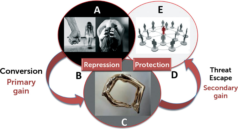
A number of psychological conceptualizations exist for FND, including psychodynamic, cognitive, and stress diathesis among others (18). Building on the earlier work of Briquet (19) and Janet (20), Freud and Breuer (1893–1895) elaborated and popularized the concept that stressful or traumatic events trigger conversion disorder in their classic treatise, Studies on Hysteria. One of the central mechanisms underlying FND in that account is the process of conversion, in which the individual inhibits or represses his or her emotional reaction to traumatic or stressful life events (e.g., childhood abuse), because it is unacceptable in some way (e.g., feelings of anger conflict with other feelings, such as love or fear of the abuser). Freud and Breuer suggested that the brain manages the resulting build-up of emotional tension by converting it into physical symptoms that represent the conflict without the individual’s having to become aware of it. Although the main function of this defense process is to reduce the anxiety generated by the conflict (primary gain of the conversion), the physical symptoms may also allow protection or escape from the stressful situation or result in other external benefits (secondary gain) (see box with clinical vignette and Figure 1).

Clinical vignette: illustration of primary and secondary gains in conversiona

A 23-year-old woman developed acute paraplegia the day that she received an invitation to a wedding of an old schoolmate, which was scheduled to take place in her hometown. She had left her hometown 5 years ago and had never returned. All of her family reunions since then had occurred outside this town. She had been sexually abused by a neighbor in the town as a child, but she kept these events secret. Because she knew that this neighbor would also attend the wedding, the invitation evoked traumatic memories and created an intrapsychic conflict. She wanted to attend the wedding of her friend, but at the same time she could not fathom the risk of facing her aggressor. The onset of physical symptoms solved the internal conflict, protecting her from potentially being confronted by the aggressor (social threat) and avoiding the guilt from declining the invitation despite her sincere desire to attend the wedding.
a The link between this potential trauma and the physical symptom may not necessarily be identified at the time of diagnosis. Here, our goal is to illustrate the power and influence of how a seemingly minor event (receiving an invitation) may be relevant in the unconscious processes in functional neurological disorder/conversion disorder. The significance of such stressors is likely to become clear only with thorough recollection during psychotherapy.
FIGURE 1. Schematic illustration of the psychodynamic conversion modela
a Psychological stressor-intrapsychic conflict (panel A) triggers a defense mechanism of repression (B), accompanied by a conversion of this tension into a physical symptom (panel C). This reduces the anxiety generated by the conflict and provides a primary gain. The symptom itself (panel C) can also serve the purpose of unconsciously protecting (D) the individual, by allowing him or her to escape from a threatening social situation (panel E), which provides a secondary gain.
In these respects, conversion can be thought of as serving a protective function from the emotional and environmental stressor (and can thus be viewed as an “adaptive” mechanism). The downside of this process is that it renders the individual unable to recognize, acknowledge, or process the underlying conflict, resulting in a disabling, often chronic, functional symptom (making it a “maladaptive” mechanism). Subsequent developments of the conversion model have emphasized this inability to symbolize (i.e., mentally represent) the affect associated with adverse events as an important feature of people with functional neurological symptoms, so-called alexithymia (literally translated as “no words for feelings”). Although Freud subsequently abandoned the original incest etiological model, the remaining concept has nevertheless had a profound influence on subsequent thinking and practice in this area (thus FNDs have been classified as conversion disorders in DSM). In this review, we respond to recent concerns about the empirical basis for the conversion model (e.g., 21) by exploring what neurosciences reveal with respect to this approach.

Neuroscientific Evidence for the Conversion Model

Role of Childhood Adversities and Recent Life Events

A recent meta-analysis of 34 case-control studies, including a total of 1,405 patients (22), confirmed that rates of childhood sexual abuse, emotional neglect, and physical abuse are substantially higher in patients with FND, compared with healthy individuals and both neurological and psychiatric control patients. The same meta-analysis also confirmed that recent adult adverse life events are more frequent in patients with FND. The meta-analysis clearly showed that trauma and abuse are reported by some patients with FND. However, the meta-analysis also found that a substantial proportion of these patients (15%−77% across 13 studies) do not report a history of abuse, trauma, or significant life events. It is important to keep in mind that the methodology used in these studies considered only specific types of traumas, which have to be remembered and reported by patients.
It may be that some patients do not readily recall or recognize adverse life experiences; others who accurately report no history of abuse may have experienced other significant life events or stressors that are not captured by typical trauma measures or that they do not personally characterize as trauma. These events include losses of family or friends (or even significant pets for some) through deaths, divorces or deployments; bullying; illnesses; neglect; parents with substance abuse or conflict; and so on. Many times, these are remote events that occurred during developmental periods that are not recalled during symptom-focused consultations with neurologists or psychiatrists but are revealed only when a comprehensive developmental history is taken.
Several studies have investigated the links between life stressors and clinical presentation. Childhood physical abuse was found to be associated with a larger number of conversion symptoms in one study (23), and a history of sexual abuse was linked to a different semiological presentation of psychogenic nonepileptic seizures (PNES) in another study (semiologically expressed with more emotional triggers, urinary incontinence, self-injury, nocturnal attacks, internal experiences, flashbacks, and convulsions) (24). Likewise, a neuroimaging study demonstrated a correlation between symptom severity and childhood abuse burden and volumetric changes in the insula in women with FND (25).
Separate from research citing trauma and abuse, other research highlights a link between physical stressors and the onset of FND. A systematic review of 133 studies encompassing 869 patients found that 37% of patients with FND reported physical events immediately prior to FND symptom onset (including injuries of the soft tissues or fractures, flu-like infectious illnesses, a neurological illness such as Bell’s palsy or migraine, acute pain, drug reactions, surgery, or syncope), increasing to 80% of patients reporting events in the 3 months before symptom onset (26).
Evidently, antecedent stress is implicated in many cases of FND, but the nature of that stress is variable, and major life events are not an absolute criterion for the etiology, which is most likely multifactorial.
In contemporary psychological thinking, however, the nature of the stressor may be much more subtle than overt physical, sexual, or emotional abuse and often more difficult to identify with life event checklists. Indeed, using the detailed Life Events and Difficulties Schedule (LEDS) (27), a study found that significant stressors were identifiable in 91% of patients with FND, even though 88% of these events were not mentioned in the clinical notes, suggesting that they were not seen as relevant by the referring physician or not mentioned by the patient (28). One caveat of this study was that it included patients from a particular area of London with lower socioeconomical resources and may not be representative of all FND patients. This study also found that 53% of patients with FND had, according to blind raters, experienced life events from which physical symptoms might offer some kind of escape, compared with only 13% of a depressed control group. Such a finding is difficult to explain from a purely cognitive or biological perspective and points to a role for conversion processes in at least a proportion of patients with FND. However, it is important to note that although psychosocial stressors may be present in the majority of FND cases, specific stressors, such as remote or recent trauma, are likely to affect some but not all of the neurobiological processes underlying FND.

Repression of Adverse Memories in FND

According to the conversion model, a key function of the defenses is to prevent the recall of memories associated with the distressing events in question. Neuropsychology research has investigated this phenomenon of memory suppression and verified whether it is possible to voluntarily and actively forget unwanted memories with a think–no-think task (29). In this paradigm, subjects have to learn pairs of words (such as ordeal-roach or steam-train). Participants are then asked to either remember (think) the second word (roach) when presented with a cue (ordeal) or try not to remember (no think) the second word (train) when presented with the cue (steam). Finally, they are asked to recall all of the word pairs. Evidence showing that the think pairs (ordeal-roach) are better remembered than the no-think pairs (steam-train) supports the view that there are cognitive processes that allow us to forget some memories. A subsequent study looked at which regions of the brain were activating during this process, and a network involving increased dorsolateral prefrontal cortex (DLPFC) and decreased hippocampus activity was found (30).
With the aim of studying this network in FND, an fMRI task compared active recall of autobiographic memories in FND patients and healthy controls (31). First, a LEDS interview (as discussed above) was conducted to identify stressful memories. Then a panel of experts, blinded to whether a patient or a healthy control was being assessed, judged the severity of the event (0–4 score) as well as the escape/secondary gain that could theoretically be obtained if a physical conversion/functional symptom followed the traumatic event. This allowed two kinds of severe events to be classified: escape events (with secondary gain) and severe events (without secondary gain). Patients were also asked to subjectively rate the severity of these events. Even though they were of equal objective severity (as assessed by the blinded panel), the escape events were perceived as significantly less severe by FND patients. This is compatible with the conversion theory, which suggests that the primary gain of alleviating the emotional load attached to the autobiographical memory worked for this particular type of events.
The two regions identified as relevant in the suppression of unwanted memories (DLPFC and hippocampus as discussed above) were then chosen as a priori regions of interest for the fMRI recall task (31) (see Figure S1 in the online supplement). The recall of escape events elicited increased DLPFC and decreased hippocampal activity in FND patients (but not in healthy controls), suggesting the suppression of unwanted memories in this group. It seems that some traumatic autobiographical events are indeed processed differently in the brains of FND patients, consistent with the conversion model.

Neural Correlates of the Conversion Mechanism

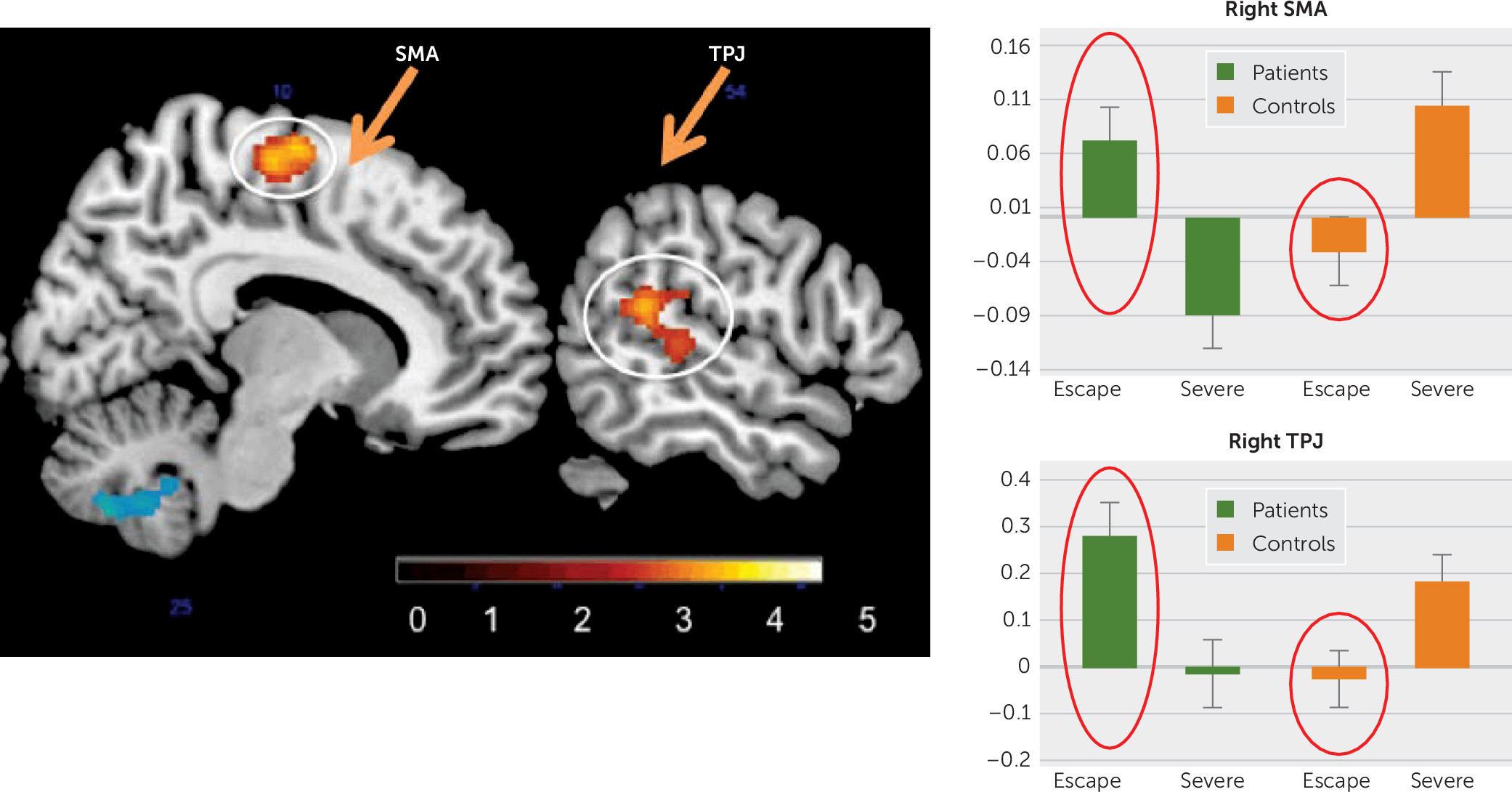
One of the challenges of the conversion model has been “measurement” of the unconscious. In the past, we have had to look at the issue indirectly. Clinically, the unconscious has been “tapped” and identified through decades of hypnosis research (3234). With functional neuroimaging, a third result from the above-mentioned fMRI task investigating recall of traumatic life events (31) is directly related to a possible conversion mechanism. In the whole-brain analysis, compared with controls, FND patients had a significant increase of activity in a cluster that included the right supplementary motor area (SMA) and another that included the right temporoparietal junction (TPJ) when they were recalling escape events more than when they were recalling other severe events (Figure 2). These are important findings for three reasons. First, the task was not targeting motor or sensory function, and yet it elicited peak responses in the SMA and the TPJ (two regions relevant in the production of physical FND symptoms). Second, this pattern was not found in all events but only in the ones with escape–secondary gain potential. Third, this was specific to FND patients and not found in healthy controls. These findings are compatible with the theory of conversion that some memories may influence brain regions implicated in sensorimotor (neurological) symptoms. The SMA is central in motor execution and inhibition (35) of conscious (36) and unconscious (37) prepotent responses. The TPJ is a key region implicated in multisensory integration, in the sense of self-agency and in making sensorimotor predictions (12). Both the SMA and the TPJ, among other networks, are key in FND symptom production, as shown by the mechanistic line of research (10, 11, 38, 39).
FIGURE 2. A putative neural signal of a “conversion” process in functional neurological disordersa
a The recall of events with secondary gain (escape events) elicited a higher activity in the right supplementary motor area (SMA) and right temporo-parietal junction (TPJ) in patients (green) compared with control subjects (orange). This suggests a “short-circuit” between limbic-memory processing regions and sensori (TPJ)-motor (SMA) regions underlying a putative neural signal of a “conversion” process in some patients with FND.

Protection Against a Threat

Other research suggests that patients with FND respond differently to psychological stressors, compared with healthy controls. For example, an experiment studied patients with nonepileptic seizures using an emotional Stroop test where participants had to name colors on which subliminal emotional faces (happy or angry) were superimposed (40). It was found that FND patients took significantly longer than healthy controls to name colors superimposed on angry faces, suggesting that the threat value of the faces was drawing attention away from the primary task in this group, even though they were not accessible to consciousness. Consistent with this, another study found that children and adolescents with FND were disproportionately quicker to identify sad faces than happy faces, compared with healthy controls, suggesting that the FND group was more efficient when the task was to identify social threat cues (41). A related finding was obtained in a study comparing healthy controls to patients with motor FND (42). In this study, participants had to maintain a constant force by pinching a force-measuring device between their thumb and index finger over 6 seconds when viewing pictures with or without threatening content (e.g., a snake or gun). Although healthy controls showed a decay in force over time for both picture types, the FND group maintained an abnormally high force in response to the threatening pictures, also suggesting altered threat processing in this group.

Animal defense mechanisms.

Some commentators have proposed examining animal threat response behaviors as a way of understanding bodily reactions to threat in patients with FND (43). Traditionally, fight-or-flight models have been used to describe both animal and human behavior under stress (44). However, sometimes neither fight nor flight are adequate or acceptable (e.g., in the case of social threat), which may explain the existence of other protective mechanisms, such as tonic immobility (45) and distraction displays (46, 47).
One possibility is that the symptoms of FND originate in innate defensive behaviors and manifest in response to adverse developmental or other experiences (48). Three examples of defensive behavior present in animals are sometimes cited as potential representations of animal models of FND.
Distraction displays are one example of defensive behavior in animals. One animal model often cited with reference to FND is the distraction displays observed in some birds (killdeers and plovers) protecting their nests. For example, killdeers (Charadrius vociferous) display “injury-feigning” or “broken-wing” behavior in the absence of real injury (46, 47) as a way of distracting the attention of predators when they approach the nest. This is meant to attract the predator away to a safe distance from the nest, after which the bird can escape. However, videos of the killdeer show the bird looking around to see whether the predator threat is clear before resuming normal function, suggesting some degree of intentionality (49). Therefore, we suggest caution before interpreting a distraction display as akin to the unconscious defensive process postulated in the conversion model of FND.
Tonic immobility—also known as thanatosis—is another antipredator behavior adopted by prey, which occurs late in an attack sequence when the fight behavior has failed and the escape possibilities are gone (50). This is a last-resort option (51), displaying motor inhibition, hypertonicity, and relative unresponsiveness (52)—mimicking death in order to avoid being eaten. In studies of opossums threatened by human or dog vigorous tactile stimulation, reductions of heart rate, respiratory rate, and body temperature occurred during death feigning, despite the animal’s being fully conscious (53). Although this behavior may resemble the atonic dialeptic semiology of nonepileptic seizures in humans, it is unclear whether there are other similarities between this phenomenon and FND in humans.
Freezing is an unlearned defensive response to threat, present in many species, including cats, whose main features are immobility, increased muscle tonus, and bradycardia (54). In contrast to tonic immobility, which is characterized by a relative unresponsiveness and occurs late in the threat sequence as a last-resort behavior, freezing is thought to confer survival advantages by avoiding detection by the predator, enhancing perception and attention and preparing the animal for escape (54). Freezing also exists in humans (55), and its study in FND patients will be discussed below.

Conversion disorder in humans: the relevance of metacognition and rerepresentation.

Unlike the animal behaviors noted above, FND symptom formation in humans can arise in the absence of a physical, visible, immediate threat. It can be hypothesized that a remote stressor, such as a memory-reminiscence of an actual or imagined threat can have similar psychobiological consequences. This would be in line with the imaging study described above (31), showing that a traumatic memory can induce a “short-circuit” in the brain and interact with normal motor and sensory function (Figure 2). Whether this short-circuit uses the same mechanisms as the one discussed in animals still needs to be established.
One obvious question when it comes to comparing animal and human behavior is the role of more advanced forms of cognition found only in humans, such as metacognition (i.e., awareness and understanding of one's own thought processes, or “thinking about one’s thinking”) and re-representation (i.e., “systematic transformation of the psychological encoding of a stimulus [e.g., scene, situation, object, narrative] from an initial set of elements representing the meaning of the stimulus to a subsequent, different set of elements,” or “the action of representing a person or thing again” [56]). Although some comparative psychologists argue that these processes are uniquely human characteristics (57), others suggest that dolphins and chimpanzees, for example, have analogs of human metacognition (58). Rerepresentation allows humans to reflect beyond the self-evident story (e.g., immediately observable life-threatening stimulus) to multilayered narratives, and metacognition allows them to be aware of their own thinking.
A psychological model of conversion (FND) could be that both of these higher-order cognitive processes are cognitively required to be able to develop FND—i.e., both are present in (and possibly unique to) humans—and that these processes are disordered in patients with FND. This may explain how an inner thought or memory or intrapsychic conflict could have an effect similar to an external threat, and we propose that both of these higher-order cognitive processes (59) play a role in the development of FND.

Neural correlates of defense behavior.

There is evidence to suggest that the neural correlates of threat processing in the limbic system, and particularly the amygdala, are different in patients with FND, compared with individuals without FND. For example, an incidental affective task was conducted in motor FND patients and healthy controls undergoing fMRI, in which participants had to determine the gender of neutral, angry, or happy faces (60). Although healthy controls showed only increased amygdala activation to angry faces, compared with neutral faces, the FND group showed this effect for both angry and happy faces. This suggests that FND patients have a hypersensitivity to emotional stimuli in general, perhaps suggesting that all social cues are perceived as potentially threatening.
A similar effect was seen in a study that found increased activity in the left amygdala in FND patients, compared with heathy controls, in response to both sad and angry faces (61). In this study, the left amygdala also became more activated over time in the FND group in response to fearful faces, compared with sad faces, seemingly suggesting sensitization, rather than habituation, to the fearful stimuli. This study also found that patients with motor FND had greater activity than controls in the periaqueductal gray area (PAG) when processing negatively valanced emotional stimuli (both sad and fearful faces) (61). This region of the brainstem is key in the network underlying the freeze response, as shown both in animal research (62, 63) and in studies of humans (64).
Another recent study found changes in brain connectivity between the amygdala and the PAG in FND patients (65). To test whether patients with FND exhibited an abnormal freeze response, a behavioral study measured postural changes during the Trier social stress test, in which participants are asked to simulate a job interview in front of two examiners (66). In this study, healthy controls showed a reduction of movements of the trunk in the second part of the interview (suggesting a normal freeze response), whereas patients with FND failed to show this behavior. These are preliminary data from a small sample and did not include measures of bradycardia, which would have helped in making inferences about the complete freeze response. What is suggested, though, is that patients have an abnormal postural response to stress. This finding cannot be generalized to all subtypes of FND, because even though there are dialeptic (motionless unresponsiveness) semiological presentations of PNES, most seizures and motor symptoms are hyperkinetic, not akinetic. More research is needed to elucidate why some patients have hyperkinetic and other patients have hypokinetic responses in FND.
In summary, there is currently convincing evidence that FND patients have abnormal threat processing resulting in a measurable abnormal body response. If a parallel animal body reaction to threat existed, it could argue for the “innate” nature of the conversion-body response. On the other hand, arguments exist for an “acquired” process, in which events and experiences become threatening because of the personal meaning they have for the individual in the context of his or her specific learning history. At this stage, more research is needed, perhaps taking an interdisciplinary approach with ethologists and sociologists to bring a fresh perspective on how and why an emotion or a painful thought can be transformed into a physical symptom.

Biological Stress and Stressors

Stressors are not the same as stress. Some patients experience events and symptoms when not stressed. Evidence, however, for an elevated stress response in patients with FND has been found in biomarker studies. In a study using the emotional Stroop test, for example, the authors found a significant positive correlation between FND participants’ reaction times to angry (i.e., threatening) faces and their basal cortisol level (67). This effect was confirmed by findings of elevated diurnal basal salivary cortisol levels in FND patients with nonepileptic seizures (68) and those with motor FND (69). The latter study also found increased salivary amylase levels (a protein secreted in response to adrenergic activation) in FND patients, compared with controls. This implicates both the hypothalamic-pituitary-adrenal axis (which is responsible for heightened cortisol levels) and a second stress pathway involving rapid adrenergic responses in FND (heightened amylase levels). Other evidence of abnormal biological stress regulation in FND comes from studies of the autonomic system. Impaired vagal tone, as measured by several heart rate variability indices, has been found in both adults (70, 71) and children (72) with FND.
Whether abnormal biological stress regulation plays a causal role in FND or is secondary to the disorder cannot be resolved yet, and further studies are needed. An association with childhood adversities has been found: basal cortisol levels were higher in FND patients, compared with controls, and even higher in a subgroup of FND patients who experienced childhood sexual abuse (68). A correlation between the number and impact of life events and basal cortisol levels was also found in a group of FND patients but not in healthy controls (69). This suggests a link between life stressors and biological measures that is specific to patients with FND. A possible hypothesis is that FND patients have a vulnerability to adverse events, which leads to abnormal stress regulation at both a biological and a neural level. Further research on these links might better explain why and how a stressor can be linked to a physical symptom as implied by the conversion model.
At present, there is a lack of evidence concerning the role of genetic factors in FND, with only a small number of familial cases having been published (73). There might, however, be a genetic vulnerability that predisposes some individuals to react to the environment—and in particular to stressors—in a different way, leading to FND symptoms. A preliminary study identified increased methylation rates on the promotor of the oxytocin receptor gene in a group of mixed FND patients (74). The gene is implicated in stress regulation, and broader studies testing several genes might shed light on the gene-environment interactions in FND.
The Conversion Model and Modern Views

The studies described above highlight the potential role of the conversion process in FND and illustrate plausible neurobiological and pathophysiological mechanisms that might underpin this process. They do not, however, explain how a psychological stressor can be associated with a specific sign or symptom, such as limb weakness, loss of consciousness, or tremors. Nevertheless, there is growing evidence that psychological stressors (in childhood and adulthood and either remembered or repressed or forgotten) are linked to biological changes as measured by stress biomarkers (cortisol, amylase, heart rate, variability, brain activity, and epigenetic changes) (75, 76), and that neural signals may reflect neuropathophysiology associated with FND (77).
Does this mean that FND is a “psychogenic” or a “neurogenic” disorder? In our view, the answer is: both. To suggest otherwise, even to frame this as an “either-or” question, implies a dualist position that FND is either all psychological—that is “in the mind”—or all biological—that is “in the body.” The research reviewed here demonstrates that this is faulty logic. Ultimately, psychological processes have physiological substrates (78), and a comprehensive account of FND has to describe both. Historically, the conversion model has focused on the psychological level of explanation; more recent approaches have utilized experimental models to examine neuropathophysiology.
Our brief review suggests that both approaches are appropriate, because they are both looking at the same entity from different angles, using different lenses and methods. To focus on one at the expense of the other, is to lose sight of the harmony between these accounts and the potential insights that each has to offer the other. The psychological level of explanation identifies the conversion process as one defense against specific mental contents, which applies in some but potentially not all patients with FND. The neuroimaging findings described by Aybek et al. (31) are specific to the types of memories being studied, identifying potential neural representations of psychological processes.
The neuroscientific findings provide clues to the pathophysiological signals associated with FND. Indeed, evidence from a number of sources (both neurobiological and psychological) suggests that a pure conversion model is insufficient to account for what is known about FND. In particular, it is evident that a single causal link between stressful or traumatic events and FND is unlikely; rather, the cause is much more likely to be multifactorial and involves a range of different predisposing, precipitating, and perpetuating factors (79, 80). A number of other psychological and neurobiological formulations exist for FND, including learning theory, family systems, and stress-allostasis, among others, which were beyond the scope of this review. Ours is not a review of all models, however; rather, we have focused on a specific psychological model to illustrate possible neurobiological substrates of unconscious processes in FND (81).
One conclusion is that psychological models explaining how the human mind and body deal with a psychosocial stressor can very well be reconciled with modern neuroscience findings. An area where this has been clearly and repeatedly demonstrated is in FND treatment trials. For patients in clinic or who have entered prospective randomized clinical trials with symptoms (movement or seizure disorders), having the opportunity to talk through their history of past and current stressors in psychotherapy enables many people to recognize the role that these experiences have played in their FND, giving them greater mastery over their stress responses and thereby symptom resolution (82). Likewise, patients with predominant motor symptoms can have symptom resolution with physical therapy and psychotherapy (83), which often include explanatory models that demonstrate the potential for normal function and how physical and psychological triggers may affect their symptoms.
Witnessing this phenomenon of symptomatic improvement through healthy verbal expression of emotional and psychological distress, making the unconscious conscious by addressing core issues, and expressing the implicit as explicit provides hope for both patients and clinicians alike. It also underscores the need for future research to improve our understanding of the brain-mind relationships in FND and ultimately the care of these patients.

Supplementary Material

File (appi.neuropsych.19040089.ds001.pdf)

References

1.
Espay AJ, Aybek S, Carson A, et al: Current concepts in diagnosis and treatment of functional neurological disorders. JAMA Neurol 2018; 75:1132–1141
2.
Stone J, Carson A, Duncan R, et al: Who is referred to neurology clinics? The diagnoses made in 3781 new patients. Clin Neurol Neurosurg 2010; 112:747–751
3.
Carson A, Lehn A: Epidemiology. Handb Clin Neurol 2016; 139:47–60
4.
Voon V, Cavanna AE, Coburn K, et al: Functional neuroanatomy and neurophysiology of functional neurological disorders (conversion disorder). J Neuropsychiatry Clin Neurosci 2016; 28:168–190
5.
Stone J, Vuilleumier P, Friedman JH: Conversion disorder: separating “how” from “why”. Neurology 2010; 74:190–191
6.
Goldstein LH, Mellers JDC: Psychologic treatment of functional neurologic disorders. Handb Clin Neurol 2016; 139:571–583
7.
Nielsen G: Physical treatment of functional neurologic disorders. Handb Clin Neurol 2016; 139:555–569
8.
van Poppelen D, Saifee TA, Schwingenschuh P, et al: Attention to self in psychogenic tremor. Mov Disord 2011; 26:2575–2576
9.
Edwards MJ, Adams RA, Brown H, et al: A Bayesian account of “hysteria”. Brain 2012; 135:3495–3512
10.
Voon V, Gallea C, Hattori N, et al: The involuntary nature of conversion disorder. Neurology 2010; 74:223–228
11.
Maurer CW, LaFaver K, Ameli R, et al: Impaired self-agency in functional movement disorders: a resting-state fMRI study. Neurology 2016; 87:564–570
12.
Hallett M: Volitional control of movement: the physiology of free will. Clin Neurophysiol 2007; 118:1179–1192
13.
Keynejad RC, Frodl T, Kanaan R, et al: Stress and functional neurological disorders: mechanistic insights. J Neurol Neurosurg Psychiatry 2019; 90:813–821
14.
Pick S, Goldstein LH, Perez DL, et al: Emotional processing in functional neurological disorder: a review, biopsychosocial model and research agenda. J Neurol Neurosurg Psychiatry 2019; 90:704–711
15.
Diagnostic and Statistical Manual of Mental Disorders, 5th ed. Arlington, VA, American Psychiatric Association, 2013
16.
Stone J, Edwards MJ: How “psychogenic” are psychogenic movement disorders? Mov Disord 2011; 26:1787–1788
17.
Stone J, LaFrance WC Jr, Brown R, et al: Conversion disorder: current problems and potential solutions for DSM-5. J Psychosom Res 2011; 71:369–376
18.
LaFrance JW, Bjornaes H: Designing treatment plans based on etiology of psychogenic nonepileptic seizures; in Gates and Rowan’s Nonepileptic Seizures. Edited by LaFrance JW, Schachter S. New York, Cambridge University Press, 2018
19.
Briquet P: Clinical and Therapeutic Treatise on Hysteria [in French]. Paris, Baillière et Fils, 1859
20.
Janet P: Psychological Automatism [in French]. Paris, Félix Alcan, 1889
21.
Roelofs K, Spinhoven P: Trauma and medically unexplained symptoms: towards an integration of cognitive and neuro-biological accounts. Clin Psychol Rev 2007; 27:798–820
22.
Ludwig L, Pasman JA, Nicholson T, et al: Stressful life events and maltreatment in conversion (functional neurological) disorder: systematic review and meta-analysis of case-control studies. Lancet Psychiatry 2018; 5:307–320
23.
Roelofs K, Keijsers GP, Hoogduin KA, et al: Childhood abuse in patients with conversion disorder. Am J Psychiatry 2002; 159:1908–1913
24.
Selkirk M, Duncan R, Oto M, et al: Clinical differences between patients with nonepileptic seizures who report antecedent sexual abuse and those who do not. Epilepsia 2008; 49:1446–1450
25.
Perez DL, Matin N, Barsky A, et al: Cingulo-insular structural alterations associated with psychogenic symptoms, childhood abuse and PTSD in functional neurological disorders. J Neurol Neurosurg Psychiatry 2017; 88:491–497
26.
Stone J, Carson A, Aditya H, et al: The role of physical injury in motor and sensory conversion symptoms: a systematic and narrative review. J Psychosom Res 2009; 66:383–390
27.
Brown GW, Harris TO, Peto J: Life events and psychiatric disorders: 2. nature of causal link. Psychol Med 1973; 3:159–176
28.
Nicholson TR, Aybek S, Craig T, et al: Life events and escape in conversion disorder. Psychol Med 2016; 46:2617–2626
29.
Anderson MC, Green C: Suppressing unwanted memories by executive control. Nature 2001; 410:366–369
30.
Anderson MC, Ochsner KN, Kuhl B, et al: Neural systems underlying the suppression of unwanted memories. Science 2004; 303:232–235
31.
Aybek S, Nicholson TR, Zelaya F, et al: Neural correlates of recall of life events in conversion disorder. JAMA Psychiatry 2014; 71:52–60
32.
Spiegel H, Spiegel D (eds): Trance and Treatment: Clinical Uses of Hypnosis, 2nd ed. Arlington, VA, American Psychiatric Publishing, 2004
33.
Raz A, Shapiro T: Hypnosis and neuroscience: a cross talk between clinical and cognitive research. Arch Gen Psychiatry 2002; 59:85–90
34.
Moene FC, Spinhoven P, Hoogduin KA, et al: A randomized controlled clinical trial of a hypnosis-based treatment for patients with conversion disorder, motor type. Int J Clin Exp Hypn 2003; 51:29–50
35.
Cai W, George JS, Chambers CD, et al: Stimulating deep cortical structures with the batwing coil: how to determine the intensity for transcranial magnetic stimulation using coil-cortex distance. J Neurosci Methods 2012; 204:238–241
36.
Nachev P, Wydell H, O’Neill K, et al: The role of the pre-supplementary motor area in the control of action. Neuroimage 2007; 36(suppl 2):T155–T163
37.
Sumner P, Nachev P, Morris P, et al: Human medial frontal cortex mediates unconscious inhibition of voluntary action. Neuron 2007; 54:697–711
38.
Voon V, Brezing C, Gallea C, et al: Aberrant supplementary motor complex and limbic activity during motor preparation in motor conversion disorder. Mov Disord 2011; 26:2396–2403
39.
Nahab FB, Kundu P, Maurer C, et al: Impaired sense of agency in functional movement disorders: an fMRI study. PLoS One 2017; 12:e0172502
40.
Bakvis P, Roelofs K, Kuyk J, et al: Trauma, stress, and preconscious threat processing in patients with psychogenic nonepileptic seizures. Epilepsia 2009; 50:1001–1011
41.
Kozlowska K, Brown KJ, Palmer DM, et al: Specific biases for identifying facial expression of emotion in children and adolescents with conversion disorders. Psychosom Med 2013; 75:272–280
42.
Blakemore RL, Sinanaj I, Galli S, et al: Aversive stimuli exacerbate defensive motor behaviour in motor conversion disorder. Neuropsychologia 2016; 93(pt A):229–241
43.
Gilbert P: Evolutionary approaches to psychopathology: the role of natural defences. Aust N Z J Psychiatry 2001; 35:17–27
44.
Cannon WB: Bodily Changes in Pain, Hunger, Fear and Rage: An Account of Recent Researches Into the Function of Emotional Excitement. New York, Appleton, 1915. http://archive.org/details/cu31924022542470
45.
Bracha HS, Ralston TC, Matsukawa JM, et al: Does “fight or flight” need updating? Psychosomatics 2004; 45:448–449
46.
Gochfeld M: Antipredator behavior: aggressive and distraction displays of shorebirds, in Shorebirds: Breeding Behavior and Populations. Edited by Olla JBBL. Boston, Springer, 1984
47.
Brunton D: The effects of nesting stage, sex, and type of predator on parental defense by killdeer (Charadrius vociferous): testing models of avian parental defense. Behav Ecol Sociobiol 1990; 26:181–190
48.
Kozlowska K: The developmental origins of conversion disorders. Clin Child Psychol Psychiatry 2007; 12:487–510
49.
Ristau C (ed): Aspects of the Cognitive Ethology of an Injury-Feigning Bird, the Piping Plover. Hillsdale, NJ, Erlbaum; 1991
50.
Humphreys RK, Ruxton GD: A review of thanatosis (death feigning) as an anti-predator behaviour. Behav Ecol Sociobiol 2018; 72:22
51.
Rogers SM, Simpson SJ: Thanatosis. Curr Biol 2014; 24:R1031–R1033
52.
Gallup GG: Tonic immobility: the role of fear and predation. Psychol Rec 1977; 27:41–61
53.
Gabrielsen GW, Smith EN: Physiological responses associated with feigned death in the American opossum. Acta Physiol Scand 1985; 123:393–398
54.
Hagenaars MA, Oitzl M, Roelofs K: Updating freeze: aligning animal and human research. Neurosci Biobehav Rev 2014; 47:165–176
55.
Roelofs K, Spinhoven P, Sandijck P, et al: The impact of early trauma and recent life-events on symptom severity in patients with conversion disorder. J Nerv Ment Dis 2005; 193:508–514
56.
Kurtz K: Re-representation in comparison: building an empirical case. J Exp Theor Artif Intell 2005; 17:447–459
57.
Crystal JD, Foote AL: Metacognition in animals. Comp Cogn Behav Rev 2009; 4:1–16
58.
Smith JD, Zakrzewski AC, Church BA: Formal models in animal-metacognition research: the problem of interpreting animals’ behavior. Psychon Bull Rev 2016; 23:1341–1353
59.
Bègue I, Blakemore R, Klug J, et al: Metacognition of visuomotor decisions in conversion disorder. Neuropsychologia 2018; 114:251–265
60.
Voon V, Brezing C, Gallea C, et al: Emotional stimuli and motor conversion disorder. Brain 2010; 133:1526–1536
61.
Aybek S, Nicholson TR, O’Daly O, et al: Emotion-motion interactions in conversion disorder: an FMRI study. PLoS One 2015; 10:e0123273
62.
Kim JJ, Rison RA, Fanselow MS: Effects of amygdala, hippocampus, and periaqueductal gray lesions on short- and long-term contextual fear. Behav Neurosci 1993; 107:1093–1098
63.
Vianna DM, Graeff FG, Brandão ML, et al: Defensive freezing evoked by electrical stimulation of the periaqueductal gray: comparison between dorsolateral and ventrolateral regions. Neuroreport 2001; 12:4109–4112
64.
Hermans EJ, Henckens MJ, Roelofs K, et al: Fear bradycardia and activation of the human periaqueductal grey. Neuroimage 2013; 66:278–287
65.
Diez I, Ortiz-Terán L, Williams B, et al: Corticolimbic fast-tracking: enhanced multimodal integration in functional neurological disorder. J Neurol Neurosurg Psychiatry 2019; 90:929–938
66.
Zito GA, Apazoglou K, Paraschiv-Ionescu A, et al: Abnormal postural behavior in patients with functional movement disorders during exposure to stress. Psychoneuroendocrinology 2019; 101:232–239
67.
Bakvis P, Spinhoven P, Roelofs K: Basal cortisol is positively correlated to threat vigilance in patients with psychogenic nonepileptic seizures. Epilepsy Behav 2009; 16:558–560
68.
Bakvis P, Spinhoven P, Giltay EJ, et al: Basal hypercortisolism and trauma in patients with psychogenic nonepileptic seizures. Epilepsia 2010; 51:752–759
69.
Apazoglou K, Mazzola V, Wegrzyk J, et al: Biological and perceived stress in motor functional neurological disorders. Psychoneuroendocrinology 2017; 85:142–150
70.
Maurer CW, Liu VD, LaFaver K, et al: Impaired resting vagal tone in patients with functional movement disorders. Parkinsonism Relat Disord 2016; 30:18–22
71.
Ponnusamy A, Marques JL, Reuber M: Heart rate variability measures as biomarkers in patients with psychogenic nonepileptic seizures: potential and limitations. Epilepsy Behav 2011; 22:685–691
72.
Kozlowska K, Palmer DM, Brown KJ, et al: Reduction of autonomic regulation in children and adolescents with conversion disorders. Psychosom Med 2015; 77:356–370
73.
Stamelou M, Cossu G, Edwards MJ, et al: Familial psychogenic movement disorders. Mov Disord 2013; 28:1295–1298
74.
Apazoglou K, Adouan W, Aubry JM, et al: Increased methylation of the oxytocin receptor gene in motor functional neurological disorder: a preliminary study. J Neurol Neurosurg Psychiatry 2018; 89:552–554
75.
Teicher MH, Samson JA: Childhood maltreatment and psychopathology: a case for ecophenotypic variants as clinically and neurobiologically distinct subtypes. Am J Psychiatry 2013; 170:1114–1133
76.
Dannlowski U, Kugel H, Huber F, et al: Childhood maltreatment is associated with an automatic negative emotion processing bias in the amygdala. Hum Brain Mapp 2013; 34:2899–2909
77.
Szaflarski JP, LaFrance WC Jr: Psychogenic nonepileptic seizures (PNES) as a network disorder: evidence from neuroimaging of functional (psychogenic) neurological disorders. Epilepsy Curr 2018; 18:211–216
78.
Andreasen NC: Linking mind and brain in the study of mental illnesses: a project for a scientific psychopathology. Science 1997; 275:1586–1593
79.
Reuber M, Howlett S, Khan A, et al: Non-epileptic seizures and other functional neurological symptoms: predisposing, precipitating, and perpetuating factors. Psychosomatics 2007; 48:230–238
80.
LaFrance WCJ Jr, Devinsky O: Treatment of nonepileptic seizures. Epilepsy Behav 2002; 3(suppl):19–23
81.
LaFrance JW, Bjønaes H: Designing treatment plans based on etiology of psychogenic nonepileptic seizures; in Gates and Rowan’s Nonepileptic Seizures, 4th ed. Edited by LaFrance WCSS Jr. New York, Cambridge University Press, 2018
82.
LaFrance WCJ Jr, Baird GL, Barry JJ, et al: Multicenter pilot treatment trial for psychogenic nonepileptic seizures: a randomized clinical trial. JAMA Psychiatry 2014; 71:997–1005
83.
Nielsen G, Stone J, Matthews A, et al: Physiotherapy for functional motor disorders: a consensus recommendation. J Neurol Neurosurg Psychiatry 2015; 86:1113–1119

Information & Authors

Information

Published In

Go to The Journal of Neuropsychiatry and Clinical Neurosciences
The Journal of Neuropsychiatry and Clinical Neurosciences
Pages: 24 - 32
PubMed: 31619119

History

Received: 9 April 2019
Revision received: 24 August 2019
Accepted: 30 August 2019
Published online: 17 October 2019
Published in print: Winter 2020

Keywords

  1. Functional Neurological Disorder
  2. Conversion Disorder
  3. Psychodynamic Neurobiology
  4. Neurology
  5. Somatoform Disorders

Authors

Details

Alexandre Cretton, Clin.Psy.
The Department of Neurology, University Hospital Bern and University of Bern, Bern, Switzerland (Cretton, Aybek); the School of Health Sciences, University of Manchester, Manchester, United Kingdom (Brown); Manchester Academic Health Sciences Centre, Greater Manchester Mental Health National Health Service Foundation Trust, Manchester, United Kingdom (Brown); and the Departments of Psychiatry and Neurology, Rhode Island Hospital, Brown University, Providence, R.I. (LaFrance).
Richard J. Brown, Clin.Psy.D.
The Department of Neurology, University Hospital Bern and University of Bern, Bern, Switzerland (Cretton, Aybek); the School of Health Sciences, University of Manchester, Manchester, United Kingdom (Brown); Manchester Academic Health Sciences Centre, Greater Manchester Mental Health National Health Service Foundation Trust, Manchester, United Kingdom (Brown); and the Departments of Psychiatry and Neurology, Rhode Island Hospital, Brown University, Providence, R.I. (LaFrance).
W. Curt LaFrance, Jr., M.D., M.P.H.
The Department of Neurology, University Hospital Bern and University of Bern, Bern, Switzerland (Cretton, Aybek); the School of Health Sciences, University of Manchester, Manchester, United Kingdom (Brown); Manchester Academic Health Sciences Centre, Greater Manchester Mental Health National Health Service Foundation Trust, Manchester, United Kingdom (Brown); and the Departments of Psychiatry and Neurology, Rhode Island Hospital, Brown University, Providence, R.I. (LaFrance).
Selma Aybek, M.D. [email protected]
The Department of Neurology, University Hospital Bern and University of Bern, Bern, Switzerland (Cretton, Aybek); the School of Health Sciences, University of Manchester, Manchester, United Kingdom (Brown); Manchester Academic Health Sciences Centre, Greater Manchester Mental Health National Health Service Foundation Trust, Manchester, United Kingdom (Brown); and the Departments of Psychiatry and Neurology, Rhode Island Hospital, Brown University, Providence, R.I. (LaFrance).

Notes

Send correspondence to Dr. Aybek ([email protected]).

Competing Interests

The authors report no financial relationships with commercial interests.

Funding Information

Schweizerischer Nationalfonds zur Förderung der Wissenschaftlichen Forschunghttp://dx.doi.org/10.13039/501100001711: PP00P3_176985
Supported by the Swiss National Science Foundation (grant PP00P3_176985 to Dr. Aybek).

Metrics & Citations

Metrics

Citations

Export Citations

If you have the appropriate software installed, you can download article citation data to the citation manager of your choice. Simply select your manager software from the list below and click Download.

For more information or tips please see 'Downloading to a citation manager' in the Help menu.

Format
Citation style
Style
Copy to clipboard

View Options

View options

PDF/EPUB

View PDF/EPUB

Login options

Already a subscriber? Access your subscription through your login credentials or your institution for full access to this article.

Personal login Institutional Login Open Athens login
Purchase Options

Purchase this article to access the full text.

PPV Articles - Journal of Neuropsychiatry and Clinical Neurosciences

PPV Articles - Journal of Neuropsychiatry and Clinical Neurosciences

Not a subscriber?

Subscribe Now / Learn More

PsychiatryOnline subscription options offer access to the DSM-5-TR® library, books, journals, CME, and patient resources. This all-in-one virtual library provides psychiatrists and mental health professionals with key resources for diagnosis, treatment, research, and professional development.

Need more help? PsychiatryOnline Customer Service may be reached by emailing [email protected] or by calling 800-368-5777 (in the U.S.) or 703-907-7322 (outside the U.S.).

Media

Figures

Other

Tables

Share

Share

Share article link

Share