Full access
Clinical and Research Reports
Published Online: 5 June 2020

Topography of Cholinergic Changes in Dementia With Lewy Bodies and Key Neural Network Hubs

Publication: The Journal of Neuropsychiatry and Clinical Neurosciences

Abstract

Objectives:

The authors investigated the topography of cholinergic vulnerability in patients with dementia with Lewy bodies (DLB) using positron emission tomography (PET) imaging with the vesicular acetylcholine transporter (VAChT) [18F]-fluoroethoxybenzovesamicol ([18F]-FEOBV) radioligand.

Methods:

Five elderly participants with DLB (mean age, 77.8 years [SD=4.2]) and 21 elderly healthy control subjects (mean age, 73.62 years [SD=8.37]) underwent clinical assessment and [18F]-FEOBV PET.

Results:

Compared with the healthy control group, reduced VAChT binding in patients with DLB demonstrated nondiffuse regionally distinct and prominent reductions in bilateral opercula and anterior cingulate to mid-cingulate cortices, bilateral insula, right (more than left) lateral geniculate nuclei, pulvinar, right proximal optic radiation, bilateral anterior and superior thalami, and posterior hippocampal fimbria and fornices.

Conclusions:

The topography of cholinergic vulnerability in DLB comprises key neural hubs involved in tonic alertness (cingulo-opercular), saliency (insula), visual attention (visual thalamus), and spatial navigation (fimbria/fornix) networks. The distinct denervation pattern suggests an important cholinergic role in specific clinical disease-defining features, such as cognitive fluctuations, visuoperceptual abnormalities causing visual hallucinations, visuospatial changes, and loss of balance caused by DLB.
The cholinergic system plays an important role in human cognition. Evidence of degeneration of this system has been reported by postmortem and in vivo imaging studies in neurodegenerative disorders, especially Alzheimer’s disease (AD) and dementia with Lewy bodies (DLB).
The cerebral cholinergic system that is involved in the regulation of attention and higher order cognitive processing is grouped into eight distinct nuclear groups (cholinergic cell groups Ch1–Ch8) based on the efferent projection to their anatomical targets. Efferents from the Ch1 (medial septal nucleus) and Ch2 (vertical limb of the diagonal band of Broca) provide major innervation for the hippocampus via the fornix; Ch3 (horizontal limb of the diagonal band of Broca) provides innervation for the olfactory bulb; Ch4 (nucleus basalis of Meynert) delivers major cholinergic input to the neocortical mantle and amygdala; and Ch5–6 (pedunculopontine and laterodorsal tegmental nucleus) provide cholinergic projections to the thalamus, basal ganglia, basal forebrain, other brainstem structures, and spinal cord (14). Efferents from Ch7 (medial habenula) and Ch8 (parabigeminal nucleus) provide innervation for the interpeduncular nucleus and superior colliculus, respectively (3). Because these systems support a number of cognitive, neurobehavioral, and motor functions, cholinergic losses are thought to play an important role in cognitive and neurobehavioral changes in AD, Parkinson’s disease (PD) and DLB (58). We have previously shown that cortical (Ch4) cholinergic losses were more severe in DLB and PD dementia compared with AD and PD without dementia (9), and they were correlated with the degree of cognitive impairment (7). It is plausible that the earlier manifestation of neurobehavioral symptoms in DLB compared with AD may, in part, reflect more severe or more extensive cholinergic losses that are not limited to cortical changes but also involve subcortical changes (1).
Characterizing the severity and topographic extent of cholinergic denervation in DLB may provide better insight in the relationship between cholinergic system changes and clinical manifestations, such as cognitive fluctuations, attentional deficits, visuospatial and visuoperceptual changes, visual hallucinations, or falls in DLB (10, 11). In vivo brain imaging studies of DLB have shown nigrostriatal dopaminergic and more diffuse brain cholinergic losses (12). However, a limitation of previous cholinergic imaging studies in DLB was the use of either global cortical or large lobar volumes of interest to characterize cholinergic innervation changes in this disorder. This may have resulted in missed recognition of small-sized regional cholinergic changes due to dilution when computing average brain binding changes in large lobar or global cortical volumes of interest. The purpose of the present study was to explore a more granular topographic assessment of regional cholinergic binding differences between DLB and control subjects using a spatially nonbiased whole brain voxel-based analysis of vesicular acetylcholine transporter (VAChT) [18F]-fluoroethoxybenzovesamicol ([18F]-FEOBV) positron emission tomography (PET). We hypothesized that cholinergic brain changes do not follow a globally diffuse denervation pattern but that cholinergic denervation changes in DLB may depend on specific regional brain functions. Here, we report the findings of [18F]-FEOBV PET obtained from five participants with DLB and 21 healthy control subjects. [18F]-FEOBV whole brain and volume-of-interest PET data from a subset of these participants have been previously reported (1).

Methods

FEOBV PET data from a subset of the participants in this study were previously reported in a FEOBV PET methods study (1). The present study is based on the previously described PET quantification method but has a different (i.e., voxel-based) group PET comparison approach that enables subregional granular brain assessment.

Participants

The patients were recruited at the Cognitive Disorders Clinic in the University of Michigan Health system. Third International Consensus criteria were used to diagnose probable DLB (13). The control group was acquired from our poll of the existing normal control elderly PET database matched with our patients’ age and gender. At the time of the PET scans, the normal controls had a normal neurological examination with no history of neurological or psychiatric diseases. Any participants who had evidence of large vessel strokes or other intracranial lesions were excluded from the current study. Written informed consent (or assent) was collected from the participant (or legal representative) before study participation. The study was approved by the University of Michigan Medical Institutional Review Board and in compliance with the Declaration of Helsinki guidelines. All participants underwent brain MRIs and delayed acquisition (3–3.5 hours, scanned every 5 minutes for a total of six frames) [18F]-FEOBV (bolus intravenous of 8 mCi) PET. T1-weighted imaging was performed on a 3-T Philips Achieva system (Philips, Best, the Netherlands). A three‐dimensional inversion recovery‐prepared turbo field echo was performed in the sagittal plane (repetition time/echo time/inversion time=9.8/4.6/1,041 ms; turbo factor=200; single average; field of view=240×200×160 mm; acquired matrix=240×200×160 slices) and reconstructed to 1‐mm isotropic resolution. PET imaging was performed in three-dimensional imaging mode with a Siemens ECAT Exact HR+tomograph, as previously reported (14). [18F]-FEOBV was prepared in high radiochemical purity (>95%) (15). Delayed dynamic imaging was performed over 30 minutes (in six 5-minute frames) starting 3 hours after an intravenous bolus dose injection of 8 mCi [18F]-FEOBV (3). The PET imaging frames were spatially coregistered within subjects with a rigid-body transformation to reduce the effects of subject motion during the imaging session.

Spatial Preprocessing

The Freesurfer software suite was used to create a supratentorial white matter mask. The mask included only voxels from the Freesurfer white matter segmentation that was above the ventricle. The mask was further eroded using a morphological filter to avoid partial volume effects from the cortical areas and include only the core voxels of supratentorial white matter voxels. The remaining voxels after this procedure comprised the reference region. The mean image from the six delayed-imaging frames was normalized using the reference region to create parametric images that reflected the distribution volume ratios (1). All parametric PET images and the registered structural MR images were processed using high-dimensional DARTEL registration; spatial normalization to a template space in Montreal Neurological Institute (MNI) space; and segmentation into gray matter, white matter, and cerebrospinal fluid using the SPM12 software package. The parametric PET images were corrected for partial volume effects using the Müller-Gartner (16) method before being normalized into MNI space. The normalized images were spatially smoothed (full width at half maximum=8 mm) to remove random noise.

Statistical Analysis

Group comparison of demographic and clinical data of the DLB and normal control subjects was performed using Student’s t or chi-square testing (where group classification was the independent variable and the demographic and clinical measures were the dependent variables). Using SPM12, we applied voxel-based two-sample t tests comparing the corrected and spatially normalized parametric images between groups. The between-group statistical parametric mapping results were thresholded at voxel level (p<0.001) and corrected for whole-brain comparisons using peak-level false discovery rate (p<0.005). Clusters that survived the peak-level false discovery rate were interpreted as significant.

Results

This cross-sectional study consisted of five participants with DLB (females, N=3; males, N=2), with an average age of 77.8 years (SD=4.2), an average disease duration of 4.5 years (SD=2.1; median=6; range=4), and a mean Mini-Mental State Examination (MMSE) (17) score of 18.6 (SD=4.8). The core clinical criteria of the participants with DLB are presented in Table 1.
TABLE 1. Demographic and clinical characteristics of participants with dementia with Lewy bodies (DLB)
CharacteristicDLB patient 1DLB patient 2DLB patient 3DLB patient 4DLB patient 5
Age (years)7575787685
GenderFemaleFemaleMaleMaleFemale
Duration (years)2.52666
Mini-Mental State Examination score2419121622
FluctuationsYesYesYesYesYes
HallucinationsNoNoYesYesYes
ParkinsonismYesYesNoYesYes
MedicationDonepezil, carbidopa-levodopa, citalopram, buspironeDonepezil, carbidopa-levodopaClonazepam, memantine, quetiapineDonepezil, carbidopa-levodopa, quetiapineDonepezil, venlafaxine
Demographic and clinical characteristics of the DLB and control groups are presented in Table 2. Twenty-one elderly healthy control subjects (females, N=13; males, N=8) were examined, with an average age of 73.62 years (SD=8.37) years and a mean MMSE score of 28.67 (SD=1.35).
TABLE 2. Demographic group differences between participants with dementia with Lewy bodies (DLB) and healthy control subjects
 DLBHealthy control
CharacteristicaMeanSDMeanSDStatistical comparison
     tp
Age (years)77.84.273.628.37–1.0720.294
Mini-Mental Status Examination score18.64.828.671.354.6700.009
Education (years)11.80.4517.622.335.4680.000
a
The male:female ratio for the DLB group was 2:3, and the male:female ratio for the healthy control group was 8:13; χ2=0.006, p=0.937.
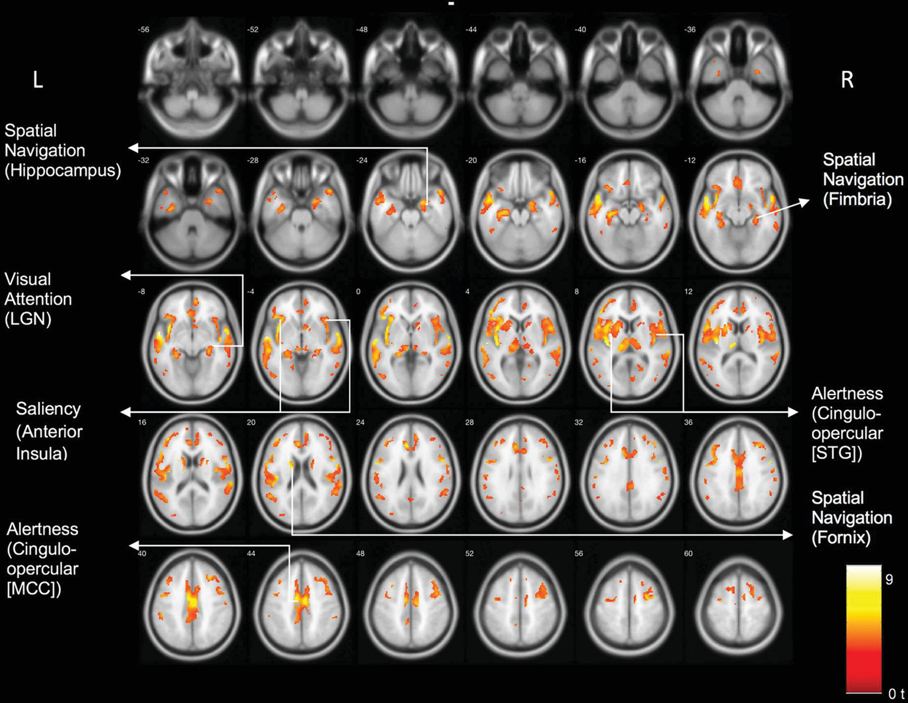
Results of the voxel-based comparison between the DLB and control group are shown in Figure 1. There were significant cholinergic binding reductions in the DLB compared with the control group in the following areas: the bilateral opercula, anterior to mid-cingulate cortices, bilateral insula, right more than left lateral geniculate nuclei, pulvinar, right proximal optic radiation, bilateral anterior and superior thalami, and bilateral posterior hippocampal fimbria and fornices. Table 3 lists the main clusters and their peak MNI coordinates and associated network hubs. We repeated this analysis with years of education included as a covariate. This analysis yielded similar results (see Figure S1 in the online supplement).
FIGURE 1. Clusters of reduced cholinergic binding in dementia of Lewy bodiesa
a The image shows a statistical parametric voxel-based analysis of VAChT binding compared with normal controls projected on a mean normalized structural image (Montreal Neurological Institute) in the axial plane. Results show reductions in key areas associated with visual attention (visual thalamus), salience (insula), spatial navigation (fimbria/fornix), and alertness (cingulo-opercular) networks. LGN=lateral geniculate nucleus, MCC=mid-cingulate cortex, STG=superior temporal gyrus, VAChT=vesicular acetylcholine transporter.
TABLE 3. Significant clusters with reduced cholinergic binding in patients with dementia with Lewy bodies (DLB) compared with normal elderly patientsa
Clusterb (voxels)pcPeak MNI coordinates (x, y, z)ztPeak voxel locationdPredominant neural network hube
2,172<0.001–28, –18, 184.846.18Left hippocampus peak with cluster extending into LGNSpatial navigation and visual attention networks
6,116<0.001–58, –6, –46.249.482Left BA 22 peak with cluster extending into insulaCingulo-opercular and saliency networks
7,726<0.00152, 4, 345.507.566Right BA 6 peak with cluster extending into insulaCingulo-opercular and saliency networks
274<0.001–4, 32, –83.904.642Left BA 32 (anterior cingulum)Cingulo-opercular network
213<0.00119, 2, 183.914.684Right caudate with cluster extending into fornixSpatial navigation network
a
BA=Brodmann’s area, LGN=lateral geniculate nucleus, MNI=Montreal Neurological Institute.
b
Significant cluster extending to key areas.
c
Corrected peak-wise false discovery rate.
d
Brain region with peak t values and the extending key regions for the cluster.
e
Key large-scale neural network hubs that overlap with the significant clusters.

Exploratory Post Hoc Cognitive Correlation Analysis

We performed an exploratory voxel-based regression analysis of the PET data and the MMSE as an outcome parameter within the group of the DLB participants. Results are shown in Figure S2 in the online supplement. Clusters (uncorrected p<0.05) were seen in the following regions: bilateral lateral geniculate nucleus, bilateral lingual gyri/optic radiations, left medial occipital, left superior posterior parietal, right superior parietal, right posterior frontal, right superior temporal cortices, and the right hippocampus. No clusters meeting this criterion were found in a similar voxel-based PET-MMSE regression analysis in the normal control group.

Discussion

Voxel-based whole brain analysis of VAChT PET identified a distinct topographic cholinergic denervation pattern rather than diffuse cerebral losses in DLB compared with the elderly control group. Regions showing cholinergic denervation overlapped with key hubs of neural networks responsible for visual attention (visual thalamus) (18), salience (insula) (19), spatial navigation (fimbria/fornix) (20), and maintenance of alertness (cingulo-opercular) networks (21). Specifically, we found cholinergic changes in the following hubs of each network: maintenance of alertness (cingulo-opercular) network: insula, operculum, cingulum, thalamus; salience network: anterior insula, anterior cingulum; visual attention network: visual thalamus, including the lateral geniculate nucleus; spatial navigation network: fornix, fimbria, hippocampus, and anterior thalamus.
Graph theory analysis of functional connectivity resting state MRI identified two distinct top-down task-control networks (22, 23). The dorsolateral prefrontal cortex and intraparietal sulcus are hubs within the frontoparietal task control (FPTC) network. The FPTC network is important for start-cue and error-related activity and may initiate and adapt control on a more rapid and trial-by-trial basis. The cingulo-opercular task control (COTC) network encompasses medial superior frontal cortex, dorsal anterior cingulate, frontal operculum, anterior insula, and thalamic regions (22). Effectively, the COTC graph may include parts of both the large-scale distributed cingulo-opercular network and the salience networks (23). The topography of cholinergic reductions in our patients with DLB suggests preferential cholinergic involvement of the COTC. The COTC network controls the goal-directed behavior more on a set maintenance mode using a longer duration time scale, such as needed for maintaining alertness. Clinically, this may be reflected in a lack of maintenance of a functional operative state and difficulties staying on task in persons with DLB in their daily life functions, resulting in cognitive fluctuations. Our findings are also compatible with the emerging notion of an important role of the cingulo-opercular network in task-switching from a passive task to an active task (24).
Another interesting observation was the asymmetry of visual thalamic region findings, such as right more than left lateral geniculate nuclei, pulvinar, and right proximal optic radiation changes. These findings may reflect associations with the ventral attention network, which has a right-hemispheric dominance (25). The ventral attention network is involved in the detection of bottom-up salient and behaviorally relevant stimuli in the environment, especially when the stimuli are initially unattended. This network is highly integrated with the visual system and includes the ventral part of the supramarginal gyrus, ventral frontal cortex, posterior part of the superior temporal sulcus and gyrus, inferior frontal gyrus, middle frontal gyrus, frontal operculum, and anterior insula, which acts as a “network breaker” that interrupts attention in the dorsal system and reorients attention toward a stimulus-driven object (26). These observations agree with our previous findings that lower cholinergic activity in the thalamus was associated with decreased saliency processing in patients with PD (27). This is likely mediated by the right lateral geniculate nucleus, where decreased cholinergic innervation may be associated with compromised visual attention, perceptual abnormalities, and possibly hallucinations in patients with DLB. We also recently reported on an association between reduced cholinergic integrity of the right lateral geniculate nucleus and falls in patients with PD (28). A similar association may exist in patients with DLB; however, this will require further confirmation.
The hippocampus plays an important role in spatial navigation (20), which is a key function for safely walking. Our findings show evidence of reduced cholinergic transporter binding in the hippocampal fimbria and fornices. Cholinergic deficits in these regions may compromise the integration of cognitive and sensorimotor functions while walking. Clinically, these regional deficits may hypothetically be associated with wandering behavior in the patients with DLB.
Exploratory findings of a voxel-based whole brain regression analysis of the cholinergic PET data, using the MMSE as the outcome parameter, showed several clusters that overlapped with several of the topographic cholinergic denervation regions. A notable finding was involvement of the visual system, especially the bilateral lateral geniculate nuclei and adjacent visual lobe regions. This may possibly be associated with visual processing dependent cognitive and/or neurobehavioral changes in DLB. Other regions showed overlap with the COTC and spatial navigation networks. No clusters meeting this criterion were found for a similar analysis in the normal control group, suggesting disease-specific cognitive correlates in the DLB group. These preliminary findings need to be confirmed in a larger sample size.
Identification of neural network changes underlying the clinical symptom manifestations in patients with DLB would augur network neurostimulation approaches as a complementary treatment strategy to existing pharmacological and behavioral interventions. Furthermore, our study has identified hubs of key neural networks that may contribute to the clinical symptom manifestation of DLB. Future studies comparing in vivo imaging findings directly with postmortem data may be very informative to advance our understanding of the neurodegenerative mechanisms underlying DLB. For example, findings of cholinergic changes in the lateral geniculate nucleus in DLB are very novel, but at present there is a critical gap of knowledge to explain the cholinergic vulnerability of the lateral geniculate nucleus in DLB. Another topic of future in vivo-ex vivo correlation research would be to assess for pathological changes in hubs corresponding to regional network vulnerability in postmortem data.
There are several limitations of this study. One limitation is the small sample size. Further studies using a larger sample sizes are needed to validate these topographic VAChT findings and associate them with clinical symptoms. The second limitation is the cross-sectional design, which lacks longitudinal information about the cholinergic binding areas and how cholinergic vesicular transporters may change in relationship to the temporal manifestation of specific clinical symptoms. Third, we do not have detailed neuropsychological testing data to validate the clinical diagnosis of probable DLB. Lastly, our neural network interpretation of the topography of neuroimaging data in the absence of neuropsychological metrics, functional MRI, or electrophysiological measures remains speculative.

Conclusions

Cholinergic changes are not diffuse in DLB but have a topographical distinct pattern of vulnerability that overlaps with hubs of important large-scale functional networks. Therefore, cholinergic network changes may explain some of the disease-defining features of DLB.

Supplementary Material

File (appi.neuropsych.19070165.ds001.pdf)

References

1.
Nejad-Davarani S, Koeppe RA, Albin RL, et al: Quantification of brain cholinergic denervation in dementia with Lewy bodies using PET imaging with [18F]-FEOBV. Mol Psychiatry 2019; 24:322–327
2.
Mesulam MM: Cholinergic circuitry of the human nucleus basalis and its fate in Alzheimer’s disease. J Comp Neurol 2013; 521:4124–4144
3.
Arciniegas DB: Cholinergic dysfunction and cognitive impairment after traumatic brain injury. Part 1: the structure and function of cerebral cholinergic systems. J Head Trauma Rehabil 2011; 26:98–101
4.
Selden NR, Gitelman DR, Salamon-Murayama N, et al: Trajectories of cholinergic pathways within the cerebral hemispheres of the human brain. Brain 1998; 121:2249–2257
5.
Pinto T, Lanctôt KL, Herrmann N: Revisiting the cholinergic hypothesis of behavioral and psychological symptoms in dementia of the Alzheimer’s type. Ageing Res Rev 2011; 10:404–412
6.
Perry EK, Curtis M, Dick DJ, et al: Cholinergic correlates of cognitive impairment in Parkinson’s disease: comparisons with Alzheimer’s disease. J Neurol Neurosurg Psychiatry 1985; 48:413–421
7.
Bohnen NI, Kaufer DI, Hendrickson R, et al: Cognitive correlates of cortical cholinergic denervation in Parkinson’s disease and parkinsonian dementia. J Neurol 2006; 253:242–247
8.
Hilker R, Thomas AV, Klein JC, et al: Dementia in Parkinson disease: functional imaging of cholinergic and dopaminergic pathways. Neurology 2005; 65:1716–1722
9.
Bohnen NI, Kaufer DI, Ivanco LS, et al: Cortical cholinergic function is more severely affected in parkinsonian dementia than in Alzheimer disease: an in vivo positron emission tomographic study. Arch Neurol 2003; 60:1745–1748
10.
McKeith IG, Boeve BF, Dickson DW, et al: Diagnosis and management of dementia with Lewy bodies: fourth consensus report of the DLB Consortium. Neurology 2017; 89:88–100
11.
McKeith IG, Wesnes KA, Perry E, et al: Hallucinations predict attentional improvements with rivastigmine in dementia with lewy bodies. Dement Geriatr Cogn Disord 2004; 18:94–100
12.
Bohnen NI, Müller MLTM, Frey KA: Molecular imaging and updated diagnostic criteria in Lewy body dementias. Curr Neurol Neurosci Rep 2017; 17:73
13.
Ballard C, Ziabreva I, Perry R, et al: Differences in neuropathologic characteristics across the Lewy body dementia spectrum. Neurology 2006; 67:1931–1934
14.
Bohnen NI, Müller ML, Kotagal V, et al: Heterogeneity of cholinergic denervation in Parkinson’s disease without dementia. J Cereb Blood Flow Metab 2012; 32:1609–1617
15.
Shao X, Hoareau R, Hockley BG, et al: Highlighting the versatility of the Tracerlab synthesis modules: Part 1: fully automated production of [F]labelled radiopharmaceuticals using a Tracerlab FX(FN). J Labelled Comp Radiopharm 2011; 54:292–307
16.
Müller-Gärtner HW, Links JM, Prince JL, et al: Measurement of radiotracer concentration in brain gray matter using positron emission tomography: MRI-based correction for partial volume effects. J Cereb Blood Flow Metab 1992; 12:571–583
17.
Folstein MF, Folstein SE, McHugh PR: “Mini-Mental State”: A practical method for grading the cognitive state of patients for the clinician. J Psychiatr Res 1975; 12:189–198
18.
Saalmann YB, Kastner S: Cognitive and perceptual functions of the visual thalamus. Neuron 2011; 71:209–223
19.
Menon V, Uddin LQ: Saliency, switching, attention and control: a network model of insula function. Brain Struct Funct 2010; 214:655–667
20.
Sutherland RJ, Rodriguez AJ: The role of the fornix/fimbria and some related subcortical structures in place learning and memory. Behav Brain Res 1989; 32:265–277
21.
Coste CP, Kleinschmidt A: Cingulo-opercular network activity maintains alertness. Neuroimage 2016; 128:264–272
22.
Dosenbach NU, Fair DA, Cohen AL, et al: A dual-networks architecture of top-down control. Trends Cogn Sci 2008; 12:99–105
23.
Power JD, Cohen AL, Nelson SM, et al: Functional network organization of the human brain. Neuron 2011; 72:665–678
24.
Sadaghiani S, D’Esposito M: Functional characterization of the cingulo-opercular network in the maintenance of tonic alertness. Cereb Cortex 2015; 25(9):2763–2773
25.
Shulman GL, Pope DL, Astafiev SV, et al: Right hemisphere dominance during spatial selective attention and target detection occurs outside the dorsal frontoparietal network. J Neurosci 2010; 30:3640–3651
26.
Corbetta M, Shulman GL: Control of goal-directed and stimulus-driven attention in the brain. Nat Rev Neurosci 2002; 3:201–215
27.
Kim K, Müller MLTM, Bohnen NI, et al: Thalamic cholinergic innervation makes a specific bottom-up contribution to signal detection: Evidence from Parkinson’s disease patients with defined cholinergic losses. Neuroimage 2017; 149:295–304
28.
Bohnen NI, Kanel P, Zhou Z, et al: Cholinergic system changes of falls and freezing of gait in Parkinson’s disease. Ann Neurol 2019; 85:538–549

Information & Authors

Information

Published In

Go to The Journal of Neuropsychiatry and Clinical Neurosciences
The Journal of Neuropsychiatry and Clinical Neurosciences
Pages: 370 - 375
PubMed: 32498602

History

Received: 22 July 2019
Revision received: 13 December 2019
Accepted: 20 March 2020
Published online: 5 June 2020
Published in print: Fall 2020

Keywords

  1. Dementia
  2. Neurology
  3. Cognition
  4. Dementia With Lewy Bodies
  5. Saliency

Authors

Details

Prabesh Kanel, Ph.D. [email protected]
Department of Radiology (Kanel, Müller, Sanchez-Catasus, Koeppe, Frey, Bohnen) and Department of Neurology (Frey, Bohnen), University of Michigan, Ann Arbor; Neurology Service and Geriatric Research Education and Clinical Center, Veterans Administration Ann Arbor Healthcare System, Ann Arbor, Mich. (Bohnen); Morris K. Udall Center of Excellence for Parkinson’s Disease Research, University of Michigan, Ann Arbor (Kanel, Müller, Sanchez-Catasus, Bohnen); and Department of Neurology, University of Groningen, University Medical Center Groningen, the Netherlands (van der Zee).
Martijn L.T.M. Müller, Ph.D.
Department of Radiology (Kanel, Müller, Sanchez-Catasus, Koeppe, Frey, Bohnen) and Department of Neurology (Frey, Bohnen), University of Michigan, Ann Arbor; Neurology Service and Geriatric Research Education and Clinical Center, Veterans Administration Ann Arbor Healthcare System, Ann Arbor, Mich. (Bohnen); Morris K. Udall Center of Excellence for Parkinson’s Disease Research, University of Michigan, Ann Arbor (Kanel, Müller, Sanchez-Catasus, Bohnen); and Department of Neurology, University of Groningen, University Medical Center Groningen, the Netherlands (van der Zee).
Sygrid van der Zee, M.S.
Department of Radiology (Kanel, Müller, Sanchez-Catasus, Koeppe, Frey, Bohnen) and Department of Neurology (Frey, Bohnen), University of Michigan, Ann Arbor; Neurology Service and Geriatric Research Education and Clinical Center, Veterans Administration Ann Arbor Healthcare System, Ann Arbor, Mich. (Bohnen); Morris K. Udall Center of Excellence for Parkinson’s Disease Research, University of Michigan, Ann Arbor (Kanel, Müller, Sanchez-Catasus, Bohnen); and Department of Neurology, University of Groningen, University Medical Center Groningen, the Netherlands (van der Zee).
Carlos A. Sanchez-Catasus, Ph.D.
Department of Radiology (Kanel, Müller, Sanchez-Catasus, Koeppe, Frey, Bohnen) and Department of Neurology (Frey, Bohnen), University of Michigan, Ann Arbor; Neurology Service and Geriatric Research Education and Clinical Center, Veterans Administration Ann Arbor Healthcare System, Ann Arbor, Mich. (Bohnen); Morris K. Udall Center of Excellence for Parkinson’s Disease Research, University of Michigan, Ann Arbor (Kanel, Müller, Sanchez-Catasus, Bohnen); and Department of Neurology, University of Groningen, University Medical Center Groningen, the Netherlands (van der Zee).
Robert A. Koeppe, Ph.D.
Department of Radiology (Kanel, Müller, Sanchez-Catasus, Koeppe, Frey, Bohnen) and Department of Neurology (Frey, Bohnen), University of Michigan, Ann Arbor; Neurology Service and Geriatric Research Education and Clinical Center, Veterans Administration Ann Arbor Healthcare System, Ann Arbor, Mich. (Bohnen); Morris K. Udall Center of Excellence for Parkinson’s Disease Research, University of Michigan, Ann Arbor (Kanel, Müller, Sanchez-Catasus, Bohnen); and Department of Neurology, University of Groningen, University Medical Center Groningen, the Netherlands (van der Zee).
Kirk A. Frey, M.D., Ph.D.
Department of Radiology (Kanel, Müller, Sanchez-Catasus, Koeppe, Frey, Bohnen) and Department of Neurology (Frey, Bohnen), University of Michigan, Ann Arbor; Neurology Service and Geriatric Research Education and Clinical Center, Veterans Administration Ann Arbor Healthcare System, Ann Arbor, Mich. (Bohnen); Morris K. Udall Center of Excellence for Parkinson’s Disease Research, University of Michigan, Ann Arbor (Kanel, Müller, Sanchez-Catasus, Bohnen); and Department of Neurology, University of Groningen, University Medical Center Groningen, the Netherlands (van der Zee).
Nicolaas I. Bohnen, M.D., Ph.D.
Department of Radiology (Kanel, Müller, Sanchez-Catasus, Koeppe, Frey, Bohnen) and Department of Neurology (Frey, Bohnen), University of Michigan, Ann Arbor; Neurology Service and Geriatric Research Education and Clinical Center, Veterans Administration Ann Arbor Healthcare System, Ann Arbor, Mich. (Bohnen); Morris K. Udall Center of Excellence for Parkinson’s Disease Research, University of Michigan, Ann Arbor (Kanel, Müller, Sanchez-Catasus, Bohnen); and Department of Neurology, University of Groningen, University Medical Center Groningen, the Netherlands (van der Zee).

Notes

Send correspondence to Dr. Kanel ([email protected]).

Competing Interests

Dr. Müller has received research support from the Department of Veterans Affairs, the Michael J. Fox Foundation, and NIH. Dr. Koeppe has received grant support from NIH. Dr. Frey has received research support from AVID Radiopharmaceuticals (Eli Lilly subsidiary), GE Healthcare, and NIH; he has served as a consultant to AVID Radiopharmaceuticals, Bayer-Schering, GE Healthcare, and MIMVista; and he is a shareholder with Bristol-Myers, GE Healthcare, Merck, and Novo-Nordisk. Dr. Bohnen has received research support from the Department of Veterans Affairs, EIP Pharma, Eisai, the Michael J. Fox Foundation, and NIH. The other authors report no financial relationships with commercial interests.

Funding Information

Supported by NIH (grants P01 NS015655, RO1 NS070856, and P50 NS091856).

Metrics & Citations

Metrics

Citations

Export Citations

If you have the appropriate software installed, you can download article citation data to the citation manager of your choice. Simply select your manager software from the list below and click Download.

For more information or tips please see 'Downloading to a citation manager' in the Help menu.

Format
Citation style
Style
Copy to clipboard

View Options

View options

PDF/EPUB

View PDF/EPUB

Get Access

Login options

Already a subscriber? Access your subscription through your login credentials or your institution for full access to this article.

Personal login Institutional Login Open Athens login
Purchase Options

Purchase this article to access the full text.

PPV Articles - Journal of Neuropsychiatry and Clinical Neurosciences

PPV Articles - Journal of Neuropsychiatry and Clinical Neurosciences

Not a subscriber?

Subscribe Now / Learn More

PsychiatryOnline subscription options offer access to the DSM-5-TR® library, books, journals, CME, and patient resources. This all-in-one virtual library provides psychiatrists and mental health professionals with key resources for diagnosis, treatment, research, and professional development.

Need more help? PsychiatryOnline Customer Service may be reached by emailing [email protected] or by calling 800-368-5777 (in the U.S.) or 703-907-7322 (outside the U.S.).

Media

Figures

Other

Tables

Share

Share

Share article link

Share