Full access
Windows to the Brain
Published Online: 1 January 2012

Neuroanatomy of Dopamine: Reward and Addiction

Publication: The Journal of Neuropsychiatry and Clinical Neurosciences

Abstract

Abnormalities of the physiological reward system are believed to play a major part in many serious psychiatric disorders, such as substance abuse, pathological gambling, and major depression. Motivation that involves liking, wanting, craving, and learning about pleasurable events and behavior is heavily influenced by the circuitry of the dopamine reward system, which plays a major role in both normal reward behavior and pathological behavior and addiction. In this Windows to the Brain article, the authors elucidate the mechanisms of this system.
Abnormalities in the reward system are believed to play a role in many psychiatric disorders (for example, substance abuse, pathological gambling, major depression, schizophrenia, attention-deficit hyperactivity disorder, Parkinson's disease, Alzheimer's disease), so understanding the functional neuroanatomy of reward is important in neuropsychiatry.3,5 Reward is not a unitary concept. Major aspects include liking (e.g., pleasure, hedonia), wanting (e.g., motivation for reward, incentive salience), and learning (e.g., past experiences predicting future rewards).6 Primary (fundamental) rewards are naturally-occurring things or events that are essential for species survival and reproduction (e.g., food, sex). Secondary (higher-order) rewards are more abstract cognitive representations (e.g., monetary, artistic, altruistic, transcendent).3,6 This review will focus on the contributions of the dopamine (DA) system to reward. Many other neurotransmitter systems also participate in aspects of reward.79

Reward

Research on brain areas important for reward began with the observation by Olds and Milner in the 1950s that rats will expend great effort in order to obtain electrical stimulation of multiple brain areas, including small regions within the brainstem, diencephalon, and cortex.3,4,1013 This work was foreshadowed by earlier studies in patients with schizophrenia that focused on the septal area, in which positive immediate responses (e.g., euphoria) were reported to occur after brain stimulation.14
The medial forebrain bundle in the lateral hypothalamus was a common target for electrode placement in animal studies, as stimulation in this area evoked very robust behaviors (e.g., self-stimulation to the point of physical exhaustion, willingness run across an aversive shock grid to obtain stimulation).4,1113 Several lines of evidence suggested that rewarding electrical stimulation activated the dopamine (DA) projection from the ventral tegmental area (via the medial forebrain bundle) to nucleus accumbens, one part of what is now termed the ventral striatum.10,15 The ability of DA antagonists to decrease the effectiveness of rewarding electrical stimulation was particularly important. Subsequent studies indicated that the rewarding effect was not due to activation of the small, unmyelinated ascending DA fibers, but, rather, to large, myelinated fibers descending to brainstem.10,11,13
Animal studies indicate that brainstem DA neurons have a baseline level of activity (tonic mode, steady activation) that enables normal downstream functioning and is modulated by both positive and negative reward-related events (phasic mode, fast activation).12,1620 DA neurons increase activity in response to unexpected rewards and to stimuli that predict receipt of a reward (expectation or anticipation). During conditioned-learning, the increased activity in DA neurons shifts from the time of reward-receipt to the time of the reward-predicting stimulus. Activity is only increased by rewards if they are greater than predicted (positive prediction error). DA neurons also decrease activity when reward-expectation is not met (negative prediction error). If receipt of a reward is delayed, activity in these neurons decreases at the time the reward was expected, but did not occur, and increases when the reward is actually received. Much of behavior is guided by prediction of the future, based on past experiences. Phasic changes in DA activity, by signaling that something unexpected relating to reward has occurred, help to optimize goal-directed behavior. DA neurons also are sometimes responsive to other types of stimuli (e.g., stressful, aversive, alerting), perhaps because of their motivational salience. Functional MRI (fMRI) studies in humans have confirmed increased activation in the area of brainstem containing DA neurons during anticipation of both primary and secondary rewards.4
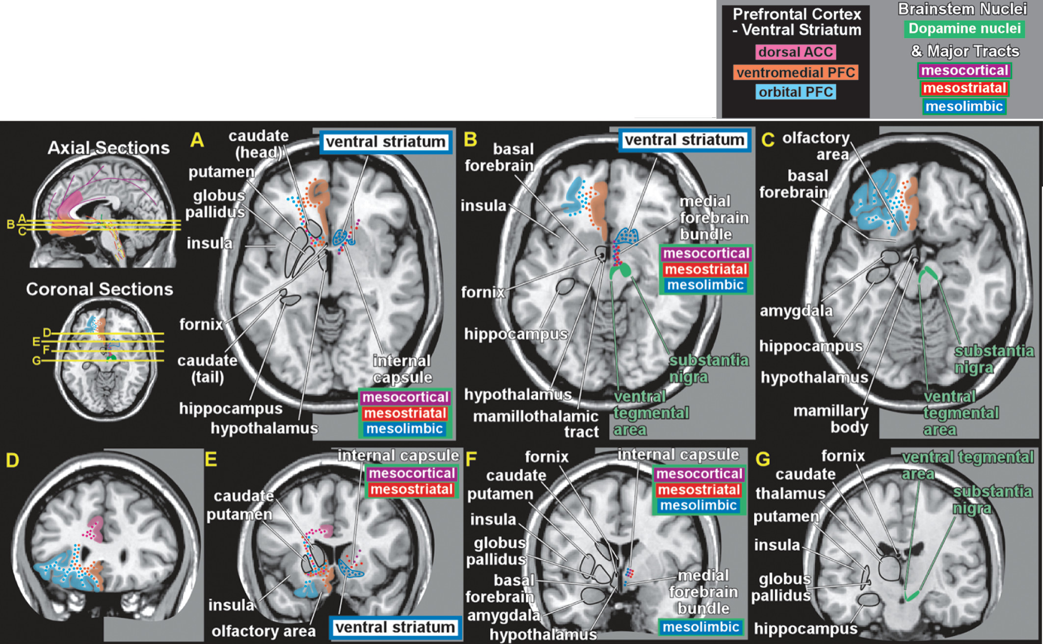
Early studies suggested three distinct ascending DA projection systems from the brainstem (Figure 1). Originally, it was thought that the limbic and cortical projections arose from the ventral tegmental area, with the substantia nigra giving rise to the projection to sensorimotor striatum (caudate and putamen); hence, the nigrostriatal name for this tract. It is now clear that, although the pathways are anatomically and functionally distinct, their cells of origin are intermixed.4,21,22
COVER and FIGURE 1. Approximate portions of the prefrontal cortex (PFC), important for reward, are color-coded (dorsal anterior cingulate cortex [ACC]: pink; ventral PFC: orange; orbital PFC: blue) on the left side of axial (A–C) and coronal (D–G) MRIs. Approximate extent and locations of major midbrain dopamine nuclei important for reward (dark green) and the major dopaminergic tracts (mesocortical: purple; mesostriatal: red; mesolimbic: dark blue) are color-coded on the right side of axial (A–C) and coronal (D–G) MRIs.
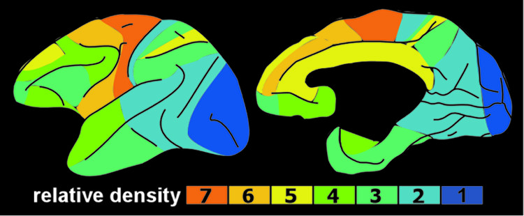
Two of these DA pathways are particularly important for the reward system.3,4 The mesocortical DA pathway projects to multiple cortical areas and is important for many aspects of reward-processing, including hedonic evaluation, comparative valuation, and option-assessment. This pathway projects primarily to prefrontal, cingulate, and entorhinal cortices in rodents, but to the entire cortical mantle in primates (Figure 2).1,2,21,22 The mesolimbic DA pathway projects primarily to the ventral striatum, but, also, to other limbic areas (e.g., amygdala, olfactory tubercle, septum).21 This pathway is important for the positive reinforcing effects of both natural rewards and drugs of abuse.4 Ventral striatum also receives strong projections from orbitofrontal, ventral medial prefrontal, and anterior cingulate cortices, as well as limbic-related subcortical areas (Figure 3).4
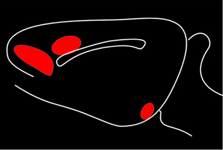
FIGURE 2. The cortical projections of brainstem dopamine (DA) neurons are much more extensive in humans (and other primates) than in rodents. LEFT: DA transporter mapping in human brain (colored by relative density) indicates innervation of the entire cortical mantle.1 RIGHT: DA terminal mapping in rat brain (red) indicates that innervation is largely confined to areas of the frontal, cingulate, and entorhinal cortices.2
FIGURE 3. Simplified Summary of the Reward Circuitry, Color-Coded to Match Figure 1.3,4
Functional imaging in humans has shown that rewards increase DA release in ventral striatum and that increasing striatal DA (by amphetamine administration) enhances rewards.4,23 Activation in ventral striatum is more strongly associated with the anticipation of reward than the actual receipt, and activation level correlates with the magnitude of the expected reward and with the effort expended to gain the reward.4,20,23 Some studies have reported decreased activation in ventral striatum when an expected reward is not received (reward prediction error).4,20 The ventral striatum contains multiple functional areas, and it is quite possible that different aspects of reward are associated with specific subregions.
Although a valuable approach, most of the studies utilizing electrical stimulation were not designed to address which aspects of reward (liking, wanting, and learning) were involved.24 The development of methods that allowed intracranial drug self-administration made it possible to more clearly identify regions participating in reward and to determine the nature of the influence (studies done primarily in rodents).6,11 Areas of the brain that endow a sensation (e.g., sweetness) with hedonic value (pleasure or liking) are generally identified by their ability to enhance liking of sensory rewards when stimulated.6 Areas presently believed to contain “hedonic hotspots” include both ventral striatum and ventral pallidum, brainstem (e.g., ventral tegmental area, parabrachial nucleus), and frontal cortex (e.g., orbitofrontal, cingulate, medial prefrontal and insular cortices). Some of these same areas are also important for endowing a sensation with motivational value (incentive-salience or wanting). The nature of the stimulation is important. Thus, there are areas within ventral striatum that evoke both liking and wanting when activated by opioids, but only wanting when activated by DA.25 Recent studies suggest that DA is very important for the motivational value of rewards.24

Addiction

The economic costs (direct and indirect) of drug abuse are immense, estimated at $180.9 billion for the United States in 2002 alone.26 Addiction to various substances is found across cultures worldwide, and animals will voluntarily self-administer drugs-of-addiction in laboratory settings.12,13 Within the last decade, it has been recognized that behavioral addictions (e.g., gambling, pathological internet use, food) share the same core features as substance addictions. These include craving, tolerance, withdrawal, and compulsive use, despite occupational, interpersonal, and financial adversity. Although pathological levels of motivation (incentive-salience theory), learned compulsive behaviors (learning theory, habit theory), and avoidance of the negative aspects of withdrawal (negative reinforcement theory, opponent process theory) are likely all involved, the role each plays in the development and maintenance of addictions is a matter of much debate.12,23,27
A three-stage cycle (binge/intoxication, withdrawal/negative emotional state, preoccupation/anticipation) has been proposed, in which a shift from impulsive to compulsive behaviors occurs as addiction develops.12,13 From a neurobiological standpoint, addiction is a disorder of brain reward mechanisms that are crucial for survival.9,12,13,23,2830 Although the reinforcing value of drugs and the development of addiction involve multiple areas and neurotransmitter systems that differ by drug-of-abuse, the DA system is of central importance to all. The mesolimbic DA system is activated by all major drugs of abuse, with the ventral striatum a key structure. In animal studies, the brain stimulation required for reward (reward threshold) is reduced by acute administration of drugs of abuse. Activation in the ventral striatum is thought to be important in the reward-driven binge/intoxication stage, with engagement of dorsal striatum for the habit-formation that is believed to underlie progression to compulsive use. It has been proposed that drugs-of-abuse induce larger and more prolonged activations than natural stimuli, promoting habit-formation that is quite robust and resistant to change. Intrinsically below-normal functioning in the DA system has been proposed as a risk factor for development of addiction (reward-deficiency hypothesis). Reward thresholds increase (sensitivity to rewards decrease) during protracted withdrawal after chronic drug administration, suggesting compromise of the DA system. The anhedonia and motivational deficits present during the withdrawal/negative emotional state stage may be due to decreased DA function (reward-deficiency). It has been proposed that continued drug use at this stage is more to restore a normal DA level to (“get straight”) than to evoke a large DA increase to (“get high”). Alterations in the brain's stress systems also occur, and may be important for aversive stimulus effects and/or heightened anxiety. Altered activity in areas of prefrontal cortex and perturbations in their modulation of limbic-related subcortical areas (particularly ventral striatum, amygdala, and hippocampus) are present in the preoccupation/anticipation stage and may give rise to the deficits in executive functions (e.g., self-control, salience-attribution), and memory commonly present in addiction.
CONCLUSION

Although there is much yet be understood regarding the neurobiology of reward and its circuitry, it is certainly clear that abnormalities in these pathways can have profound effects on human behavior and on some psychiatric illnesses. As scientists are more able to map these pathways and understand the relationships with other neurotransmitters, it is anticipated that improved clinical interventions will be developed to lessen the long-term course of addiction.

References

1.
Lewis DA, Melchitzky DS, Sesack SR, et al.: Dopamine transporter immunoreactivity in monkey cerebral cortex: regional, laminar, and ultrastructural localization. J Comp Neurol 2001; 432:119–136
2.
Lindvall O, Bjorklund A, Moore RY, et al.: Mesencephalic dopamine neurons projecting to neocortex. Brain Res 1974; 81:325–331
3.
Chau DT, Roth RM, Green AI: The neural circuitry of reward and its relevance to psychiatric disorders. Curr Psychiatry Rep 2004; 6:391–399
4.
Haber SN, Knutson B: The reward circuit: linking primate anatomy and human imaging. Neuropsychopharmacology 2010; 35:4–26
5.
Der-Avakian A, Markou A: The neurobiology of anhedonia and other reward-related deficits. Trends Neurosci 2012; 35:68–77
6.
Kringelbach ML, Berridge KC: Toward a functional neuroanatomy of pleasure and happiness. Trends Cogn Sci 2009; 13:479–487
7.
Le Merrer J, Becker J, Befort K, et al.: Reward-processing by the opioid system in the brain. Physiol Rev 2009; 89:1379–1412
8.
Kranz GS, Kasper S, Lanzenberger R: Reward and the serotonergic system. Neuroscience 2010; 166:1023–1035
9.
Lajtha A, Sershen H: Heterogeneity of reward mechanisms. Neurochem Res 2010; 35:851–867
10.
Wise RA: Forebrain substrates of reward and motivation. J Comp Neurol 2005; 493:115–121
11.
Ikemoto S: Brain reward circuitry beyond the mesolimbic dopamine system: a neurobiological theory. Neurosci Biobehav Rev 2010; 35:129–150
12.
Koob GF, Volkow ND: Neurocircuitry of addiction. Neuropsychopharmacology 2010; 35:217–238
13.
Gardner EL: Addiction and brain reward and anti-reward pathways. Adv Psychosom Med 2011; 30:22–60
14.
Baumeister AA: The Tulane electrical brain stimulation program: a historical case study in medical ethics. J Hist Neurosci 2000; 9:262–278
15.
Heimer L, Van Hoesen GW: The limbic lobe and its output channels: implications for emotional functions and adaptive behavior. Neurosci Biobehav Rev 2006; 30:126–147
16.
Schultz W: Multiple dopamine functions at different time courses. Annu Rev Neurosci 2007; 30:259–288
17.
Schultz W: Multiple functions of dopamine neurons. F1000 Bio Rep 2010; 2:2
18.
Schultz W: Dopamine signals for reward value and risk: basic and recent data. Behav Brain Funct 2010; 6:24
19.
Bromberg-Martin ES, Matsumoto M, Hikosaka O: Dopamine in motivational control: rewarding, aversive, and alerting. Neuron 2010; 68:815–834
20.
Glimcher PW: Understanding dopamine and reinforcement learning: the dopamine reward prediction-error hypothesis. Proc Natl Acad Sci U S A 2011; 108:15647–15654
21.
Bjorklund A, Dunnett SB: Dopamine neuron systems in the brain: an update. Trends Neurosci 2007; 30:194–202
22.
Arias-Carrióon O, Stamelou M, Murillo-Rodriguez E, et al.: Dopaminergic reward system: a short integrative review. Int Arch Med 2010; 3:24
23.
Hommer DW, Bjork JM, Gillman JM: Imaging brain response to reward in addictive disorders. Ann N Y Acad Sci 2011; 1216:50–61
24.
Berridge K, Kringelbach ML: Building a neuroscience of pleasure and well-being. Psychol Well-Being: Theory Res Prac 2011; 1:3
25.
Smith KS, Berridge KC, Aldridge JW: Disentangling pleasure from incentive salience and learning signals in brain reward circuitry. PNAS 2011; 108:E255–E264
26.
Office of National Drug-Control Policy: The Economic Costs of Drug Abuse in the United States, 1992–2002; www.ncjrs.gov/ondcppubs/publications/pdf/economic_costs
27.
Stacy AW, Wiers RW: Implicit cognition and addiction: a tool for explaining paradoxical behavior. Annu Rev Clin Psychol 2010; 6:551–575
28.
Lingford-Hughes A, Watson B, Kalk N, et al.: Neuropharmacology of addiction and how it informs treatment. Br Med Bull 2010; 96:93–110
29.
Goldstein RZ, Volkow ND: Dysfunction of the prefrontal cortex in addiction: neuroimaging findings and clinical implications. Nat Rev Neurosci 2011; 12:652–669
30.
Schultz W: Potential vulnerabilities of neuronal reward, risk, and decision mechanisms to addictive drugs. Neuron 2011; 69:603–617

Information & Authors

Information

Published In

Go to The Journal of Neuropsychiatry and Clinical Neurosciences
The Journal of Neuropsychiatry and Clinical Neurosciences
Pages: 1 - 4
PubMed: 22450608

History

Published online: 1 January 2012
Published in print: Winter 2012
Received: 27 January 2012

Authors

Details

Katherine H. Taber, Ph.D.
Drs. Taber and Hurley are affiliated with the Veterans Affairs Mid-Atlantic Mental Illness Research, Education, and Clinical Center, and the Research and Education Service Line at the W.G. Hefner Veterans Affairs Medical Center in Salisbury, NC; Dr. Taber is affiliated with the Division of Biomedical Sciences at the Edward Via College of Osteopathic Medicine in Blacksburg, VA, and the Department of Physical Medicine and Rehabilitation at Baylor College of Medicine in Houston, TX; Dr. Black is affiliated with the Departments of Psychiatry and Neurology at the University of Vermont, College of Medicine, Burlington, VT; Dr. Porrino is Chair and Professor of the Department of Physiology and Pharmacology at Wake Forest University School of Medicine in Winston-Salem, NC; Dr. Hurley is affiliated with the Departments of Psychiatry and Radiology at Wake Forest University School of Medicine in Winston-Salem, NC, and the Menninger Department of Psychiatry and Behavioral Sciences at Baylor College of Medicine in Houston, TX. Address correspondence to Dr. Robin Hurley, Hefner VA Medical Center, Salisbury, NC.
Deborah N. Black, M.D.
Drs. Taber and Hurley are affiliated with the Veterans Affairs Mid-Atlantic Mental Illness Research, Education, and Clinical Center, and the Research and Education Service Line at the W.G. Hefner Veterans Affairs Medical Center in Salisbury, NC; Dr. Taber is affiliated with the Division of Biomedical Sciences at the Edward Via College of Osteopathic Medicine in Blacksburg, VA, and the Department of Physical Medicine and Rehabilitation at Baylor College of Medicine in Houston, TX; Dr. Black is affiliated with the Departments of Psychiatry and Neurology at the University of Vermont, College of Medicine, Burlington, VT; Dr. Porrino is Chair and Professor of the Department of Physiology and Pharmacology at Wake Forest University School of Medicine in Winston-Salem, NC; Dr. Hurley is affiliated with the Departments of Psychiatry and Radiology at Wake Forest University School of Medicine in Winston-Salem, NC, and the Menninger Department of Psychiatry and Behavioral Sciences at Baylor College of Medicine in Houston, TX. Address correspondence to Dr. Robin Hurley, Hefner VA Medical Center, Salisbury, NC.
Linda J. Porrino, Ph.D.
Drs. Taber and Hurley are affiliated with the Veterans Affairs Mid-Atlantic Mental Illness Research, Education, and Clinical Center, and the Research and Education Service Line at the W.G. Hefner Veterans Affairs Medical Center in Salisbury, NC; Dr. Taber is affiliated with the Division of Biomedical Sciences at the Edward Via College of Osteopathic Medicine in Blacksburg, VA, and the Department of Physical Medicine and Rehabilitation at Baylor College of Medicine in Houston, TX; Dr. Black is affiliated with the Departments of Psychiatry and Neurology at the University of Vermont, College of Medicine, Burlington, VT; Dr. Porrino is Chair and Professor of the Department of Physiology and Pharmacology at Wake Forest University School of Medicine in Winston-Salem, NC; Dr. Hurley is affiliated with the Departments of Psychiatry and Radiology at Wake Forest University School of Medicine in Winston-Salem, NC, and the Menninger Department of Psychiatry and Behavioral Sciences at Baylor College of Medicine in Houston, TX. Address correspondence to Dr. Robin Hurley, Hefner VA Medical Center, Salisbury, NC.
Robin A. Hurley, M.D.
Drs. Taber and Hurley are affiliated with the Veterans Affairs Mid-Atlantic Mental Illness Research, Education, and Clinical Center, and the Research and Education Service Line at the W.G. Hefner Veterans Affairs Medical Center in Salisbury, NC; Dr. Taber is affiliated with the Division of Biomedical Sciences at the Edward Via College of Osteopathic Medicine in Blacksburg, VA, and the Department of Physical Medicine and Rehabilitation at Baylor College of Medicine in Houston, TX; Dr. Black is affiliated with the Departments of Psychiatry and Neurology at the University of Vermont, College of Medicine, Burlington, VT; Dr. Porrino is Chair and Professor of the Department of Physiology and Pharmacology at Wake Forest University School of Medicine in Winston-Salem, NC; Dr. Hurley is affiliated with the Departments of Psychiatry and Radiology at Wake Forest University School of Medicine in Winston-Salem, NC, and the Menninger Department of Psychiatry and Behavioral Sciences at Baylor College of Medicine in Houston, TX. Address correspondence to Dr. Robin Hurley, Hefner VA Medical Center, Salisbury, NC.

Notes

Correspondence: [email protected] (e-mail).

Metrics & Citations

Metrics

Citations

Export Citations

If you have the appropriate software installed, you can download article citation data to the citation manager of your choice. Simply select your manager software from the list below and click Download.

For more information or tips please see 'Downloading to a citation manager' in the Help menu.

Format
Citation style
Style
Copy to clipboard

View Options

View options

PDF/EPUB

View PDF/EPUB

Get Access

Login options

Already a subscriber? Access your subscription through your login credentials or your institution for full access to this article.

Personal login Institutional Login Open Athens login
Purchase Options

Purchase this article to access the full text.

PPV Articles - Journal of Neuropsychiatry and Clinical Neurosciences

PPV Articles - Journal of Neuropsychiatry and Clinical Neurosciences

Not a subscriber?

Subscribe Now / Learn More

PsychiatryOnline subscription options offer access to the DSM-5-TR® library, books, journals, CME, and patient resources. This all-in-one virtual library provides psychiatrists and mental health professionals with key resources for diagnosis, treatment, research, and professional development.

Need more help? PsychiatryOnline Customer Service may be reached by emailing [email protected] or by calling 800-368-5777 (in the U.S.) or 703-907-7322 (outside the U.S.).

Media

Figures

Other

Tables

Share

Share

Share article link

Share